Digital Poster
Advanced Contrast Mechanisms: Diffusion & Electric Tissue Properties
ISMRM & ISMRT Annual Meeting & Exhibition • 03-08 June 2023 • Toronto, ON, Canada

Digital Poster
Advanced Contrast Mechanisms: Diffusion & Electric Tissue Properties
| Computer # | |||
|---|---|---|---|
5163.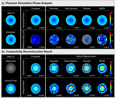 |
61 |
Implicit Regularization for Improving Phase-based EPT with
Stein’s Unbiased Risk Estimator
Chuanjiang Cui1,
Kyu-Jin Jung1,
Jun-Hyeong Kim1,
and Dong-Hyun Kim1
1Department of Electrical and Electronic Engineering, Yonsei University, Seoul, Korea, Republic of Keywords: Electromagnetic Tissue Properties, Electromagnetic Tissue Properties Phase-based EPT algorithm is extremely sensitive to noise. Although various denoising algorithms have been introduced to suppress noise amplification, residual artifact cause instability conductivity error or broadening boundary artifact. In this work, we propose a novel generative network trained with Stein’s unbiased risk estimator under the purely unsupervised learning framework, which improve the performance of phase-based conductivity reconstruction algorithms. In addition, the proposed method does not need any dataset for training neural network and not require any prior information for designing explicit regularization. |
|
5164.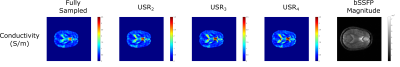 |
62 |
Investigating Variable Density Sampling Patterns in Spiral
Trajectories for use in MREPT
Safa Özdemir1,2,
Efe Ilicak1,2,
Lothar R. Schad1,2,
and Frank G. Zöllner1,2
1Computer Assisted Clinical Medicine, Medical Faculty Mannheim, Heidelberg University, Mannheim, Germany, 2Mannheim Institute for Intelligent Systems in Medicine, Medical Faculty Mannheim, Heidelberg University, Mannheim, Germany Keywords: Electromagnetic Tissue Properties, Electromagnetic Tissue Properties Magnetic Resonance Electrical Properties Tomography (MREPT) is used for obtaining conductivity by utilizing B1 phase images. In order to obtain B1 phase, different pulse sequences can be used. Among these, spiral trajectory based imaging has various advantages including high SNR efficiency and acquisition speed and was demonstrated in MREPT previously. In this work, to further improve spiral trajectory based imaging in MREPT, we investigate variable density sampling patterns. In vivo brain results indicate that conductivity images are quite robust with the choice of undersampling pattern and whole brain coverage can be completed under a minute with multiple averages. |
|
5165.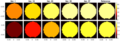 |
63 |
Simultaneous Estimation of Electrical Properties and Incident
Fields Using Global Maxwell Tomography with B1+ and MR Signal
Data
Jose E. C. Serralles1,
Ilias I. Giannakopoulos2,
Georgy D. Guryev1,
Luca Daniel1,
and Riccardo Lattanzi2
1Research Laboratory of Electronics (RLE), Department of Electrical Engineering and Computer Science, Massachusetts Institute of Technology, Cambridge, MA, United States, 2The Bernard and Irene Schwartz Center for Biomedical Imaging (CBI), Department of Radiology, New York University Grossman School of Medicine, New York, NY, United States Keywords: Electromagnetic Tissue Properties, Electromagnetic Tissue Properties We propose a novel approach to electrical property (EP) estimation that also estimates the incident fields generated by a coil, by solving a basis pursuit problem. We also propose a novel Global Maxwell Tomography (GMT) formulation that uses the MR signal instead of $$$B_1^+$$$. We tested our approach experimentally by reconstructing the average EP of a homogeneous cylinder. We obtained < 5% estimation error using only $$$B_1^+$$$ data obtained from MR Fingerprinting. We then estimated the receive sensitivity maps $$$B_1^-$$$ by using the new signal-based GMT. |
|
5166.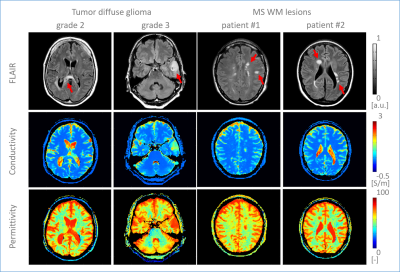 |
64 |
Brain water-content based Electrical Properties Tomography in
healthy volunteers, tumor and multiple sclerosis patients
Stefano Mandija1,2,
Sarah Jacobs3,
Jordi Kleinloog1,2,
Hongyan Liu1,2,
Oscar van der Heide1,2,
Anja van der Kolk1,4,
Alessandro Sbrizzi1,2,
and Cornelis van den Berg1,2
1Department of Radiotherapy, UMC Utrecht, Utrecht, Netherlands, 2Computational Imaging Group for MR Therapy and Diagnostics, UMC Utrecht, Utrecht, Netherlands, 3Department of Radiology, UMC Utrecht, Utrecht, Netherlands, 4Department of Medical Imaging, Radboud UMC, Nijmegen, Netherlands Keywords: Electromagnetic Tissue Properties, Electromagnetic Tissue Properties, Conductivity In this work, we first extend the validation of the water-content based Electrical Properties Tomography (wEPT) model from brain white matter to gray matter conductivity reconstructions in healthy volunteers. Secondly, we show that wEPT reconstructions calibrated on 10 healthy volunteers from an MR-STAT clinical trial dataset show a conductivity increase in pathological regions for 6 primary brain tumor and 9 multiple sclerosis (MS) patients from the same study. For diffuse glioma, a positive correlation between grade and conductivity is observed. For MS white matter lesions a clear conductivity increase is observed compared to healthy white matter. |
|
5167.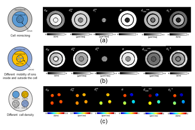 |
65 |
Measurement of Extracellular Electrical Conductivity Using
Conductivity Tensor Imaging
Bup Kyung Choi1,
Nitish Katoch1,
Ji Ae Park2,
Tae Hoon Kim3,
Young Hoe Hur4,
Jin Woong Kim5,
and Hyung Joong Kim1
1Department of Biomedical Engineering, Kyung Hee University, Seoul, Korea, Republic of, 2Division of Applied RI, Korea Institute of Radiological and Medical Science, Seoul, Korea, Republic of, 3Medical Convergence Research Center, Wonkwang University Hospital, Iksan, Korea, Republic of, 4Department of Hepato-Biliary-Pancreas Surgery, Chonnam National University Medical School, Gwangju, Korea, Republic of, 5Department of Radiology, Chosun University Hospital and Chosun University College of Medicine, Gwangju, Korea, Republic of Keywords: Electromagnetic Tissue Properties, Electromagnetic Tissue Properties, Electrical Conductivity, Low-frequency conductivity, High-field MRI, Tissue properties, Cell density imaging Conductivity measured at low-frequency can provide information on extracellular space (ECS), which will be useful in clinical applications such as tumor imaging and bioelectromagnetic modeling. Recently proposed conductivity tensor imaging (CTI) technique provides the anisotropic low-frequency conductivity distribution extracted from high-frequency conductivity measurement from the B1 map of MRI. The extracellular conductivity measured using CTI from three phantoms was compared with an impedance analyzer. The accuracy of the CTI technique was estimated to be high enough for most clinical applications. |
|
5168.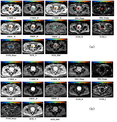 |
66 |
Simultaneously Applying Multiple Diffusion Models to Predict
Histologic Grade of Bladder Urothelial Carcinoma
Junting Guo1,
Lu Zhang1,
Shuo Li1,
Ding Li1,
Zhichang Fan1,
Meining Chen2,
Guoqiang Yang3,
Yan Li3,
Le Wang3,
Bin Wang3,
and Xiaochun Wang3
1College of Medical Imaging, Shanxi Medical University, Taiyuan, China, 2MR Scientific Marketing, Siemens Healthcare, Shanghai, China, 3Department of Radiology, The First Hospital of Shanxi Medical University, Taiyuan, China Keywords: Multi-Contrast, Cancer Tumor grading is the most important single prognostic factor for bladder urothelial carcinoma. In this study, we compared 5 diffusion models for assessing low- and high-grade in bladder urothelial carcinoma, including continuous-time random-walk (CTRW), incoherent motion within the voxel (IVIM), stretched exponential model (SEM), diffusion kurtosis imaging (DKI) and fractional-order calculus (FROC). The study found that CTRW_D, DKI_Dapp, DKI_Kapp, FROC_D, IVIM_D and SEM_DDC were significantly different between low- and high-grade bladder urothelial carcinoma and could distinguish one from the other. |
|
5169.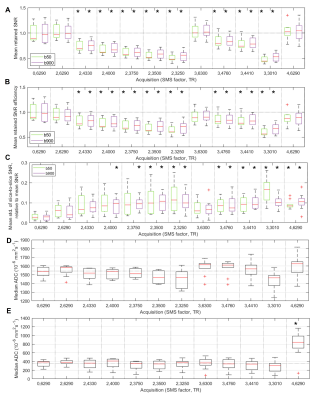 |
67 |
Determination of optimal parameters for accelerated whole-body
diffusion-weighted imaging (WB-DWI) using simultaneous
multi-slice (SMS)
Sam Keaveney1,2,
Alina Dragan1,
Julie Hughes1,
Mihaela Rata1,2,
Thomas Benkert3,
Christina Messiou1,2,
Dow-Mu Koh1,2,
and Jessica M Winfield1,2
1MRI Unit, Royal Marsden NHS Foundation Trust, Sutton, United Kingdom, 2Division of Radiotherapy and Imaging, The Institute of Cancer Research, London, United Kingdom, 3MR Application Predevelopment, Siemens Healthcare GmbH, Erlangen, Germany Keywords: Diffusion/other diffusion imaging techniques, Whole Body, Simultaneous Multi-Slice Simultaneous multi-slice (SMS) has the potential to accelerate whole-body diffusion-weighted imaging (WB-DWI). This work presents quantitative and qualitative assessment of SMS image quality in phantoms and healthy volunteers, as a strategy to determine the optimal parameters for the clinical application of a research application SMS sequence. An SMS factor of 2 and a moderately reduced TR did not introduce significant bias to ADC measurements. The reduction in SNR for these acquisitions was statistically, but not clinically, significant. These parameters offer time savings of up to 24% with respect to non-SMS settings and should be investigated further in a patient population. |
|
5170.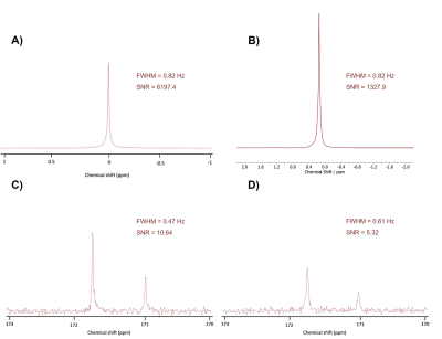 |
68 |
New biomaterial for 3D cell modeling and NMR analysis: low
signal interference and high diffusion for robust metabolic
studies in-vitro
Alba Herrero Gómez1,
Marc Azagra1,
and Irene Marco Rius1
1Institute for Bioengineering of Catalonia, Barcelona, Spain Keywords: Molecular Imaging, New Devices Due to its non-invasive nature, NMR has become a pillar as a diagnostic technique, as well as a support for biochemical assays and disease tracking both in vivo and in-vitro. However, due to its magnetic susceptibility, some of the materials used for tissue engineering and 3D cell modeling are incompatible with the technique, interfering with data acquisition and reducing its applications in-vitro. We developed a cryogel that could help bridge this gap between NMR and tissue engineering. |
|
5171.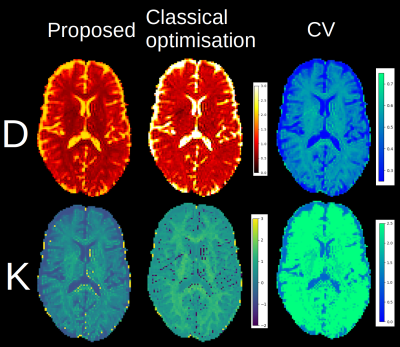 |
69 |
Fast consensus optimisation in diffusion MRI modeling
Samuel St-Jean1 and
Markus Nilsson1
1Clinical Sciences Lund, Lund University, Lund, Sweden Keywords: Diffusion/other diffusion imaging techniques, Data Analysis, diffusion modeling This work explores how to obtain parameter maps from a diffusion MRI model under 30 seconds and how to leverage multiple plausible solutions into a consensus solution using a framework similar to MR fingerprinting. The method is tested with a two compartments model and a mean kurtosis model and compared with a standard optimisation solver. Visual improvements are shown on parameter maps, showing less implausible values than the standard method, but without the use of constraints or assumptions. Access to a distribution of plausible values allows to compute most likely value from this distribution in the case of parameter degeneracies. |
|
5172.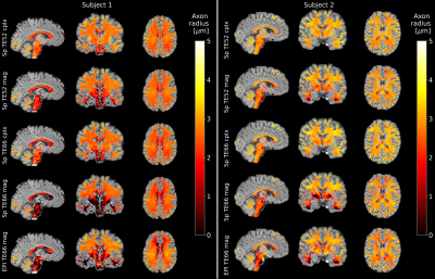 |
70 |
Spiral readout improves in vivo MR axon radius estimation in
human white matter
Marten Veldmann1,
Luke J. Edwards2,
Kerrin J. Pine2,
Philipp Ehses1,
Nikolaus Weiskopf2,3,
and Tony Stöcker1,4
1MR Physics, Deutsches Zentrum für Neurodegenerative Erkrankungen e.V., Bonn, Germany, 2Department of Neurophysics, Max Planck Institute for Human Cognitive and Brain Sciences, Leipzig, Germany, 3Felix Bloch Institute for Solid State Physics, Faculty of Physics and Earth Sciences, Leipzig University, Leipzig, Germany, 4Department of Physics & Astronomy, University of Bonn, Bonn, Germany Keywords: Diffusion/other diffusion imaging techniques, Diffusion/other diffusion imaging techniques We compared spiral and EPI diffusion imaging at ultra-high b-values for axon radius estimation in the white matter. For data acquisition, a custom multiband spiral sequence was combined with trajectory monitoring and higher order image reconstruction. The lower echo time of the spiral sequence led to increased relative SNR compared to EPI and improved estimation of axon radii. The resulting axon radius maps from spiral scans were more homogeneous especially in low-SNR regions. We also found, that denoising performed on complex data instead of magnitude data significantly improved axon radius estimation. |
|
5173.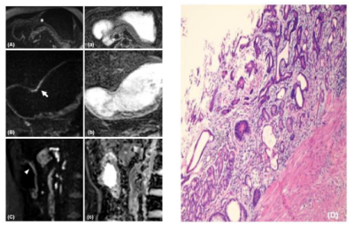 |
71 |
Multi-orientation ZOOMit diffusion-weighted imaging in the
preoperative T staging of gastric cancer
Wei-Yue Xu1,
Qiong Li1,
Ya-Jun Hou1,
Yi-Cheng Hsu2,
Thomas Benkert3,
Yu-Dong Zhang1,
and Xi-Sheng Liu1
1Department of Radiology, the First Affiliated Hospital with Nanjing Medical University, Nanjing, China, 2MR Collaboration, Siemens Healthineers Ltd, Shanghai, China, 3MR Application Predevelopment, Siemens Healthcare GmbH, Erlangen, Germany Keywords: Diffusion/other diffusion imaging techniques, Diffusion/other diffusion imaging techniques, ZOOMit diffusion-weighted imaging Magnetic resonance imaging has been demonstrated to be a useful tool in the preoperative diagnosis of gastric cancer. With improved imaging quality and flexible imaging orientation, ZOOMit diffusion-weighted imaging (DWI) is attracting attention in gastric tumor imaging. The stomach is a hollow organ with variable morphology. Thus, multi-orientation ZOOMit DWI imaging provides a complete view of the lesion compared with axial DWI, thus leading to more accurate preoperative staging in treatment strategy determination. |
|
5174.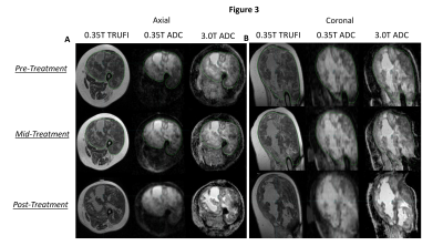 |
72 |
Diffusion imaging on a 0.35 T MRI-guided linear accelerator
providing accurate in vivo apparent diffusion coefficient maps
Joseph Weygand1,
Tess Armstrong2,
J.M. Bryant1,
Jacqueline Andreozzi1,
Ibrahim M. Oraiqat1,
Casey L. Liveringhouse1,
Kujtim Latifi1,
Kosj Yamoah1,
James R. Costello3,
Eduardo G. Moros1,
Issam M. El Naqa4,
Arash O. Naghavi1,
Stephen A. Rosenberg1,
and Gage Redler1
1Department of Radiation Oncology, Moffitt Cancer Center, Tampa, FL, United States, 2Department of Product Development, ViewRay, Oakwood Village, OH, United States, 3Department of Radiology, Moffitt Cancer Center, Tampa, FL, United States, 4Department of Machine Learning, Moffitt Cancer Center, Tampa, FL, United States Keywords: Diffusion/other diffusion imaging techniques, Radiotherapy Diffusion weighted imaging (DWI) allows for the evaluation of tumor cellularity. Its application on a 0.35 T MRI-guided linear accelerator (MRL) would facilitate integration of this information into radiotherapy planning and potentially allow for online biologically-guided plan adaption. This study demonstrates the capability of a DWI protocol both in phantom and in vivo. In particular, it is demonstrated that quantitively accurate, repeatable, and geometrically precise ADC maps can be produced in phantom on the 0.35 T MRL. Additionally, this technique was applied in vivo on one sarcoma patient receiving same-day diagnostic diffusion scans before, during, and after radiotherapy. |
|
5175.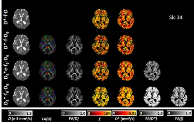 |
73 |
Integrating intravoxel incoherent motion and diffusion tensor
MRI of the brain into a single fast acquisition - a
model-selection study
Olaf Dietrich1,
Mengfei Cai2,
Anil Tuladhar2,
Mina Jacob2,
Gerald Drenthen3,
Jacobus Jansen3,4,
José Marques2,
Jens Ricke1,
Frank-Erik de Leeuw2,
Marco Duering5,
and Walter Backes3
1Department of Radiology, LMU University of Munich, Munich, Germany, 2Department of Neurology, Donders Center for Medical Neurosciences, Radboud University Medical Center, Nijmegen, Netherlands, 3Schools for Mental Health and Neuroscience (MHeNs) and Cardiovascular Diseases (CARIM), Department of Radiology and Nuclear Medicine, Maastricht University Medical Center, Maastricht, Netherlands, 4Department of Electrical Engineering, Eindhoven University of Technology, Eindhoven, Netherlands, 5Medical Image Analysis Center (MIAC AG) and qbig, Department of Biomedical Engineering, University of Basel, Basel, Switzerland Keywords: Diffusion/other diffusion imaging techniques, Diffusion Tensor Imaging, Intravoxel incoherent motion MRI; Model selection; Brain; Cerebral small vessel disease The acquisition of IVIM and DTI data of the brain can be integrated into a single measurement, which offers the possibility to determine orientation-dependent (tensorial) perfusion parameters in addition to established IVIM and DTI parameters. The purpose of this study was to evaluate the feasibility of such an integrated IVIM-DTI protocol with a clinically feasible scan time below 6 minutes and to establish the maximum number of DTI and IVIM tensor parameters that can reliably be determined with this approach by comparing 17 different IVIM-diffusion models with 4 to 19 model parameters. |
|
5176. |
74 |
Soft-Tissue Sarcomas demonstrate Fractional Anisotropy in
Diffusion Tensor Imaging and Fractional Anisotropy Values Change
After Treatment
Imogen Thrussell1,2,
Jessica Winfield1,2,
Sadiq Usman2,
Jennifer Newman2,
Georgina Hopkinson2,
Shane Zaidi1,2,
Aisha Miah1,2,
Christina Messiou1,2,
and Matthew Blackledge1,2
1The Institute of Cancer Research, London, United Kingdom, 2The Royal Marsden NHS Foundation Trust, London, United Kingdom Keywords: Diffusion/other diffusion imaging techniques, Diffusion Tensor Imaging, Cancer, Fractional Anisotropy, DWI, ADC In previous studies average values of Apparent Diffusion Coefficient (ADC) have been shown to change significantly after radiotherapy in Soft-Tissue Sarcomas (STS). In this study we evaluate the Fractional Anisotropy (FA) of STS before and after treatment and demonstrate that (i) tumours can exhibit significant diffusion anisotropy, and (ii) changes in FA are inversely correlated with ADC change. Diffusion-Tensor Imaging (DTI) may therefore provide important microstructural information when interpreting change in STS after radiotherapy, and that diffusion anisotropy should be accounted for when optimising diffusion-weighted imaging (DWI) protocols for STS. |
|
5177.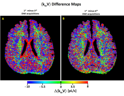 |
75 |
Does MADI Detect Temporal Brain Metabolic Activity Changes?
Martin M. Pike1,
Xin Li1,
Eric Baetscher1,
Thomas M. Barbara1,
Manoj K. Sammi1,
Alexander A. Stevens1,
and Charles S. Springer1
1Advanced Imaging Research Center, Oregon Health & Science University, Portland, OR, United States Keywords: Molecular Imaging, Metabolism, Activity Metabolic Activity Diffusion Imaging [MADI] maps on-going sodium pump metabolic flux. Here, we investigate the question as to whether MADI can detect temporal changes of this activity in sequential acquisitions from the healthy human brain. |
|
5178.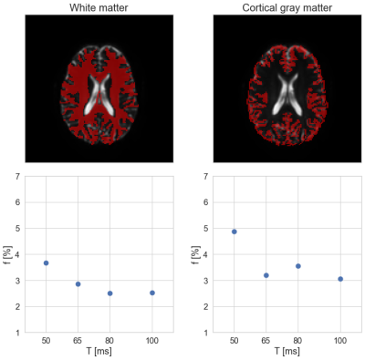 |
76 |
Time-dependence of perfusion fraction with flow-compensated
intravoxel incoherent motion MRI in the brain
Louise Rosenqvist1,
Mikael Montelius1,
Maria Ljungberg1,2,
and Oscar Jalnefjord1,2
1Department of Radiation Physics, Institute of Clinical Sciences, Sahlgrenska Academy, University of Gothenburg, Gothenburg, Sweden, 2Department of Medical Physics and Biomedical Engineering, Sahlgrenska University Hospital, Gothenburg, Sweden Keywords: Diffusion/other diffusion imaging techniques, Perfusion, IVIM Using IVIM imaging for completely non-invasive perfusion MRI is gaining popularity. IVIM perfusion assessment depends not only on capillary network characteristics, but also on diffusion encoding-time. In this study, a flow-compensated pulse sequence with variable encoding-time was implemented and validated through phantom measurements, and subsequently used to study the encoding-time dependence of IVIM perfusion fraction in human brain tissue. Initial findings indicate a decrease in perfusion fraction as encoding-time increases. |
|
 Back to Meeting Home
 Back to the Program-at-a-Glance
The International Society for Magnetic Resonance in Medicine is accredited by the Accreditation Council for Continuing Medical Education to provide continuing medical education for physicians.
 Back to Meeting Home
 Back to the Program-at-a-Glance
 View Presentation Video