Digital Poster
Oxygenation & Preclinical fMRI
ISMRM & ISMRT Annual Meeting & Exhibition • 03-08 June 2023 • Toronto, ON, Canada

| Computer # | |||
|---|---|---|---|
4014.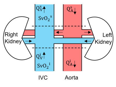 |
81 |
Quantification of bilateral kidney oxygen consumption with
MOTIVE-bSSFP
Rajiv S Deshpande1,
Michael C Langham1,
Cheng-Chieh Cheng1,
and Felix W Wehrli1
1University of Pennsylvania, Philadelphia, PA, United States Keywords: Oxygenation, Kidney, metabolic rate of oxygen Kidney oxygen consumption rate increases during the early stages of diabetes, ultimately leading to hypoxia of renal tissue. Thus, a reliable non-invasive approach to quantify kidney metabolism has significant clinical potential. Here, an approach to quantify the kidney oxygen utilization of both kidneys is reported. The method relies on the MOTIVE-bSSFP pulse sequence to measure SvO2 and blood flow rate in the inferior vena cava, above and below the branching of the renal vessels. Conservation of mass yields an expression to compute bilateral renal metabolic rate of oxygen. |
|
4015.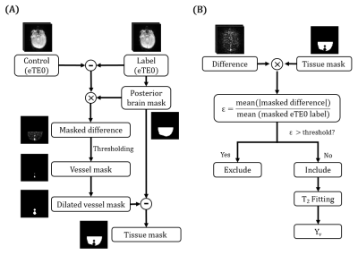 |
82 |
Motion-corrected quantification of cerebral venous oxygenation
in vulnerable populations: from neonates to older adults
Yifan Gou1,
Christopher Golden2,
Zixuan Lin3,
Jennifer Shepard2,
Aylin Tekes3,
Zhiyi Hu3,
Xin Li3,
Kumiko Oishi4,
Marilyn Albert5,
Hanzhang Lu3,
Peiying Liu3,6,
and Dengrong Jiang3
1Department of Biomedical Engineering, Johns Hopkins University, Baltimore, MD, United States, 2Department of Pediatrics, Johns Hopkins University School of Medicine, Baltimore, MD, United States, 3Department of Radiology, Johns Hopkins University School of Medicine, Baltimore, MD, United States, 4Center for Imaging Science, Johns Hopkins University Whiting School of Engineering, Baltimore, MD, United States, 5Department of Neurology, Johns Hopkins University School of Medicine, Baltimore, MD, United States, 6Department of Diagnostic Radiology and Nuclear Medicine, University of Maryland School of Medicine, Baltimore, MD, United States Keywords: Oxygenation, Oxygenation Cerebral venous oxygenation (Yv) is a biomarker in various brain diseases such as hypoxic-ischemic-encephalopathy in neonates and Alzheimer's disease in the elderly. T2-Relaxation-Under-Spin-Tagging (TRUST) MRI is a widely used technique to measure global Yv level. However, subject motion during TRUST scan can cause considerable errors in Yv quantification. This work developed an automatic algorithm for motion-corrected quantification of Yv, which showed improved precision of Yv estimation in both neonates and elderly adults. |
|
4016.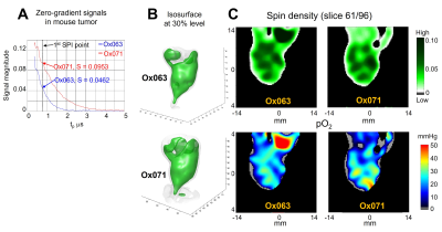 |
83 |
Deuterated trityl spin probe for in vivo pO2 imaging with
improved precision using Time-Domain EPR
SHUN KISHIMOTO1,
Nallathamby Devasahayam1,
Kota Yamashita1,
Kazumasa Horie1,
Kazutoshi Yamamoto1,
Jeffrey R Brender1,
and Murali C Krishna1
1NCI, Bethesda, MD, United States Keywords: Oxygenation, Cancer Due to the narrow linewidth and slower signal decay, it was expected that Ox071 is suitable for wide range pO2 estimation. In this work, we present our first 3D in vivo EPR oximetry using Ox071 in comparison with Ox063. EPR imaging of MIA Paca-2 tumor and healthy kidneys were performed on successive days by using either Ox071 or Ox063, resulting in similar spin density, pO2 maps, and pO2 histograms in the tumor regions and more homogeneous pO2 profile with Ox071 in kidney compared with Ox063. The result suggested that Ox071 is applicable for oximetry in tissue at higher pO2. |
|
4017.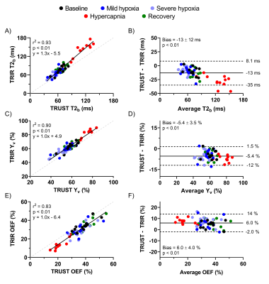 |
84 |
Venous blood oxygenation measurements using TRUST and T2-TRIR
during hypoxic and hypercapnic gas challenges
Koen P.A. Baas1,
Chau Vu2,
Jian Shen2,
Bram F. Coolen3,
Gustav J. Strijkers3,
John C. Wood2,4,
and Aart J. Nederveen1
1Radiology and Nuclear Medicine, Amsterdam UMC, Amsterdam, Netherlands, 2Biomedical Engineering, University of Southern California, Los Angeles, CA, United States, 3Biomedical Engineering and Physics, Amsterdam UMC, Amsterdam, Netherlands, 4Division of Cardiology, Children's Hospital Los Angeles, University of Southern California, Los Angeles, CA, United States Keywords: Oxygenation, Oxygenation TRUST and T2-TRIR can be used to measure venous blood oxygenation (Yv) and oxygen extraction fraction (OEF). T2-TRIR also offers simultaneous blood T1 (T1b) measurements for hematocrit calculation in lieu of invasive blood samples. Here, TRUST and T2-TRIR were compared across a broad Yv range using hypoxic and hypercapnic challenges in healthy volunteers, while T2-TRIR-derived hematocrit estimates were compared with venipuncture results. T2-TRIR- and TRUST-derived Yv and OEF values exhibited a small constant bias (-5.4±3.5% and 6.0±4.0% respectively, both p<0.01) across all stimuli. Hematocrit by venipuncture and from T1b were unbiased but had broad confidence intervals (1.7±6.3%, p=0.20). |
|
4018.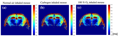 |
85 |
Study of Oxidative Stress due to X-ray Irradiation in Mouse
Brain using TOLD MRI Imaging
Raj Kumar Parajuli1,
Megumi Ueno2,
Saaya Suzuki1,
Akira Sumiyoshi1,
Takayuki Obata1,
Ichio Aoki1,
and Ken-ichiro Matsumoto2
1Department of Molecular Imaging and Theranostics, National Institutes for Quantum Science and Technology (QST), Chiba, Japan, 2Department of Radiation Regulatory Science Research, National Institutes for Quantum Science and Technology (QST), Chiba, Japan Keywords: Oxygenation, Brain, Oxidative stress, TOLD, X-ray irradiation This study aims to evaluate the metabolic changes caused on the brain tissues due to radiation exposure to healthy tissues while tumor treatment. The tissue oxidization and reduction mechanism represented by redox imaging could be a suitable index to evaluate in vivo oxidative stress in living animals that could evaluate metabolic damages due to irradiation. We adopted tissue oxygen level dependent (TOLD) MRI imaging to analyze TOLD signal for the control group mice and the irradiated mice. The result shows the distinct oxygen consumption levels in the control and the irradiated mice. |
|
4019.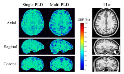 |
86 |
Oxygen extraction fraction (OEF) mapping by using multi-delay
arterial spin labeling based on QSM+qBOLD
Xiaoyi Liu1,
Yayan Yin1,
Juan Wei2,
and Jie Lu1
1Radiology and Nuclear Medicine, Xuanwu Hospital, Capital Medical University, Beijing 100053, China, Beijing, China, 2GE Healthcare, MR Research China, Beijing, Beijing, China Keywords: Arterial spin labelling, Metabolism, oxygen extraction fraction We aimed to improve the accuracy of OEF mapping by using multi-delay arterial spin labeling based on quantitative susceptibility mapping plus quantitative blood oxygen level-dependent magnitude (QSM+qBOLD, or QQ). 3D multi-echo gradient-echo and ASL sequence were performed in 7 healthy subjects. ROIs analysis and paired t-test compared QQ with single-delay ASL and QQ with multi-delay ASL. QQ with multi-delay ASL shows smaller OEFs than QQ with single-delay ASL method. Corresponding OEFs were 38.9±3.4% and 39.6±3.4% (P<0.01) in gray matter, 39.7±2.9% and 40.2±2.9% in whole brain(P<0.01), respectively. Our study suggests that QQ with multi-delay ASL provides more accurate OEF than QQ with single-delay ASL, which may have potential in the use of clinical diseases. |
|
4020.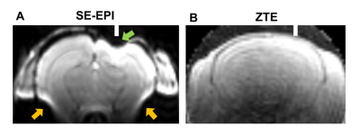 |
87 |
Zero-echo time (ZTE) pulse sequence enables EEG-fMRI with
suppression of susceptibility artifacts.
Ayako Imamura1,2,3,
Rikita Araki4,
Yukari Takahashi3,
Koichi Miyatake2,
Fusao Kato3,
Sakiko Honjoh2,
and Tomokazu Tsurugizawa3,5,6
1Ph. D. Program in Humanics, University of Tsukuba, Tsukuba, Japan, 2International Institute for Integrative Sleep Medicine (WPI-IIIS), University of Tsukuba, Tsukuba, Japan, 3Department of Neuroscience, The Jikei University School of Medicine, Tokyo, Japan, 4Bruker Japan K.K., Yokohama, Japan, 5Human Informatics and Interaction Research Institute, National Institute of Advanced Industrial Science and Technology (AIST), Tsukuba, Japan, 6Faculty of Engineering,, University of Tsukuba, Tsukuba, Japan Keywords: fMRI, fMRI Simultaneous recording of fMRI and EEG is a promising approach to realize functional brain imaging with high temporal and spatial resolution. However, using standard echo planar imaging in conventional fMRI studies, implantation of electrodes on the cortical surface induces strong magnetic susceptibility artifacts in fMRI images. The zero-echo time (ZTE) sequences show a remarkable reduction in sensitivity to magnetic susceptibility artifacts and motion-derived artifacts. In this study, we showed that the ZTE sequence suppressed the susceptibility artifact by electrodes and air in the ear canal in mice. Furthermore, ZTE showed the typical functional connectivity in the mice at resting state. |
|
4021.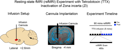 |
88 |
Pharmacological Inactivation of the Inhibitory Zona Incerta
Decreased Resting-state Functional MRI Connectivity
Junjian Wen1,2,
Xunda Wang1,2,
Teng Ma1,2,3,
Linshan Xie1,2,
Alex T. L. Leong1,2,
and Ed X. Wu1,2,4
1Department of Electrical and Electronic Engineering, The University of Hong Kong, Hong Kong SAR, China, 2Laboratory of Biomedical Imaging and Signal Processing, The University of Hong Kong, Hong Kong SAR, China, 3Department of Diagnostic Radiology, The University of Hong Kong, Hong Kong SAR, China, 4School of Biomedical Sciences, The University of Hong Kong, Hong Kong SAR, China Keywords: fMRI (resting state), Neuroscience In the recent decade, resting-state functional MRI (rsfMRI) has emerged as the most invaluable, non-invasive imaging technique to map long-range, brain-wide functional connectivity networks. Despite the enormous potential inherent in this technique, our present knowledge of the neural underpinnings of rsfMRI connectivity remains generally incomplete given the lack of studies examining the role of the inhibitory neural population, which is the counterpart of the excitatory neurons. In this study, we directly examine the role of zona incerta, which is one of the major source of inhibitory drive to the cortex. |
|
4022.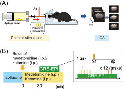 |
89 |
Whole-brain fMRI study of mice under medetomidine/ketamine
anesthesia to identify brain regions activated by musk odors
Sosuke Yoshinaga1,
Yumiko Tsubakihara1,
Takayuki Fukumoto1,
Shotaro Maita1,
Mitsuhiro Takeda1,
and Hiroaki Terasawa1
1Faculty of Life Sciences, Kumamoto University, Kumamoto, Japan Keywords: New Devices, Preclinical Muscone, a major component of musk odor, attracts male mice. The odor-evoked behaviors are mediated by neural circuits activated by the stimulation. We have been working on whole-brain BOLD fMRI studies of mice, using a method that employs periodic odor stimulation and independent component analysis. In this study, we investigated the brain regions activated by muscone in mice under medetomidine and low-dose ketamine anesthesia. Ketamine reportedly exhibits disinhibition of glutamate signaling at a low dose. As the result, we identified numerous muscone-evoked activated regions located in the olfactory pathways and higher-order regions in the cerebrum. |
|
4023.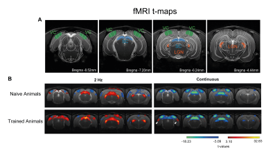 |
90 |
Behavioural Relevance modulates BOLD-fMRI responses in the rat
Visual Cortex
Ana Mafalda Valente1,
Rita Gil1,
and Noam Shemesh1
1Champalimaud Research, Champalimaud Foundation, Lisbon, Portugal Keywords: fMRI, Multimodal, NEURO Neuronal responses are shaped by experiences. Plasticity can occur at single neuron level or at the full population level. Here, we established a novel behavioural task requiring rats to distinguish between continuous and flickering lights. We then performed fMRI in trained vs naive animals and investigated BOLD-fMRI responses along the visual pathway. When light flashes become meaningful in trained animals, the BOLD activation patterns are significantly modulated compared to naive counterparts, in particular in higher visual cortex and associative areas. BOLD-fMRI signals are thus capable of deciphering plasticity arising from strong associations with actions and rewards. |
|
4024.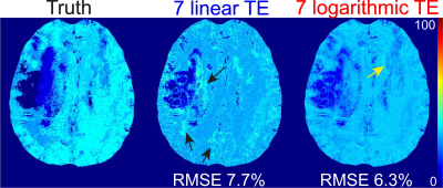 |
91 |
An improved data acquisition for robust oxygen extraction
fraction (OEF) mapping using an integrative model of QSM and
qBOLD (QSM+qBOLD=QQ)
Junghun Cho1,
Pascal Spincemaille2,
Thanh D. Nguyen 2,
and Yi Wang2
1Biomedical Engineering, SUNY Buffalo, Buffalo, NY, United States, 2Weill Cornell Medicine, New York, NY, United States Keywords: Oxygenation, Quantitative Imaging Oxygen extraction fraction (OEF) is critical to evaluate brain tissue viability and function in neurologic disorders. An integrated model of QSM and qBOLD (QSM+qBOLD or QQ) has been developed to map OEF utilizing a routine multi-echo gradient echo (mGRE) acquisition without impractical vascular challenges. This study proposes a novel mGRE acquisition with logarithmic echo spacing that acquires more data points in short echo time regime, which is critical to decouple the model parameters in QQ. The proposed novel mGRE provided more accurate OEF in two simulations, compared to the conventional mGRE. |
|
4025.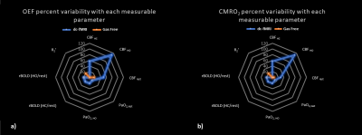 |
92 |
Uncertainties in baseline cerebral oxidative metabolism mapping
by gas-free and dual-calibrated fMRI: a simulation-based
comparison
Xiaole Zhong1,2 and
J. Jean Chen1,2,3
1Department of Medical Biophysics, University of Toronto, Toronto, ON, Canada, 2Rotman Research Institute, Baycrest Hospital, Toronto, ON, Canada, 3Institute of Biomedical Engineering, University of Toronto, Toronto, ON, Canada Keywords: fMRI, Metabolism Calibrated BOLD fMRI is becoming a method of choice in assessing baseline cerebral oxidative metabolism (CMRO2), with gas-free calibrated fMRI being an non-invasive alternative to the more established gas-calibrated fMRI methods. However, the relative repeatabilities of these two approaches are unknown, each depending on a unique set of input measurements. This study uses analytical simulations to investigate the sensitivity of baseline OEF and CMRO2 estimates to the measured inputs of each method. Our results suggest that gas-free calibrated fMRI is substantially less sensitive to input variations than gas-calibrated fMRI, demonstrating the potential for simulation-informed methodological selection. |
|
4026.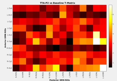 |
93 |
Default Mode Network is Not Related to Similarly Vigilance
Dependent Slow Rhythms
Vahid Khalilzad Sharghi1,
Eric Maltbie1,
Wen-Ju Pan1,
Shella Keilholz1,
and Kaundinya Gopinath1
1Emory University, Atlanta, GA, United States Keywords: fMRI (resting state), Brain Connectivity Several studies point to brain slow rhythms as the basis of rsfMRI signal. We recently reported that only vigilance/arousal dependent components of fMRI signal that are not specific to brain function networks (BFNs) decrease after suppression of slow rhythms, while BFNs increase in apparent strength. Default mode network (DMN) is a BFN that exhibits similar dependence on vigilance as slow rhythms. In this study, we examined the effects of slow rhythm suppression on the integrity of DMN. Our results show that DMN is not related to these rhythms and behaves just like other BFNs upon their suppression. |
|
4027.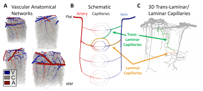 |
94 |
Computing hemodynamic point-spread functions with biophysical
simulations of microvascular networks: effects of trans-laminar
capillaries
Grant Hartung1,2,
Avery J. L. Berman1,2,
Divya Varadarajan1,2,
Jingyuan Chen1,2,
and Jonathan R. Polimeni1,2,3
1Athinoula A. Martinos Center For Biomedical Imaging, MGH, Charlestown, MA, United States, 2Department of Radiology, Harvard Medical School, Boston, MA, United States, 3Health Sciences and Technology, Massachusetts Institute of Technology, Cambridge, MA, United States Keywords: fMRI, Blood vessels, High-Resolution fMRI, Biophysical Simulation Here we attempted to estimate a “point-spread function” of blood flow and oxygenation in the cerebral cortex through biophysical simulations based on a reconstructed vascular anatomical network. We stimulated individual pre-capillary arterioles and computed the downstream capillary flow and oxygenation based on fluid dynamics and oxygen transport through blood and tissue. We quantified this spread in the directions radial and tangential to the cortex. While several biases in spread were observed, we found a roughly 400 μm spread in the radial direction and generally narrower spread in the tangential direction, although both varied with cortical depth. |
|
4028.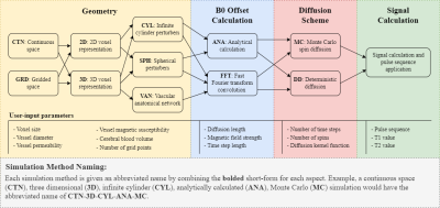 |
95 |
BOLDSwimSuite: A Python-based tool suite for numerical
simulations of transverse relaxation in the presence of diverse
field perturbers
Jacob Chaussé1,
Avery J. L. Berman2,3,
and J. Jean Chen1,4
1Rotman Research Institute, North York, ON, Canada, 2Department of Physics, Carleton University, Ottawa, ON, Canada, 3University of Ottawa Institute of Mental Health Research, Ottawa, ON, Canada, 4Department of Medical Biophysics, University of Toronto, Toronto, ON, Canada Keywords: Simulations, Software Tools Numerical simulations are valuable tools for understanding the transverse relaxation process, but their use is impeded by computational complexities and a lack of standard in simulation pipelines. We present a new software suite (named BOLDSwimSuite) to address these challenges, making numerical simulations accessible to a wider community of researchers. Among its many features, this toolbox is able to accommodate arbitrary magnetic-field perturbers as well as simulate different MRI sequences. This toolbox is useful for investigating BOLD fMRI contrast and MR vascular fingerprinting among other applications. |
|
4029.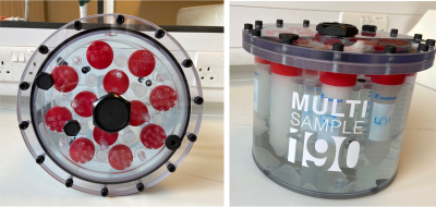 |
96 |
Development of a quantitative BOLD phantom for validation of
acquisition strategies.
Ahlam Alzaidi1,2,
Rafal Panek3,
and Nicholas P Blockley1
1School of Life Sciences, University of Nottingham, Nottingham, United Kingdom, 2Radiology Department, Taif University, Taif, Saudi Arabia, 3Medical Physics and Clinical Engineering, Nottingham University Hospitals NHS Trust, Nottingham, United Kingdom Keywords: Relaxometry, Phantoms A phantom to produce R2' (reversible transverse relaxation rate) contrast was developed by inducing subvoxel magnetic susceptibility variations that mimic the quantitative Blood Oxygenation Level Dependant (qBOLD) MRI signal. Microscopic glass bubbles were used for this purpose and offer a cheaper option for making large volume phantoms than existing phantoms that use precision polystyrene microspheres. A linear relationship between R2' and the glass bubble volume fraction was found. Comparisons of multiparametric qBOLD acquisitions between two MR vendors were also made. In the future, phantoms such as this could facilitate quality assurance for qBOLD acquisition strategies and assist with multicentre harmonization. |
|
4030.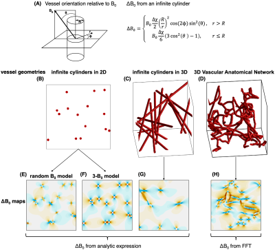 |
97 |
Simulating BOLD fMRI transverse relaxation at 3 T: How accurate
is the infinite cylinder model of blood vessels?
Avery J.L. Berman1,2,
Jacob Chausse3,
Grant Hartung4,5,
Jonathan R. Polimeni4,5,6,
and J. Jean Chen3,7
1Department of Physics, Carleton University, Ottawa, ON, Canada, 2University of Ottawa Institute of Mental Health Research, Ottawa, ON, Canada, 3Rotman Research Institute, Baycrest Health Sciences, North York, ON, Canada, 4Athinoula A. Martinos Center for Biomedical Imaging, Massachusetts General Hospital, Charlestown, MA, United States, 5Department of Radiology, Harvard Medical School, Boston, MA, United States, 6Harvard-MIT Division of Health Sciences and Technology, Massachusetts Institute of Technology, Cambridge, MA, United States, 7Department of Medical Biophysics, University of Toronto, Toronto, ON, Canada Keywords: Simulations, fMRI Simulations of the BOLD signal have provided invaluable insights into the biophysical underpinnings of BOLD contrast. Typically, infinite cylinders are used to represent the vasculature, although, in reality the vasculature is much more complex. In this study, we compared several biophysical modelling techniques for simulating the BOLD effect, including using Vascular Anatomical Networks (VANs) to model the capillary bed with realistic geometry. We found the majority of the different simulation approaches produced relatively consistent results with each other. The VAN simulations were consistent with the infinite cylinders, suggesting that infinite cylinders are a reasonable approximation to more realistic vascular models. |
|
 Back to Meeting Home
 Back to the Program-at-a-Glance
The International Society for Magnetic Resonance in Medicine is accredited by the Accreditation Council for Continuing Medical Education to provide continuing medical education for physicians.
 Back to Meeting Home
 Back to the Program-at-a-Glance
 View Presentation Video