Digital Poster
Qualitative Muscle Imaging
ISMRM & ISMRT Annual Meeting & Exhibition • 03-08 June 2023 • Toronto, ON, Canada

| Computer # | |||
|---|---|---|---|
4051.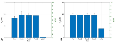 |
121 |
Increase in Fast-relaxing Sodium After a Brief Plantarflexion
Exercise Detected by Multi-Echo UTE MRI and Biexponential
Regression
Kwan-Jin Jung1,
Hsin-Yu Fang2,
Brad Sutton3,
and Kenneth Wilund2
1Biomedical Imaging Center, Beckman Institute, University of Illinois at Urbana-Champaign, Urbana, IL, United States, 2Department of Kinesiology and Community Health, University of Illinois at Urbana-Champaign, Urbana, IL, United States, 3Department of Bioengineering, University of Illinois at Urbana-Champaign, Urbana, IL, United States Keywords: Muscle, Relaxometry, Sodium, Muscle, Exercise We measured the exercise-induced change in fast and slow T2-relaxing sodium in calf muscles separately using a UTE sequence with multiple echoes. Three minutes of plantarflexion exercise resulted in an increase in the fast-relaxing sodium and no change in the slow-relaxing component. Based on the known physiology that muscle intracellular sodium increases during muscle contraction, the measured fast-relaxing sodium might represent muscle intracellular sodium. This finding is valuable in studying intra- and extracellular sodium separately. |
|
4052.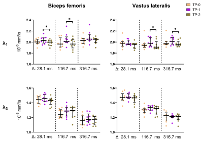 |
122 |
Investigating muscle micro-trauma with time-dependent diffusion
and the random permeable barrier model
Susanne S. Rauh1,
Donnie Cameron2,
Oliver J. Gurney-Champion3,
Mario Maas3,
Martijn Froeling4,
Hermien E. Kan2,
Aart Nederveen3,
Gustav Strijkers1,
and Melissa T. Hooijmans3
1Department of Biomedical Engineering and Physics, Amsterdam Movement Sciences, Amsterdam UMC, location AMC, Amsterdam, Netherlands, 2C.J. Gorter MRI Center, Department of Radiology, Leiden University Medical Center, Leiden, Netherlands, 3Department of Radiology and Nuclear Medicine, Amsterdam UMC, location AMC, Amsterdam, Netherlands, 4Department of Radiology, Utrecht UMC, Utrecht, Netherlands Keywords: Muscle, Diffusion Tensor Imaging Repetitive muscle micro-trauma can result in severe muscle injuries. Diffusion tensor imaging can detect small but significant changes due to muscle micro-trauma, but the sensitivity is limited. Longer and multiple diffusion times can potentially increase the sensitivity to micro-trauma as they facilitate muscle fiber diameter and permeability estimations with the random permeable barrier model (RPBM). In this study, we demonstrated that a diffusion time of 116.7ms showed largest percentage change in DTI indices suggesting an increased sensitivity to exercise-induced muscle micro-trauma after running a marathon. No effect was found in the RPBM-derived membrane permeability and fiber diameter. |
|
4053.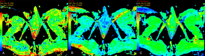 |
123 |
Axial diffusivity and kurtosis derived based on DWI reflect
microstructure changes in patients with in stress urinary
incontinence patients
Kun Ou1,
Weiyn Vivian Liu2,
and Kun Zhang1,3
1Department of Radiology, First Affiliated Hospital of Hunan University of Chinese Medicine, Changsha, China, 2MR Research, GE Healthcare, Beijing, China, 3College of Integrated Traditional Chinese and Western Medicine, Hunan University of Chinese Medicine, ChangSha, China Keywords: Muscle, Diffusion/other diffusion imaging techniques, Diffusion kurtosis imaging Stress urinary incontinence (SUI) is the most common form of urinary incontinence. However, in patients with SUI, the pelvic floor muscles generally have no obvious radiographic abnormalities. This study applied diffusion-weighted imaging with different analysis models and found that patients with SUI had significantly lower FA and Da values based on gaussian-distributed water movement and higher values in the Ka based on non-gaussian-distributed water movement compared to the healthy volunteers. This study showed both DTI and DKI derived parameters can detect microstructure changes in the the pubic visceral muscle of the SUI patients. |
|
4054.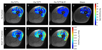 |
124 |
In Vivo Mapping of Muscle Fibrosis: The Influence of Signal
Model Inputs
Mary Elizabeth Hall1,
Yael Vainberg1,
Garry Gold1,
and Valentina Mazzoli1
1Radiology, Stanford University, Stanford, CA, United States Keywords: Muscle, Aging, Fibrosis, Sarcopenia Fibrosis could be an early sign of declining muscle quality. Short T2*-signal fraction, which can be calculated by fitting a 3-exponential model to UTE data, has been shown to be correlated with collagen content, which is related to fibrosis. However, this type of signal model has 8 fitting parameters, which could result in overfitting to a limited number of echoes. We constrained the signal model 3 different ways, and compared the resulting short T2*-signal fraction values. There were significant differences between methods, demonstrating that these choices can affect parameter values and diagnostic sensitivity of short T2*-signal fraction for fibrosis detection. |
|
4055.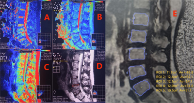 |
125 |
Intervertebral disc’s quantitative measurements from synthetic
MR correlates with its degeneration and bone marrow fat fraction
Xiaochun Yuan1,
Ze Li1,
Min Fu1,
Xiaochen Wei2,
and Junrong Chen3
1Chengdu Sport University, Chengdu, China, 2GE Healthcare China, Beijing, China, 3Sichuan Province Orthopedic Hospital, Chengdu, China Keywords: Muscle, Fat, Quantitative imaging, Lumbar disc herniation The conventional Pfirrmann grading currently used to assess the degree of disc degeneration is a semi-quantitative assessment criterion. And the relationship between vertebral body bone marrow fat and disc degeneration needs further clarification. In this study, quantitative synthetic MR technique as well as fat quantification MR technique are used to investigate relationship between imaging markers and degeneration levels. Results indicates intervertebral discs’ quantitative T2, T1 and PD values are significantly different among Pfirrmann grading groups. Significant negative correlations are found between Pfirrmann grading and quantitative values. T1 and PD values have weak correlation with bone marrow fat fraction. |
|
4056.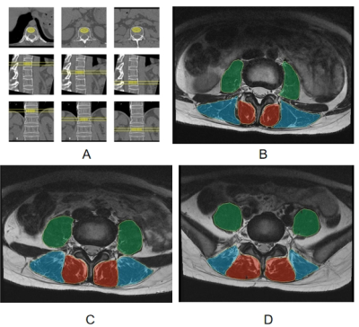 |
126 |
Relationship between the properties of paraspinal muscles and
bone mineral density in patients with lumbar disc herniation
Ze Li1,
Min Fu1,
Xiaochun Yuan1,
Huilou Liang2,
and Junrong Chen3
1Chengdu Sport University, Chengdu, China, 2GE Healthcare China, Beijing, China, 3Sichuan Province Orthopedic Hospital, Chengdu, China Keywords: Muscle, Fat, Quantitative imaging, Paraspinal muscle Paraspinal muscles and vertebral bodies are important to maintain spinal stability. Muscles and bones are functional tissues that interact with each other. We investigated the relationship between the properties of paraspinal muscles obtained from MR imaging and bone mineral density of vertebral bodies measured from quantitative CT in patients with lumbar disc herniation (LDH). Our results showed that the fat fraction of paraspinal muscles was negatively correlated with bone mineral density of vertebral bodies in LDH patients, and the fat fraction of multifidus muscle at the L3/L4 lumbar disc level may be the optimal predictor of bone mineral density changes. |
|
4057.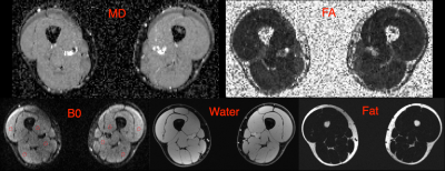 |
127 |
Time-dependent diffusion in healthy adult human thigh muscle in
a clinically feasible acquisition
Amy McDowell1,
Thorsten Feiweier2,
Carolynne Doherty1,
Stephen J Wastling1,
Matt G Hall3,
Chris A Clark4,
Tarek Yousry5,
Mary Reilly1,
and John S Thornton1
1Queen Square Centre for Neuromuscular Diseases, University College London, London, United Kingdom, 22Siemens Healthcare GmbH, Erlangen, Germany, 3Medical Radiation Physics, National Physical Laboratory, London, United Kingdom, 4Developmental Imaging and Biophysics, University College London, London, United Kingdom, 5Queen Square Institute of Neurology, University College London, London, United Kingdom Keywords: Muscle, Diffusion Tensor Imaging Muscle fibres are larger than axons with a complex hierarchical internal structure causing a decrease in the diffusion coefficient with diffusion time. Acquisition of larger field-of-view thigh-level images is challenged by increased distortion, higher B0 inhomogeneity, incomplete fat saturation and longer acquisition times. We optimised a protocol to study the time-dependence of diffusion indices in healthy human thigh muscle in a clinically feasible scan time. MD and FA showed strong time dependence but slight disparities from previous work in the calf, possibly due to differences in the musculature of the thigh, male/female ratio, or changes in the acquisition with optimisation. |
|
4058.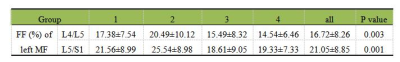 |
128 |
Association Between Modic Change and Paraspinal Muscle Fat
Infiltration Based on MRI
Min Fu1,
Ze Li1,
Xiaochun Yuan1,
Xiaocheng Wei2,
and Junrong Chen3
1Chengdu Sport University, Chengdu, Sichuan, China, 2GE Healthcare China, Beijing, China, 3Sichuan Province Orthopedic Hospital, Chengdu, Sichuan, China Keywords: Muscle, Fat, Quantitative imaging, Modic change Paraspinal muscles play an important role in maintaining the stability of lumbar spine. The influencing factors of paraspinal muscle degeneration are still controversial. IDEAL-IQ MR sequence can provide quantitative fat fraction map to objectively and accurately evaluate the fat infiltration in paraspinal muscles. By using IDEAL-IQ MR sequence, this study investigated the influence of Modic change (MC) on quantitative propertied of paraspinal muscles in patients with low back pain (LBP). Our results showed that MC can aggravate the degeneration of paraspinal muscles, especially the multifidus, in LBP patients. |
|
4059.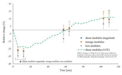 |
129 |
Evaluation of viscoelastic changes of the psoas major muscle
after exercise using MR elastography
Tetsushi Habe1,2,
Tomokazu Numano1,3,
Daiki Ito1,2,
Kouichi Takamoto4,
and Hisao Nishijo5
1Department of Radiological Sciences, Graduate School of Human Health Sciences, Tokyo Metropolitan University, Tokyo, Japan, 2Office of Radiation Technology, Keio University Hospital, Tokyo, Japan, 3Human Technology Research Insutitue, National Institute of Advanced Industrial Science and Techonology, Ibaraki, Japan, 4Department of Sport and Health Sciences, Faculty of Human Sciences, University of East Asia, Yamaguchi, Japan, 5System Emotional Science, Faculty of Medicine, University of Toyama, Toyama, Japan Keywords: Muscle, Elastography The shear modulus of the psoas major muscle (PM) after exercise has been evaluated using magnetic resonance elastography (MRE) previously, however, a separate evaluation of elasticity and viscosity has not been performed. We evaluate storage modulus representing elasticity and loss modulus representing viscosity of the PM using MRE after exercise in this study. Comparing the before and immediately after exercise, the loss modulus showed a greater rate of decrease than the storage modulus, suggesting that the loss modulus, which reflects viscosity, is more useful for detecting changes of muscle tissue immediately after exercise. |
|
4060.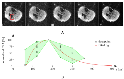 |
130 |
Imaging of the Contraction Pattern of Spontaneous Muscular
Activities using a Multiple-Point Diffusion-Weighted Stimulated
Echo Sequence
Martin Schwartz1,2,
Petros Martirosian1,
Guenter Steidle1,
Bin Yang2,
and Fritz Schick1
1Section on Experimental Radiology, University Hospital of Tuebingen, Tuebingen, Germany, 2Institute of Signal Processing and System Theory, University of Stuttgart, Stuttgart, Germany Keywords: Muscle, Muscle Spontaneous mechanical activities in resting musculature can be visualized using stimulated-echo or spin-echo diffusion-weighted sequences. Temporal resolution is limited to the sampling of one point in time of the contraction pattern. Therefore, a concept for multiple points in time imaging of spontaneous activities is proposed in this work. The implemented MR sequence was validated by measurements from a water phantom and of the resting lower leg musculature of healthy subjects. |
|
4061.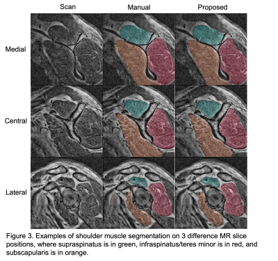 |
131 |
Feasibility of Automated Segmentation of 3D Shoulder Muscle
Volume via Deep Learning for Rotator Cuff Repair Patients
Mingrui Yang1,
Bong-Jae Jun1,
Tammy Owings1,
Joshua Polster1,
Carl Winalski1,
Kathleen Derwin1,
Eric Ricchetti1,
and Xiaojuan Li1
1Cleveland Clinic, Cleveland, OH, United States Keywords: Muscle, Machine Learning/Artificial Intelligence It has been shown that muscle volume and fat fraction play significant roles in musculoskeletal disorder diagnosis and prognosis. Reliable clinical tools for their evaluation, however, are currently missing. One hurdle is the challenging and laborious manual segmentation process on MR images. We proposed here a deep learning based automated tool for 3D shoulder muscle volume segmentation and achieved accurate segmentation results on clinical MR images from rotator cuff repair patients. The proposed model can be a valuable tool for shoulder muscle volume quantification and subsequent fat fraction analysis to further understand their association with clinical outcomes following shoulder procedures. |
|
4062.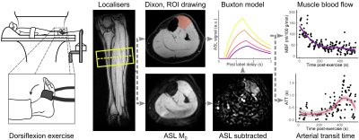 |
132 |
Towards patient-friendly arterial-spin-labeling MRI in muscle:
Effect of exercise intensity and standardisation on perfusion
parameters
Donnie Cameron1,
Esther J. Schrama2,
Linde Boogaarts1,
Thom T.J. Veeger1,
Celine Baligand3,
Lydiane Hirschler1,
Matthias J.P. van Osch1,
and Hermien E. Kan1
1C.J. Gorter MRI Center, Department of Radiology, Leiden University Medical Center, Leiden, Netherlands, 2Department of Neurology, Leiden University Medical Center, Leiden, Netherlands, 3Laboratoire des Maladies Neurodégénératives, Université Paris-Saclay, Fontenay-aux-Roses, France Keywords: Muscle, Perfusion, Exercise Perfusion is fundamental to muscle function, and is limited in muscular dystrophies—representing a promising biomarker for clinical trials. However, in-magnet exercise paradigms for measuring ASL perfusion are seldom standardised and require high-intensity exercise, which is impracticable in patients. In volunteers, we tested the acceptability of three protocols: two standardised paradigms with different intensities, and a non-standardised paradigm at a single intensity. Exercise standardisation improved SNR. In general, muscle blood flow, T2*, and SNR decreased, while arterial transit time increased with time after exercise; T2* was higher after high-intensity exercise. SNR at lower exercise-intensity was insufficient for robust perfusion-parameter estimation. |
|
4063.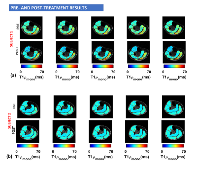 |
133 |
The effects of exercise on ankle mobility and lower leg
hyaluronan distribution in type 2 diabetes
Rajiv G Menon1,
Smita Rao2,
Preeti Raghavan3,
and Ravinder Regatte1
1Radiology, NYU Grossman School of Medicine, New York, NY, United States, 2Department of Physical Therapy, New York University, New York, NY, United States, 3Johns Hopkins University School of Medicine, Baltimore, MD, United States Keywords: Muscle, Diabetes, exercise intervention Type 2 Diabetes Mellitus (T2DM) is a major cause of a number of systemic pathological processes, including muscle stiffness. In this study, we examine the effects of exercise on peak ankle dorsiflexion, and ankle stiffness and investigate hyaluronan distribution with 3D-T1ρ MRI in 10 individuals with T2DM. Our findings suggest that participation in a 10-week moderate intensity exercise intervention may alter hyaluronan distribution in the compartments of the lower leg calf muscle, and is accompanied by increased dorsiflexion, reduced stiffness in individuals with diabetes, and a reduction in MRI based T1ρ measures |
|
4064.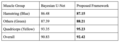 |
134 |
Variational Model Augmented Deep Learning for Small Training
Data MRI Thigh Muscle Segmentation
Paramjyoti Mohapatra1,
Weihong Guo1,
Mingrui Yang2,
Richard Lartey2,
and Xiaojuan Li2
1Mathematics, Applied Mathematics and Statistics, Case Western Reserve University, Cleveland, OH, United States, 2Lerner Research Institute, Cleveland Clinic, Cleveland, OH, United States Keywords: Muscle, Machine Learning/Artificial Intelligence, muscle segmentation, fusion, variational model Accurate automatic MRI thigh muscle group segmentation is essential for muscle morphology and composition which are related to osteoarthritis and sarcopenia. Due to challenges caused by lack of contrast between muscle groups, Deep Learning (DL) becomes a more natural choice than traditional model based approaches. It is however expensive to obtain a large amount of training data and DL using small training data often results in overfitting. We use a variational model based segmentation method in conjunction with a Bayesian neural network to optimize the train framework, producing about 1.6% increase in dice coefficients while working with minimal annotated data. |
|
4065.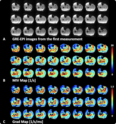 |
135 |
Large coverage, high temporal and spatial resolution lower
extremity blood oxygen level-dependent imaging
Jianxun Qu1,
Xiaoyuan Fan2,
Tianye Lin3,
Xiaoxi Yu4,
and Feng Feng2
1SIEMENS Healthineers, Beijing, China, 2Radiology Department, Peking Union Medical College Hospital, Beijing, China, 3Radiology Department, Peking University Cancer Hospital & Institute, Beijing, China, 4Vascular Surgery Department, Peking Union Medical College Hospital, Beijing, China Keywords: Muscle, Perfusion Multi echo gradient echo (GRE) sequence is usually used in lower extremity BOLD experiments. This study replaced the GRE acquisition by SMS EPI and achieved broader coverage and higher temporal and spatial resolution. The minimum ischemia value (MIV) during the ischemia session and the R2* change rate (Grad) during the reperfusion session were calculated. Both MIV and Grad present muscle group-wise distribution. This study demonstrated the feasibility of the SMS GRE-EPI sampling strategy in the skeletal muscle BOLD experiment. |
|
4066.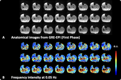 |
136 |
lower limbs skeletal muscle perfusion evaluation with the
Fourier decomposition technique
Jianxun Qu1 and
Tianye Lin2
1SIEMENS Healthineers, Beijing, China, 2Radiology Department, Peking University Cancer Hospital & Institute, Beijing, China Keywords: Muscle, Perfusion This study incorporated the Fourier decomposition (FD) technique to the lower extremity blood oxygen level-dependent, or reactive hyperemia, technique to evaluate the perfusion level of lower limbs. A periodically repeated ischemia-reperfusion paradigm was used to support the frequency analysis in FD. A characteristic frequency intensity corresponding to the ischemia-reperfusion cycle frequency was observed. The feasibility of the proposed method was demonstrated on healthy subjects. |
|
4067. |
137 |
Quiet Lumbar Spine MR Imaging: Preliminary Study with Different
Noise Reduction Factors
Jiageng Shen1,
Ailian Liu1,
Qingwei Song1,
Shuheng Zhang2,
and Guobin Li2
1the First Affiliated Hospital of Dalian Medical University, Dalian, China, 2Shanghai United Imaging Healthcare Co., Ltd, Shanghai, China Keywords: Muscle, Spinal Cord To evaluate the clinical value of quiet T1WI and T2WI sequences in lumbar spine imaging with different noise reduction factors (RFs). |
|
4068. |
138 |
Monitoring changes of the muscles around the knee joint in
amateur marathon athletes using synthetic MRI: a preliminary
study
Dantian Zhu1,
Wenhao Wu1,
Shaolin Li1,
Long Qian2,
Yajun Ma3,
and Yijie Fang1
1Department of Radiology,Fifth Affiliated Hospital, Sun Yat-Sen University, Zhuhai, China, 2MR Research, GE Healthcare, Beijing, China, 3University of California, San Diego, Department of Radiology, San Diego, CA, United States Keywords: Muscle, Muscle To investigate the value of synthetic MRI sequences for quantitative detection of the muscles around the knee joints before and after a marathon. Marathon runners were examined with Synthetic MRI sequences of both knees. Quantitative profiles of T1, T2, and PD were obtained after scanning. The differences in T1, T2, and PD values of each muscle were analyzed. Most muscle subregions had elevated T1, T2, and PD values 48 hours after the marathon compared to pre-race, and decreased after 1 month of post-race rest. The synthetic MRI sequences can be useful for detecting dynamic changes in the knee muscles. |
|
4069. |
139 |
Automatic Segmentation of Rotator Cuff Muscles on MR Images
Using Deep Learning
Ehsan Alipour1,
Majid Chalian1,
and Hesamoddin Jahanian1
1Radiology, University of Washington, Seattle, WA, United States Keywords: Muscle, MSK, Deep learning Rotator cuff (RC) injuries are a common occurrence affecting millions of people across the globe. Quantitative MRI-based evaluation of RC injuries can aid in early diagnosis and improve the treatment outcome. A crucial step towards developing a quantitative, clinically relevant methods for these patients, is developing reliable automatic techniques for segmentation of RC muscles. In this study, we developed a deep convolutional neural network model to automatically segment RC muscles on T1-weighted MR images. We showed that the proposed deep learning method provides rapid and reliable automatic segmentation of RC muscles, with an accuracy comparable with that of human raters. |
|
4070. |
140 |
The Feasibility Study of Extraocular Muscle Volume in Patients
with Myasthenia Gravis Based on 3D TOFMRA
Qin Zhou1,
Pei Chen1,
Zhiyun Yang1,
Xiaoxiao Zhao2,
Yingxi Chen1,
and Mengzhu Wang3
1The first affiliated hospital of sun yat-sen university, Guangzhou, China, 2The first affiliated hospital of sun yat-sen university, Guanzhou, China, 3MR Scientific Marketing, Siemens Healthineers Ltd, Guanzhou, China Keywords: Muscle, MR Value, myasthenia gravis, TOF MRA, extraocular muscle The study investigated the feasibility of TOF MAR sequence in evaluating extraocular muscle morphological changes in patients with myasthenia gravis. The results show that volume measurement of EOM based on TOF MRA imaging can effectively quantify atrophy of EOM in MG patients, which is significantly related to the poor response to medication and the long duration of the disease, meaning that it can be used as a non-invasive auxiliary diagnostic tool for prognosis evaluation of MG patients. |
|
 Back to Meeting Home
 Back to the Program-at-a-Glance
The International Society for Magnetic Resonance in Medicine is accredited by the Accreditation Council for Continuing Medical Education to provide continuing medical education for physicians.
 Back to Meeting Home
 Back to the Program-at-a-Glance
 View Presentation Video