Weekday Course
How to Image Inflammation in the Brain: From Tradition to Vision
ISMRM & ISMRT Annual Meeting & Exhibition • 03-08 June 2023 • Toronto, ON, Canada

Weekday Course
How to Image Inflammation in the Brain: From Tradition to Vision
| 16:00 |
 |
With PET/MRI & with PET Tracers
Bruno Stankoff
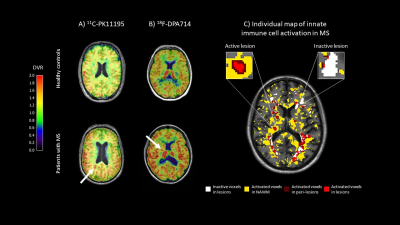
Keywords: Cross-organ: Inflammation, Neuro: Neurodegeneration, Contrast mechanisms: Molecular imaging Whereas the iron component sometimes associated with inflammation is accessible to MRI, the molecular specificity of PET is required to quantify neuroinflammation linked to innate immune cells. Tracers targeting TSPO are becoming largely available, with second generation tracers allowing an optimized sensitivity and specificity. Developing a TSPO PET imaging protocol implies the selection of an appropriate tracer, and the application of a robust and reproducible quantification model for data analysis. Following these requisites, promising results have been recently obtained in multiple sclerosis unravelling that an unexpectedly high proportion of lesions have a persistent neuroinflammatory content that drives progression. |
| 16:30 |
Image Inflammation in the Brain With Spectroscopy Tools (Proton & X-Nuclei)
Yao Li
|
|
| 17:00 |
Novel Preclinical Directions: Fluorine MRI
Sonia Waiczies
Keywords: Cross-organ: Inflammation, Physics & Engineering: Preclinical MRI, Contrast mechanisms: Molecular Imaging Inflammation is a key constituent of most neurological conditions including multiple sclerosis. Concerns related to gadolinium-based contrasts agents is limiting their use to monitor inflammation in patients. Preclinical efforts to quantify inflammation includes the development of fluorine-containing materials that can be detected with high specificity with fluorine (19F) MRI. This talk will go into the basics of 19F MRI, its strengths and weaknesses, as well as the approaches that strive to overcome those weaknesses. The idea of quantifying inflammation and anti-inflammatory treatment simultaneously will be introduced. Novel 19F reporter molecules and methods that improve 19F signal detection will be discussed. |
|
| 17:30 |
Novel Preclinical Directions: Nanoparticles/CEST
Aline Thomas
Keywords: Neuro: Neurodegeneration, Contrast mechanisms: Molecular imaging, : Preclinical/Animal Proton MRI has long been plagued by a lack of sensitivity to molecular events which historically restricted its use to monitoring gross anatomical changes. The development of and advancement in magnetization transfer-based contrast mechanisms and nanoparticle technology has dramatically expanded our ability to evaluate and monitor neuroinflammation using proton MRI at the molecular level. Many biomarkers in the former category, e.g. GluCEST, have already been translated to humans and are now being applied to a wide range of (neuro)inflammatory diseases. In contrast, most proton MRI agents that target specific molecules have just begun testing in the preclinical stages. |
 Back to Meeting Home
 Back to the Program-at-a-Glance
The International Society for Magnetic Resonance in Medicine is accredited by the Accreditation Council for Continuing Medical Education to provide continuing medical education for physicians.
 Back to Meeting Home
 Back to the Program-at-a-Glance
 View Presentation Video