Oral Session
Evaluating Therapeutic Response
ISMRM & ISMRT Annual Meeting & Exhibition • 03-08 June 2023 • Toronto, ON, Canada

| 13:45 |
0124.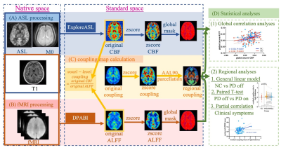 |
Effects of Levodopa Therapy on Neurovascular Coupling in
Parkinson’s Disease Patients
Chenqing Wu1,
Haoting Wu1,
Cheng Zhou1,
Jingwen Chen1,
Jiaqi Wen1,
Xiaocao Liu1,
Jianmei Qin1,
Zhengye Cao1,
Tao Guo1,
Xiaojun Guan1,
Xiaojun Xu1,
Baorong Zhang2,
and Minming Zhang1
1Department of Radiology, The Second Affiliated Hospital, Zhejiang University School of Medicine, Hangzhou, Zhejiang, China, 2Department of Neurology, The Second Affiliated Hospital, Zhejiang University School of Medicine, Hangzhou, Zhejiang, China Keywords: Parkinson's Disease, Neurodegeneration
Abnormal neurovascular coupling was found in PD
patients. These impairments were partially
normalized after levodopa therapy, and ALFF
alterations played a primary role in this
normalization effect.
|
| 13:53 |
0125.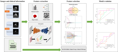 |
Deep Learning Radiomic Analysis of DCE-MRI Predicts Pathological
Complete Response to Neoadjuvant Chemotherapy in Breast Cancer
Yang Yang1,
Yaheng Fan2,
Xiaotong Xie3,
Bingsheng Huang2,
Yuting Li4,
Yan Li5,
Dinghua Xu6,
and Bihua Liu5 1Department of Radiology, Suining Central Hospital, Suining, China, 2Medical AI Lab, School of Biomedical Engineering, Health Science Center, Shenzhen University, Shenzhen, China, 3School of Life Science, South China Normal University, Guangzhou, China, 4The First Clinical Medical College, Guangdong Medical University, Zhanjiang, China, 5Department of Radiology, Dongguan People's Hospital, Dongguan, China, 6Department of Radiology, Affiliated Hospital of Guangdong Medical University, Zhanjiang, China Keywords: Breast, Treatment, dynamic contrast-enhanced magnetic resonance imaging, pathological complete response, radiomics Based on pre-treatment and early treatment dynamic contrast-enhanced magnetic resonance imaging (DCE-MRI) and clinical characteristics, we established a pathological complete response (pCR) prediction model using a deep learning radiomic (DLR) method that achieved good performance in the training and validation cohorts. The model can help clinicians evaluate whether the patient can reach pCR after neoadjuvant chemotherapy (NAC) and can provide an effective diagnostic reference for accurate medical treatment of patients receiving NAC. |
14:01 |
0126.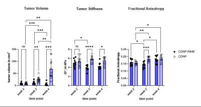 |
Magnetic Resonance Elastography reveals effects of immunotherapy
on glioma biomechanics
Yannik Streibel1,
Jessica Hunger1,2,3,
Chenchen Pan4,5,
Verena Turco2,
Manuel Fischer1,
Volker Sturm1,
Kianush Karimian-Jazi1,4,
Dennis A. Agardy2,3,5,
Giacomo Annio6,7,
Rami Mustapha8,
Christopher B. Rodell9,
Wolfgang Wick4,5,
Ralph Sinkus6,7,
Sabine Heiland1,
Frank Winkler4,5,
Michael Platten2,10,
Martin Bendszus1,
Michael O. Breckwoldt1,2,
and Katharina Schregel1,4
1Department of Neuroradiology, Heidelberg University Hospital, Heidelberg, Germany, 2Clinical Cooperation Unit Neuroimmunology and Brain Tumor Immunology, German Cancer Consortium (DTK) within the German Cancer Research Center (DKFZ), Heidelberg, Germany, 3Faculty of Biosciences, Heidelberg University, Heidelberg, Germany, 4Clinical Cooperation Unit Neurooncology, German Cancer Consortium (DTK) within the German Cancer Research Center (DKFZ), Heidelberg, Germany, 5Department of Neurology, Heidelberg University Hospital, Heidelberg, Germany, 6INSERM UMRS1148 - Laboratory for Vascular Translational Science, University Paris, Paris, France, 7School of Biomedical Engineering and Imaging Sciences, King’s College London, London, United Kingdom, 8Richard Dimbleby Laboratory of Cancer Research, School of Cancer & Pharmaceutical Sciences, King’s College London, London, United Kingdom, 9School of Biomedical Engineering, Science and Health Systems, Drexel University, Philadelphia, PA, United States, 10Department of Neurology, University Medical Center Mannheim, Heidelberg University, Mannheim, Germany Keywords: Elastography, Neuro MRI and MRE were used to monitor the effects of immunotherapy on tumor volume, FA and biomechanics of murine orthotopic glioma. Treated tumors were significantly smaller, softer and had lower FA than controls. This difference was most pronounced when comparing tumor stiffness of both groups. Controls revealed heterogeneous tumor stiffness. We hypothesize that this is caused by viable tumor cells alternated with necrotic areas and presumably immune-suppressive iba1-positive cells. In contrast, biomechanical properties of treated animals correlated better with presence of macrophages/microglia and likely reflected anti-tumorigenic inflammation. Thus, MRE could prove useful for monitoring glioma response to therapy. |
| 14:09 |
0127.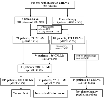 |
Transformation of the histopathological growth pattern of
colorectal liver metastases after chemotherapy predicted by an
MRI radiomics model
Shengcai Wei1,
Jing Cheng1,
and Yi Wang1
1Department of Radiology, Peking University People's Hospital, Beijing, China Keywords: Radiomics, Machine Learning/Artificial Intelligence, Histopathological growth patterns; colorectal liver metastases; transformation Our study used an MRI-based radiomics model to predict the transformation of the histopathological growth pattern (HGP) of colorectal liver metastases (CRLMs) before and after chemotherapy. After collecting data, drawing regions of interest and analyzing radiomics, we enrolled 152 patients and 299 liver metastases (99 pure desmoplastic (pdHGP) and 174 non-pdHGP). The pdHGP in the non-chemotherapy group and the post-chemotherapy group accounted for 28.3% and 42.5%, respectively (P=0.019). The fused MRI-based radiomics model demonstrated good predictive performance, and it could predict pdHGP before chemotherapy (25.3%), which was significantly different (p=0.034) compared with postoperative pdHGP after chemotherapy (39.1%). |
| 14:17 |
0128.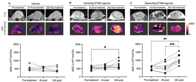 |
Acute tumour response to STING activation assessed with
multi-parametric MRI
Upasana Roy1,
Malin Pedersen1,
Carol Box1,
Jessica K.R. Boult1,
Antonio Rullan1,
Michael Schmohl2,
Sebastian Carotta3,
Kevin J. Harrington1,
and Simon P. Robinson1
1Radiotherapy and Imaging, The Institute of Cancer Research, Sutton, United Kingdom, 2Pharma GmbH & Co. KG, Boehringer Ingelheim, Biberach, Germany, 3RCV GmBH & Co KG, Boehringer Ingelheim, Vienna, Austria Keywords: Cancer, Drug Development, Preclinical, Diffusion Imaging, Relaxometry Whilst cancer immunotherapies have shown marked and durable tumour responses in some patients, the majority derive no benefit. Strategies are being exploited to enhance tumour responses to immuno-oncology agents, including pharmacological activation of the STING pathway to create a more inflamed microenvironment. Multi-parametric MRI revealed a dose-dependent increase in murine tumour ADC in response to a STING agonist that was associated with histologically confirmed increase in tumour cell death. An acute, transient increase in tumour R2*, consistent with vascular occlusion, was also found. ADC is a sensitive early imaging biomarker of tumour response to STING agonism. |
| 14:25 |
0129.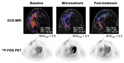 |
Multimodal serial assessment of neoadjuvant therapy response and
recurrence-free survival in locally advanced breast cancer
Anum S. Kazerouni1,
Lanell M. Peterson1,
Isaac Jenkins2,
Alena Novakova1,
Hannah M. Linden1,
David Hockenbery1,
David Mankoff3,
Jennifer Specht1,
and Savannah C. Partridge1
1University of Washington, Seattle, WA, United States, 2Fred Hutch Cancer Center, Seattle, WA, United States, 3University of Pennsylvania, Philadelphia, PA, United States Keywords: Breast, Cancer We investigated the combination of DCE-MRI and 18F-FDG PET for assessing metabolism/perfusion mismatch and predicting pathological response and recurrence-free survival (RFS) in women undergoing neoadjuvant chemotherapy (NAC) for breast cancer. Thirty-five patients with localized breast cancer were imaged with both modalities at 3 timepoints over the course of NAC. Greater mid-treatment decreases in imaging measures were predictive of pathological response and associated with improved long-term outcome. Furthermore, mid-treatment decreases in metabolism/perfusion ratios were predictive of RFS. These results indicate a complementary relationship between DCE-MRI and 18F-FDG PET metrics and potential value of metabolism/perfusion mismatch as a marker of response. |
| 14:33 |
0130.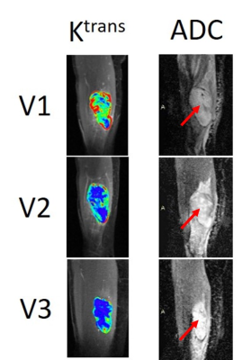 |
Multi-Parametric Quantitative MRI Assessment of Soft Tissue
Sarcoma Response to Preoperative Therapy
Wei Huang1,
Andy Kaempf1,
Brooke Beckett1,
Alina Tudorica1,
Jeong Youn Lim1,
and Christopher Ryan1
1Oregon Health & Science University, Portland, OR, United States Keywords: Cancer, Treatment, DCE-MRI, DW-MRI, Therapy Response, Ktrans, ADC Thirty patients with lower extremity soft tissue sarcoma were treated with preoperative chemoradiotherapy and underwent MRI exams including DCE- and DW-MRI pre-therapy, after the first cycle of therapy regimen, and after completion of therapy but before surgery. Ten patients had optimal and the other 20 had sub-optimal pathologic responses based on necrosis percentage of the surgical tumor specimens. Quantitative DCE-MRI markers and apparent diffusion coefficient (ADC) were superior to radiographic tumor size measurement in early prediction and evaluation of therapy response. Multi-parametric approach of combining DCE- and DW-MRI markers further improved early prediction of optimal vs. sub-optimal response. |
| 14:41 |
0131.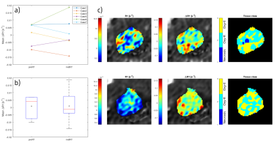 |
Feasibility of oxygen-enhanced (OE) and intravoxel incoherent
motion (IVIM) MRI for detection of HN cancer radiation therapy
induced changes
Emilia Palmér1,
Jesper Brovall2,
Oscar Jalnefjord1,2,
Karin Petruson3,
Fredrik Nordström 1,2,
Anna Karlsson1,2,
Maria Ljungberg1,2,
and Maja Sohlin1,2
1Department of Medical Radiation Sciences, Institute of Clinical Sciences, Sahlgrenska Academy, University of Gothenburg, Gothenburg, Sweden, 2Department of Medical Physics and Biomedical Engineering, Sahlgrenska University Hospital, Gothenburg, Sweden, 3Department of Oncology and Radiotherapy, Institute of Clinical Sciences, Sahlgrenska Academy, University of Gothenburg, Gothenburg, Sweden Keywords: Multi-Contrast, Radiotherapy, Diffusion/Other Diffusion Imaging Techniques Tumor oxygenation is a biomarker proposed as a predictor of radiation therapy (RT) response. Here, the feasibility of Oxygen-Enhanced MRI, intravoxel incoherent motion, and diffusion kurtosis imaging for monitoring of oxygenation changes in head and neck cancers was evaluated. Seven patients were examined pre- and mid-RT. No relative change in population mean longitudinal relaxation rate was observed following RT. A general increase was noticed in population mean diffusion and capillary perfusion fraction, and a decrease in population mean kurtosis. The implementation of these techniques was clinically feasible, and relative changes in almost every derived biomarker could be observed following RT. |
| 14:49 |
0132.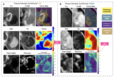 |
Self-supervised pretraining and network ensembling for spatial
mapping of treatment-effect in recurrent GBM with physiologic
MRI
Jacob Ellison1,2,3,
Nate Tran1,2,3,
Rany Hanna1,2,
Julia Cluceru1,2,
Joanna Phillips4,5,
Annette Molinaro5,
Valentina Pedoia1,2,3,
Tracy Luks1,
Anny Shai4,5,
Devika Nair1,
Javier Villanueva-Meyer1,2,
Mitchel Berger5,
Shawn Hervey-Jumper5,
Manish Aghi5,
Susan Chang5,
and Janine Lupo1,2,3 1Radiology and Biomedical Imaging, UCSF, San Francisco, CA, United States, 2Center for Intelligent Imaging, UCSF, San Francisco, CA, United States, 3Graduate Group in Bioengineering, UCSF - UC Berkeley, San Francisco, CA, United States, 4Pathology, UCSF, San Fransisco, CA, United States, 5Neurological Surgery, UCSF, San Francisco, CA, United States Keywords: Machine Learning/Artificial Intelligence, Modelling Prior characterization of treatment-effect and tumor recurrence using deep learning approaches have not optimized for spatial classification within a single lesion, which could improve surgical planning and treatment. 10mm patches of pre-surgical anatomical and physiological images surrounding the locations of histopathologically-confirmed tissue samples were used to train our models. Including physiological images, pretraining on unlabeled data in an autoencoding task, and training with an alternative cross-validation approach that enabled many networks to be ensembled, we achieved an ensembled test AUROC of 0.814 and generated spatial maps of tumor probability and model uncertainty. Performance decreased when removing any of these components. |
| 14:57 |
0133.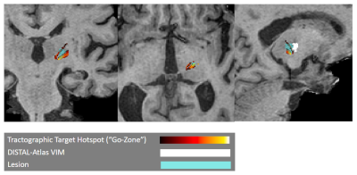 |
Clinically Feasible Patient-Specific Targeting Method for
Improved Quality-of-Life Outcomes from MRgFUS Treatment of
Essential Tremor
Anjali Datta1,
Gustavo Chau Loo Kung2,
Kristin Quah3,
Daniel Barbosa4,
Chelsea Li5,
David Purger5,
Allan Wang5,
Yosefi Chodakiewitz1,
Pejman Ghanouni1,
Vivek Buch5,
and Jennifer McNab1
1Radiology, Stanford University, Stanford, CA, United States, 2Bioengineering, Stanford University, Stanford, CA, United States, 3Electrical Engineering, Stanford University, Stanford, CA, United States, 4Neurosurgery, University of Pennsylvania, Philadelphia, PA, United States, 5Neurosurgery, Stanford University, Stanford, CA, United States Keywords: Data Analysis, Tractography & Fibre Modelling, Neurosurgical Targeting MRI-guided focused-ultrasound (MRgFUS) is an FDA-approved treatment for essential tremor. Unfortunately, the target to be ablated cannot be directly visualized on standard imaging, and its location varies interindividually. Since suboptimal lesion location can lead to side effects that impair quality of life, a method that locates personalized ablative targets that correlate with better quality-of-life outcomes (and not just with tremor reduction) could provide significant benefit. We present a clinically-feasible patient-specific probabilistic-tractography-based method for personalized targeting of MRgFUS treatment and show that similarity between the target it generates and the ablated lesion predicts QoL outcome in a dataset of 36 patients. |
| 15:05 |
0134.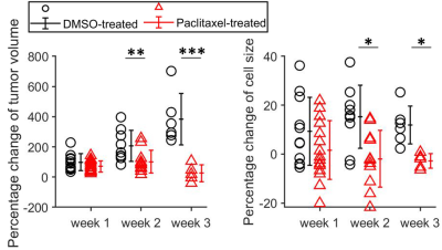 |
Non-invasive characterization of response of breast cancer to
paclitaxel chemotherapy using MR cell size imaging
xiaoyu jiang1,
Jingping Xie2,
John Gore2,
and Junzhong Xu3
1Radiology, Vanderbilt University Medical Center, nashville, TN, United States, 2Vanderbilt University Medical Center, nashville, TN, United States, 3Vanderbilt University Medical Center, Brentwood, TN, United States Keywords: Quantitative Imaging, Cancer, microstructure; diffusion; apoptosis Reliable and sensitive methods for assessing the response of breast cancer to treatment are critical for timely adjustments of therapies for individual patients, and development of novel therapies. Different from the conventional tumor-volume-based criteria, we hypothesize that more specific microstructural information on the cellular level in tumors, such as changes in cell size, may provide more accurate characterization of therapeutic response. Using both in vitro cell experiments and in vivo xenograft experiments, we demonstrated that temporal changes in MR-derived cell sizes in triple-negative MDA-MB-231 tumors treated with either drug vehicle or paclitaxel provide a new means to assess treatment response. |
| 15:13 |
0135.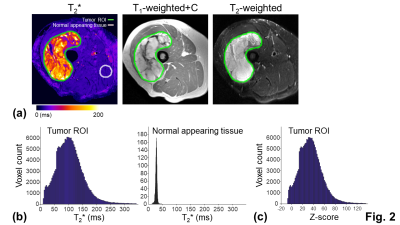 |
T2* assessment of neoadjuvant radiation therapy combined with
pharmacological ascorbate in extremity soft-tissue sarcomas: a
pilot study
Chu-Yu Lee1,
Michael S Petronek2,
Varun Monga3,
Benjamin J Miller4,
Mohammed M Milhem3,
Vincent A Magnotta1,
and Bryan G Allen2
1Department of Radiology, The University of Iowa, Iowa City, IA, United States, 2Department of Radiation Oncology, Free Radical and Radiation Biology Program, The University of Iowa, Iowa City, IA, United States, 3Department of Internal Medicine, The University of Iowa, Iowa City, IA, United States, 4Department of Orthopedics and Rehabilitation, The University of Iowa, Iowa City, IA, United States Keywords: Muscle, Tumor, Soft-tissue sarcomas; Treatment assessment; Relaxometry Soft-tissue sarcomas is commonly treated by neoadjuvant radiation therapy followed by surgical resection. Current assessment of neoadjuvant therapy relies on pathological examinations after surgery. Nonetheless, noninvasive imaging assessment can be performed before and during the treatment, offering the opportunity for predicting and early assessing treatment response. This pilot study applied T2* mapping to evaluate neoadjuvant therapy in seven patients with soft-tissue sarcomas before, during the treatment, and before surgery. The results showed strong or moderate correlations between T2* measurements and percent necrosis from pathological examinations, suggesting the potential for using T2* mapping to predict and early assess treatment response. |
| 15:21 |
0136.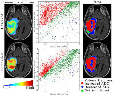 |
Effect of chemoradiation on high-grade gliomas can be forecasted
by mid-treatment images via image-driven mathematical modeling
David A Hormuth II1,2,
Maguy Farhat3,
Julianna Bronk3,
Holly Langshaw3,
Thomas E Yankeelov1,2,4,5,6,7,
and Caroline Chung3
1Oden Institute for Computational Engineering and Sciences, The University of Texas at Austin, Austin, TX, United States, 2Livestrong Cancer Institutes, The University of Texas at Austin, Austin, TX, United States, 3Radiation Oncology, The University of Texas MD Anderson Cancer Center, Houston, TX, United States, 4Biomedical Engineering, The University of Texas at Austin, Austin, TX, United States, 5Oncology, The University of Texas at Austin, Austin, TX, United States, 6Diagnostic Medicine, The University of Texas at Austin, Austin, TX, United States, 7Imaging Physics, The University of Texas MD Anderson Cancer Center, Houston, TX, United States Keywords: Tumors, Radiotherapy Technical advances in imaging and radiotherapy in the last decade have motivated the development of patient-specific treatment plans accounting for the underlying tumor biology that ultimately informs treatment efficacy. A fundamental challenge is how to leverage imaging data to optimize patient response to radiotherapy. Towards this end, we have developed an approach to forecast tumor response prior to the conclusion of therapy to enable a spatially-resolved map of response to chemoradiation. Our forecasting approach can accurately identify statistically significant changes in cell density that could potentially inform treatment adaptations. |
| 15:29 |
0137.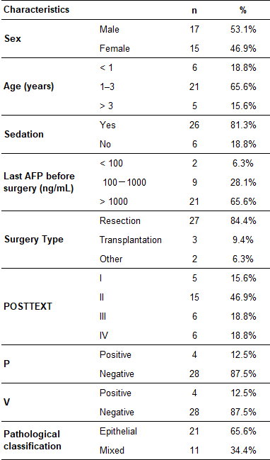 |
Pediatric Hepatoblastoma After Neoadjuvant Chemotherapy:
Diagnostic Performance of MR In Staging POSTTEXT and Vascular
Involvement.
Li Jun Qian1,
Xu Hua Gong1,
Ming Xuan Feng2,
Hai Nan Ren1,
Yan Zhou1,
Jian Rong Xu1,
Qiang Xia2,
and Yang Song3
1Radiology, Renji Hospital, Shanghai Jiao Tong University, School of Medicine, Shanghai, China, 2Liver Surgery, Renji Hospital, Shanghai Jiao Tong University, School of Medicine, Shanghai, China, 3MR Scientific Marketing, Siemens Healthineers Ltd., Shanghai, China Keywords: Cancer, Liver, Hepatoblastoma This study evaluates the diagnostic efficacy of contrast-enhanced MR imaging for preoperative POSTTEXT staging and prediction of vascular involvement in pediatric hepatoblastoma after neoadjuvant chemotherapy. The findings revealed that MR preoperative POSTTEXT staging has a good level of agreement with the reference standard. MR provides high predictive performance for identifying portal vein involvement and moderate predictive performance for identifying hepatic vein/inferior vena cava involvement. |
| 15:37 |
0138.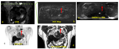 |
Detection and Assessment of Treatment Response in Multiple
Myeloma using Whole-body DETECT: Quantitative Biomarkers of MRI
and FDG-PET
Sheng Qing Lin1,
Sebastian Fonseca1,
Durga Udayakumar1,2,
Alberto Diaz de Leon1,
Orhan Oz1,
Gurbakhash Kaur3,
Aimaz Afrough3,
Larry D. Anderson Jr.3,
and Ananth J. Madhuranthakam1,2
1Radiology, UT Southwestern Medical Center, Dallas, TX, United States, 2Advanced Imaging Research Center, UT Southwestern Medical Center, Dallas, TX, United States, 3Internal Medicine, UT Southwestern Medical Center, Dallas, TX, United States Keywords: Cancer, Quantitative Imaging, Disease Biomarkers WBMRI-DETECT has been previously shown to have improved lesion conspicuity and shorter scan times compared to WBMRI-STIR and WBMRI-DWIBS in multiple myeloma. In this work, we demonstrate that quantitative biomarkers measured through WBMRI, fat fraction (FF) and apparent diffusion coefficient (ADC), have weak correlation suggesting both biomarkers to provide complementary information. Additionally, both ADC and FF showed negative correlation with the semiquantitative SUVmax from FDG-PET, demonstrating potential use of quantitative MRI biomarkers for the assessment of therapy response. |
 Back to Meeting Home
 Back to the Program-at-a-Glance
The International Society for Magnetic Resonance in Medicine is accredited by the Accreditation Council for Continuing Medical Education to provide continuing medical education for physicians.
 Back to Meeting Home
 Back to the Program-at-a-Glance
 View Presentation Video