Oral Session
MR Fingerprinting & Synthetic MRI
ISMRM & ISMRT Annual Meeting & Exhibition • 03-08 June 2023 • Toronto, ON, Canada

| 08:15 |
0421.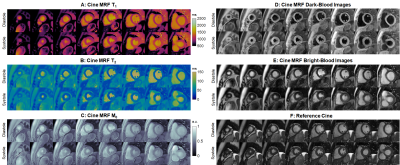 |
MR Fingerprinting with a Deep Image Prior Reconstruction for
Combined T1, T2, and M0 Mapping and Multi-Contrast Cine Imaging
Jesse Ian Hamilton1,2,
Gastao Lima da Cruz1,
Imran Rashid3,4,
Sanjay Rajagopalan3,4,
and Nicole Seiberlich1,2 1Radiology, University of Michigan, Ann Arbor, MI, United States, 2Biomedical Engineering, University of Michigan, Ann Arbor, MI, United States, 3Harrington Heart and Vascular Institute, University Hospitals Cleveland Medical Center, Cleveland, OH, United States, 4Medicine, Case Western Reserve University, Cleveland, OH, United States Keywords: MR Fingerprinting/Synthetic MR, Cardiovascular, Quantitative Imaging This work introduces a self-supervised deep learning reconstruction for cine Magnetic Resonance Fingerprinting, allowing for simultaneous cardiac phase-resolved T1, T2, and M0 mapping (without motion correction or averaging of data across different phases) and bright-blood and dark-blood cine imaging during a 10-second breathhold, with a temporal resolution (24 phases) comparable to standard cine imaging. Results are presented in simulations using the XCAT phantom and in healthy subjects, where the proposed reconstruction yielded reduced noise, undersampling artifacts, and motion blurring compared to previous low-rank and motion-corrected methods. |
| 08:23 |
0422.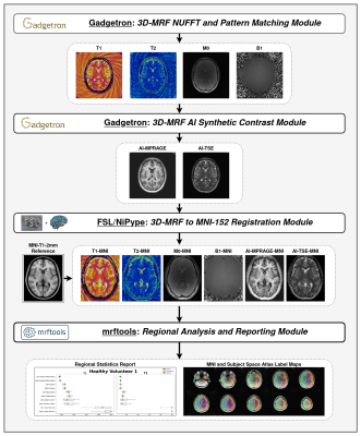 |
Fully Automated Online Reconstruction, Registration, and
Analysis Pipeline for 3D Magnetic Resonance Fingerprinting
Andrew Dupuis1,
Rasim Boyacioglu1,
Yong Chen1,
Michael Hansen2,
Kelvin Chow3,
Chaitra Badve4,
Dan Ma1,
and Mark Griswold1 1Biomedical Engineering, Case Western Reserve University, Cleveland, OH, United States, 2Microsoft Research, Redmond, WA, United States, 3Siemens Medical Solutions USA, Inc., Chicago, IL, United States, 4Radiology, University Hospitals, Cleveland, OH, United States Keywords: MR Fingerprinting/Synthetic MR, Quantitative Imaging, Gadgetron, Brian Tumor, MNI, FSL, Docker, Automated, Registration In this study, to overcome some of the clinical integration issues of MRF, we present a fully automated online reconstruction and post-processing pipeline for 3D-MRF where the quantitative maps and custom reports are returned to the scanner in real time. The whole pipeline is hosted in a Kubernetes cluster which includes Gadgetron, FSL and other custom tools in discrete Docker images. To illustrate the capability of the pipeline, 3D-MRF raw datasets of healthy and brain tumor patient datasets are reconstructed in a cloud-based Gadgetron, registered to MNI space using FSL and analyzed to compare with population based regional maps. |
08:31 |
0423.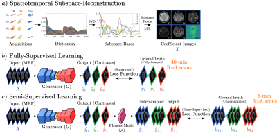 |
Semi-Supervision for Clinical Contrast-Weighted Image Synthesis
from Magnetic Resonance Fingerprinting
Mahmut Yurt1,
Cagan Alkan1,
Sophie Schauman1,2,
Xiaozhi Cao1,2,
Congyu Liao1,2,
Siddharth Iyer1,3,
Tolga Cukur4,
Shreyas Vasanawala2,
John Pauly1,
and Kawin Setsompop1,2 1Department of Electrical Engineering, Stanford University, Stanford, CA, United States, 2Department of Radiology, Stanford University, Stanford, CA, United States, 3Department of Electrical Engineering and Computer Science, Massachusetts Institute of Technology, Cambridge, MA, United States, 4Department of Electrical Engineering, Bilkent University, Ankara, Turkey Keywords: MR Fingerprinting/Synthetic MR, MR Fingerprinting Previous works have introduced deep models to synthesize clinical contrast-weighted images from magnetic resonance fingerprinting (MRF). Although these models achieve high synthesis accuracy, they demand full-supervision from fully-sampled training data of clinical contrasts which might become difficult to acquire across diverse sets due to scan costs. To eliminate undesirable reliance on full-supervision, we introduce a semi-supervised model, ssMRF, that allows training using accelerated references. ssMRF introduces a semi-supervised loss function based only on collected k-space samples of clinical contrasts, and further leverages complementary Poisson disc masks, via a multi-task learning protocol to synergistically synthesize multiple contrasts. |
| 08:39 |
0424.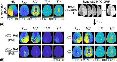 |
Only-Train-Once MR Fingerprinting for B0 and B1 Inhomogeneity
Correction in Quantitative Magnetization Transfer Contrast
Beomgu Kang1,
Munendra Singh2,
HyunWook Park1,
and Hye-Young Heo2
1School of Electrical Engineering, Korea Advanced Institute of Science and Technology, Daejeon, Korea, Republic of, 2Department of Radiology, Johns Hopkins University, Baltimore, MD, United States Keywords: MR Fingerprinting/Synthetic MR, Machine Learning/Artificial Intelligence, B0 and B1 correction Magnetization transfer contrast MR fingerprinting (MTC-MRF) enables fast reconstruction of free bulk water and semisolid macromolecules parameters. However, B0 and B1 inhomogeneities that originate from system imperfection can corrupt MR fingerprints, thereby impairing the tissue quantification. We proposed a fast, deep-learning MTC-MRF technique that simultaneously estimates multiple tissue parameters and corrects the effect of B0 and B1 variations. An only-train-once recurrent neural network was designed to perform the fast tissue parameter quantification regardless of MRF acquisition schedule. This allows a dynamic scan-wise linear calibration of the scan parameters using the measured B0 and B1 maps. |
| 08:47 |
0425.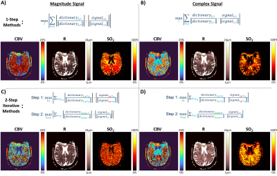 |
Optimizing an Accelerated Spin- and Gradient-Echo Sequence for
Dynamic MR Vascular Fingerprinting
Gregory J. Wheeler1,
Linh N.N. Le1,
Quimby N. Lee2,
and Audrey P. Fan1,2
1Biomedical Engineering, University of California Davis, Davis, CA, United States, 2Neurology, University of California Davis, Davis, CA, United States Keywords: MR Fingerprinting/Synthetic MR, MR Fingerprinting An accelerated spin- and gradient-echo (SAGE) pulse sequence sensitive to changes in oxygenation has been demonstrated to be suitable for MR vascular fingerprinting (MRvF), potentially enabling quantitative, multiparametric mapping of dynamic vascular physiology. This study aimed to optimize this SAGE sequence and matching algorithms used in MRvF and found that selecting shorter echo times resulted in better signal properties and sensitivity for pattern matching with lower estimation error. This optimization will enable these techniques to be used during dynamic vascular challenges and investigations into multiple, simultaneous functional cerebrovascular biomarkers. |
| 08:55 |
0426.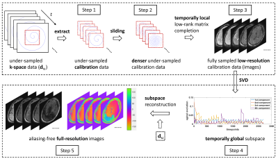 |
Simultaneous Quantification of Relaxation and Diffusion using MR
Fingerprinting with Self-Calibrated Subspace Reconstruction
Zhilang Qiu1,
Siyuan Hu1,
Walter Zhao1,
Ken Sakaie2,
Mark A. Griswold3,
Derek K. Jones4,
and Dan Ma1
1Biomedical Engineering, Case Western Reserve University, Cleveland, OH, United States, 2Imaging Institute, Cleveland Clinic, Cleveland, OH, United States, 3Radiology, Case Western Reserve University, Cleveland, OH, United States, 4Cardiff University Brain Research Imaging Centre (CUBRIC), School of Psychology, Cardiff University, Cardiff, United Kingdom Keywords: MR Fingerprinting/Synthetic MR, Diffusion/other diffusion imaging techniques We propose a self-calibrated subspace reconstruction method for multidimensional MR Fingerprinting (mdMRF) scan for simultaneous relaxation and diffusion mapping without pulsation gating. It is distortion-free, unlike EPI-based diffusion MRI. MRF images corrupted by phase errors due to bulk and physiological motions are automatically detected using an outlier detection algorithm and corrected, in order to generate artifact-free relaxation and diffusion maps. |
09:03 |
0427.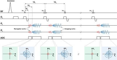 |
Improved Low-Rank and Subspace Reconstruction for Magnetic
Resonance Fingerprinting with Self-Navigating Acquisitions
Hengfa Lu1,
Huihui Ye2,3,
and Bo Zhao1,4
1Department of Biomedical Engineering, University of Texas at Austin, Austin, TX, United States, 2State Key Laboratory of Modern Optical Instrumentation, College of Optical Science and Engineering, Zhejiang University, Hangzhou, China, 3Center for Brain Imaging Science and Technology, Key Laboratory for Biomedical Engineering of Ministry of Education, College of Biomedical Engineering and Instrumental Science, Zhejiang University, Hangzhou, China, 4Oden Institute for Computational Engineering and Sciences, University of Texas at Austin, Austin, TX, United States Keywords: Quantitative Imaging, MR Fingerprinting Low-rank and subspace reconstruction methods have achieved state-of-the-art performance for MR Fingerprinting with highly-undersampled data. The existing methods learn the temporal subspace from an ensemble of magnetization evolutions generated from Bloch simulations. In this work, we present a novel self-navigating acquisition scheme for MR Fingerprinting, which utilizes a dual-echo acquisition strategy to enable subspace estimation from physically-acquired training data. The proposed acquisition substantially improves the accuracy of the low-rank and subspace reconstruction, especially when the acquisition length is short. We demonstrate the performance of the proposed method with phantom experiments and in vivo experiments. |
| 09:11 |
0428. |
Optimization of Magnetic Resonance Fingerprinting with Subspace
Reconstruction
Nan Wang1,
Xiaozhi Cao1,
Siddharth Srinivasan Iyer1,2,
Congyu Liao1,
Philiip K Lee1,
Molin Zhang2,
and Kawin Setsompop1,3
1Department of Radiology, Stanford University, Stanford, CA, United States, 2Department of Electrical Engineering and Computer Science, Massachusetts Institute of Technology, Cambridge, MA, United States, 3Department of Electrical Engineering, Stanford University, Stanford, CA, United States Keywords: Data Analysis, MR Fingerprinting Cramér-Rao Lower Bound has been used to optimize the acquisition design for MRF, but the effect from undersampling and reconstruction has not been taken into consideration. In this work, we evaluated the estimation error and standard deviation using CRLB optimized acquisition parameters with spiral undersampling trajectory and subspace reconstruction. The results demonstrated that CRLB produced lower estimation variation but is sensitive to the selected number of subspaces. A rank that is too low or two high an increase the estimation error. The dictionary match on coefficient maps provided lower estimation error and variation compared to match on whole time series. |
| 09:19 |
0429.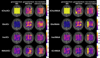 |
Generating Synthetic MR Spectroscopic Imaging using MRI and
Single-voxel MR Spectroscopy
Shuki Maruyama1 and
Hidenori Takeshima2
1Imaging Modality Group, Advanced Technology Research Department, Research and Development Center, Canon Medical Systems Corporation, Tochigi, Japan, 2Imaging Modality Group, Advanced Technology Research Department, Research and Development Center, Canon Medical Systems Corporation, Kanagawa, Japan Keywords: Machine Learning/Artificial Intelligence, Spectroscopy The authors proposed new methods to generate synthetic proton MR spectroscopic imaging (MRSI) data. The proposed methods were derived from Image-to-Image Translation with Conditional Adversarial Networks (pix2pix), taking MRI data or MRI and single-voxel MR spectroscopy (SVS) data as inputs. To integrate the features of MRI and SVS data, additional encoder and decoder networks were incorporated. The experimental results demonstrated that the proposed methods generated metabolite ratio maps with same resolution as MRI data. The synthetic maps generated from MRI+SVS were more consistent with the reference ones than those generated from MRI alone. |
| 09:27 |
0430.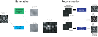 |
Synthesizing Complex Multicoil MRI Data from Magnitude-only
Images
Nikhil Deveshwar1,2,3,
Abhejit Rajagopal1,
Efrat Shimron3,
Sule Sahin1,2,
and Peder E.Z. Larson1,2
1Radiology and Biomedical Imaging, University of California, San Francisco, San Francisco, CA, United States, 2UC Berkeley - UCSF Graduate Program in Bioengineering, Berkeley and San Francisco, CA, United States, 3Electrical Engineering and Computer Sciences, University of California, Berkeley, Berkeley, CA, United States Keywords: Machine Learning/Artificial Intelligence, Machine Learning/Artificial Intelligence Obtaining paired, diverse and expert annotated medical data is extremely challenging especially for MRI reconstruction since raw data, including phase information is typically discarded from the scan leaving only the magnitude image for clinical assessment and diagnosis. Phase information contains valuable information about physiology, pathology and other tissue characteristics that are useful in developing more robust deep learning MRI reconstruction methods. In this work, we show that state of the art physics-based image reconstruction networks trained on synthetic raw MRI data consisting of synthetic phase and coil information perform comparably to image reconstruction networks trained on ground truth k-space data. |
| 09:35 |
0431.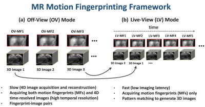 |
MR Motion Fingerprinting
Li Feng1
1Biomedical Engineering and Imaging Institute and Department of Radiology, Icahn School of Medicine at Mount Sinai, New York, NY, United States Keywords: Motion Correction, MR-Guided Interventions, real-time imaging This work proposes a new framework, called MR motion fingerprinting, for fast real-time 3D MRI with an imaging latency<500ms. MR motion fingerprinting has two stages of imaging. The first stage, called the off-view mode, acquires and reconstructs a 4D motion database consisting of hundreds of real-time 3D images and associated 2D motion fingerprints. The second step, called the live-view mode, only acquires 2D motion fingerprints, which are used to search for matched 3D images from the motion database. This new approach enables efficient, accurate and robust real-time imaging with ultralow latency, which could be used for adaptive radiotherapy on MRI-Linac. |
| 09:43 |
0432.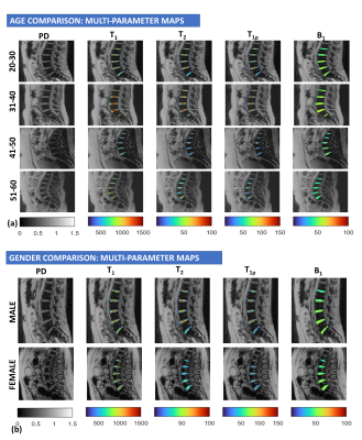 |
Characterization of age- and gender-dependent differences in the
inter-vertebral disc using MR-Fingerprinting and Textural
Analysis
Rajiv G Menon1,
Anmol Monga1,
Richard Kijowski1,
and Ravinder Regatte1
1Radiology, NYU Grossman School of Medicine, New York, NY, United States Keywords: MR Fingerprinting/Synthetic MR, Tissue Characterization, degenerative The study investigated age and gender differences in intervertebral disc (IVD) of the lumbar spine using a multiparameter MR fingerprinting (MRF) technique that simultaneously quantify T1, T2 and T1ρ. Seventeen healthy subjects were evaluated. There was a significant positive association between age and T1ρ of the IVD and between age and various gray level co-occurrence matrix features of T1 maps on texture analysis. There were no significant differences between males and females in T1, T2 and T1ρ of the IVD. The results demonstrate that multi-parameter MRF can effectively characterize early age-related IVD degeneration. |
| 09:51 |
0433.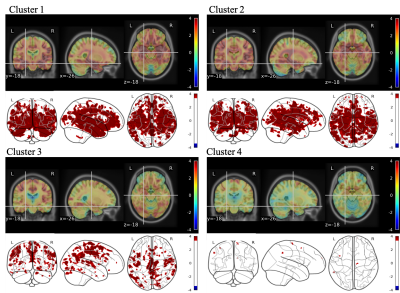 |
MRF-derived T1 and T2 map alterations in four different brain
atrophy patterns in a cohort of MCI subjects
Paolo Bosco1,
Laura Biagi1,
Matteo Cencini1,
Marta Lancione1,
Michela Matteoli2,
Simona Cintoli3,
Alessandro Sale2,
Nicoletta Berardi2,
Michela Tosetti1,
and the Train the Brain Consortium4
1IRCCS Stella Maris Foundation, Pisa, Italy, 2Institute of Neuroscience of the CNR, Pisa, Italy, 3Azienda Ospedaliero Universitaria Pisana, Pisa, Italy, 4the Train the Brain Consortium, Pisa, Italy Keywords: MR Fingerprinting/Synthetic MR, Alzheimer's Disease Brain atrophy evaluation is crucial in the assessment of neurodegenerative diseases. However, the most common MRI measures (mostly T1-weighted derived) cannot be considered as quantitative. In this study we use T1-weighted images to identify homogeneous groups of MCI subjects in terms of brain atrophy and we assessed the capability of T1 and T2 quantification through MRF technique to elucidate at voxel level the brain alterations with respect to an age-matched healthy population. Indeed, atrophy patterns can be identified in both T1 and T2 maps paving the way to innovative brain atrophy assessment in a quantitative fashion. |
| 09:59 |
0434.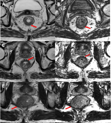 |
Combining T2-weighted and synthetic double inversion recovery
magnetic resonance imaging improved rectal cancer T staging
Zi Wang1,
Jiankun Dai2,
and Shudong Hu1
1Affiliated Hospital of Jiangnan University, wuxi, China, 2MR Reseach,GE Healthcare, Beijing, China Keywords: MR Fingerprinting/Synthetic MR, Cancer, rectal neoplasms; neoplasm staging; This study aimed at investigating the role of synthetic double inversion recovery (SyDIR) magnetic resonance imaging (MRI) in staging rectal cancer (RC). 79 pathologically confirmed RC patients were retrospectively selected. Our results showed SyDIR can provided RC anatomical features for muscular infiltration detection. Compared with using T2WI alone, the combination of T2WI and SyDIR improved the diagnose performance of muscle layer invasion and significantly increase the staging consistency between MRI and pathology for both junior and senior radiologist. Our study indicated the combination of T2WI and SyDIR would be beneficial for RC treatment selection. |
| 10:07 |
0435.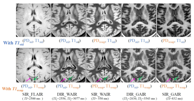 |
Water Suppression of T1 map and Synthetic Inversion Recovery
images in T2-based Water Suppression Synthetic MRI
(T2wsup-synMRI)
Tokunori Kimura1,
Natsumi Yamagishi1,
Yuki Masuda1,
and Mitsuyo Ito1
1Radiokogical engineering, Shizuoka College of Medical care Science, Hamamatsu, Japan Keywords: MR Fingerprinting/Synthetic MR, Brain The purpose of this study was to propose a new technique to provide water suppression (wsup) T1 maps by modifying our already proposed T2wsup technique, then to assess the effects of wsup-T1 on longitudinal magnetization (Mz) in synthetic MRI images of FLAIR and DIR by a simulation and an in-vivo MR study. The errors in DIR at CSF-tissue mixed portions were non-negligible with std-T1 but could be reduced with wsup-T1. The T2wsup-SynMRI technique can provide 3 kinds of water suppressed quantitative maps and the same contrasts synthetically as the acquired IR sequences of FLAIR and DIR. |
 Back to Meeting Home
 Back to the Program-at-a-Glance
The International Society for Magnetic Resonance in Medicine is accredited by the Accreditation Council for Continuing Medical Education to provide continuing medical education for physicians.
 Back to Meeting Home
 Back to the Program-at-a-Glance
 View Presentation Video