Oral Session
Flow, Perfusion & CSF
ISMRM & ISMRT Annual Meeting & Exhibition • 03-08 June 2023 • Toronto, ON, Canada

08:15 |
1174.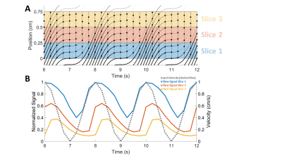 |
Modeling inflow effects in fast fMRI to quantify fluid flow
Baarbod Ashenagar 1,2,
Daniel Gomez1,2,3,
and Laura Lewis1,2
1Department of Biomedical Engineering, Boston University, Boston, MA, United States, 2Athinoula A. Martinos Center for Biomedical Imaging, Massachusetts General Hospital, Charlestown, MA, United States, 3Department of Radiology, Harvard Medical School, Boston, MA, United States Keywords: Signal Modeling, Velocity & Flow, time-of-flight, flow-enhanced fMRI signal Fast fMRI has recently been shown to be an effective method for detecting dynamic changes in cerebrospinal fluid flow, enabling measurement of a critical process for maintenance of brain health. However, while the inflow signal measured using fast fMRI is a surrogate for the underlying flow dynamics, it does not directly reflect the velocity and dynamics of flow. To understand the mapping between flow and the flow-enhanced MR signals they generate, we developed and validated a mathematical forward model that uses velocity as input and simulates dynamic fMRI inflow signal intensities for each slice of the imaging volume. |
|
08:23 |
1175.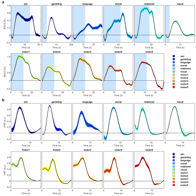 |
Towards whole brain mapping of the hemodynamic response function
Maria Guidi1,
Fabio Mangini1,2,
Marta Moraschi1,3,
Daniele Mascali1,3,
Michela Fratini3,4,
Silvia Mangia5,
Fabrizio Frezza2,
and Federico Giove1,3
1MARBILab, Enrico Fermi Research Center, Rome, Italy, 2Sapienza University, Rome, Italy, 3Fondazione Santa Lucia IRCCS, Rome, Italy, 4CNR-NANOTEC, Rome, Italy, 5CMRR, Minneapolis, MN, United States Keywords: Signal Modeling, fMRI, HRF We characterized a deconvolved haemodynamic response function (dHRF) across the whole cortex exploiting a sine series expansion in a cohort of young healthy subjects from the Human Connectome Project. We report, for different tasks and brain regions, the amplitude, latency, time-to-peak and full-width at half maximum of the fitted BOLD response and of the dHRF. We show that each of those parameters vary throughout the cortex and, to a smaller extent, across subjects. Additionally, the use of a flexible model, like the one we explored in this study, reveals that the HRF in some brain regions deviates from canonicity. |
08:31 |
1176.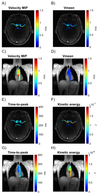 |
Integrated heart-brain 4D flow MRI evaluation of hemodynamic
coupling in healthy aging adults
Adam Richter1,
Jackson E. Moore1,
Maria Aristova1,
Ann Ragin1,
Susanne Schnell2,
Emily Rogalski3,
Michael Markl1,
and Kelly Jarvis1
1Department of Radiology, Feinberg School of Medicine, Northwestern University, Chicago, IL, United States, 2Department of Medical Physics, Institute of Physics, University of Greifswald, Greifswald, Germany, 3Psychiatry and Behavioral Sciences, Northwestern University, Chicago, IL, United States Keywords: Data Analysis, Velocity & Flow, Cerebral arteries Age-related changes in cardiovascular hemodynamics may be associated with brain changes. However, this relationship is not clear since blood flow in the heart and brain are usually imaged and analyzed in separate exams and pipelines. We have developed a technique for parameter mapping in both the aorta and intracranial vessels to allow for integrated MRI evaluation of heart-brain hemodynamic coupling. This study demonstrates a single MRI study and analysis pipeline to facilitate more comprehensive, integrated heart-brain hemodynamic analysis in clinical and research settings. |
|
08:39 |
1177.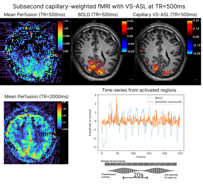 |
Capillary-weighted velocity-selective ASL for subsecond fast
fMRI
Daniel E. P. Gomez1,2,3,
Laura D. Lewis1,3,
Jonathan R. Polimeni1,2,4,
and Divya S. Bolar5 1Athinoula A. Martinos Center for Biomedical Imaging, Massachusetts General Hospital, Charlestown, MA, United States, 2Department of Radiology, Harvard Medical School, Boston, MA, United States, 3Department of Biomedical Engineering, Boston University, Boston, MA, United States, 4Division of Health Sciences and Technology, Massachusetts Institute of Technology, Cambridge, MA, United States, 5Center for Functional Magnetic Resonance Imaging, Department of Radiology, University of California San Diego, La Jolla, CA, United States Keywords: Data Acquisition, fMRI This work evaluates capillary-weighted VS-ASL for subsecond functional cerebral blood flow (CBF) mapping with high microvascular specificity. Three experiments were conducted: one to demonstrate that capillary-weighted VS-ASL is mostly free from macrovascular contamination when compared to arterial-weighted VS-ASL, one to demonstrate that functional MRI is feasible with capillary-weighted VS-ASL at moderate repetition times, and one to show that, despite lower sensitivity, capillary-weighted functional MRI can be performed at subsecond temporal resolution. Our findings suggest that capillary-weighted VS-ASL may enable the study of fast functional CBF responses with high neuronal specificity. |
| 08:47 |
1178.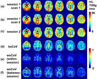 |
Test-Retest Reliability of 3D Velocity-Selective Arterial Spin
Labeling for Detecting Normal Variations of Cerebral Blood Flow
Feng Xu1,2,
Dapeng Liu1,2,
Dan Zhu1,2,
Argye E. Hillis3,
Arnold Bakker4,
Anja Soldan3,
Marilyn Albert3,
Doris D. M. Lin1,
and Qin Qin1,2
1The Russell H. Morgan Department of Radiology and Radiological Science, Johns Hopkins University, Baltimore, MD, United States, 2F.M. Kirby Research Center for Functional Brain Imaging, Kennedy Krieger Institute, Baltimore, MD, United States, 3Department of Neurology, Johns Hopkins University, Baltimore, MD, United States, 4Department of Psychiatry and Behavioral Sciences, Johns Hopkins University, Baltimore, MD, United States Keywords: Data Acquisition, Arterial spin labelling Velocity selective inversion (VSI) based velocity selective arterial spin labeling (VSASL) was recommended by a recent guideline paper. We conducted a test-retest study to evaluate the reliability of 3D VSI-VSASL. The correlations between repeated measures were 0.94/0.81 (within- /between-session) for individual absolute CBF and 0.99/0.98 for regional relative CBF. The intraclass correlation coefficients were 0.88/0.77 for absolute CBF and 0.92/0.85 for regional relative CBF. Between-subject variation in CBF was partially contributed by age and physiological parameters. VSI-VSASL demonstrates moderate to excellent reliability for detecting between-subject and between-region variations among healthy subjects, suggesting its merit in clinical applications. |
|
08:55 |
1179.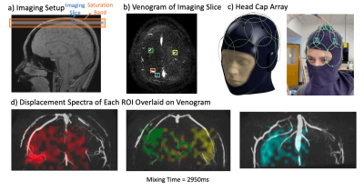 |
Advances in Post-Perfusion Venous Territories Imaging with
Displacement Spectrum Imaging (DiSpect)
Ekin Karasan1,
Jingjia Chen1,
Julian Maravilla1,
Zhiyong Zhang2,
Chunlei Liu1,3,
and Michael Lustig1
1Department of Electrical Engineering and Computer Science, University of California, Berkeley, Berkeley, CA, United States, 2School of Biomedical Engineering, Shanghai Jiao Tong University, Shanghai, China, 3Helen Wills Neuroscience Institute, Berkeley, CA, United States Keywords: Pulse Sequence Design, Perfusion DiSpect resolves the multi-dimensional displacement spectrum that spins exhibit between tagging and imaging. Previously, we showed that DiSpect can be used to trace blood draining from the capillary bed through the cerebral venous system. This work presents three innovations: 1) Validation of our results with flow phantom experiments and comparison of our in-vivo data with a detailed QSM-venogram. 2) Interleaved imaging to simultaneously probe the deep and superficial veins. 3) Use of a conformal head-cap array for imaging, with higher SNR, allowing dynamic measurement of vein drainage and observation of venous territories across large time scales. |
| 09:03 |
1180.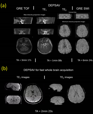 |
Modified dual-contrast multi-shot 3D EPI for distortion-free and
fast acquisition of simultaneous MR angiography and venography
(DEPSAV)
Yue Wu1,2,3,
Dehe Weng4,
Jing An4,
Rong Xue1,2,3,
Yan Zhuo1,2,3,
and Zihao Zhang1,2,5
1State Key Laboratory of Brain and Cognitive Science, Institute of Biophysics, Chinese Academy of Sciences, Beijing, China, 2University of Chinese Academy of Sciences, Beijing, China, 3The Innovation Center of Excellence on Brain Science, Chinese Academy of Sciences, Beijing, China, 4Siemens Shenzhen Magnetic Resonance Ltd, Shenzhen, China, 5Institute of Artificial Intelligence, Hefei Comprehensive National Science Center, Hefei, China Keywords: Data Acquisition, Blood GRE-based vascular imaging suffers from the low acquisition efficiency. In this study, we presented a new 3D dual-contrast multishot-EPI based acquisition method called DESPAV for simultaneous MR angiography and venography. Full flow compensation for K-space center line and novel Center-out trajectory was implemented. A reconstruction pipeline was developed to correct for inter-shot phase errors, off-resonance induced artifact, and enable distortion-free multi-shot joint reconstruction. Preliminary results showed that DEPSAV can provide fast intracranial arterial and venous vasculature depiction with comparable contrast and image quality to 3D GRE-based TOF/SWI, while achieve ~3-fold reduction in acquisition time. |
| 09:11 |
1181.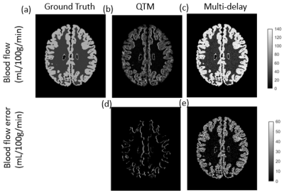 |
Quantitative transport mapping (QTM) of the brain with simulated
microvasculature model
Renjiu Hu1,2,
Qihao Zhang2,3,
Dominick J. Romano2,3,
and Yi Wang2,3
1Sibley School of Mechanical and Aerospace Engineering,, Cornell University, Ithaca, NY, United States, 2Department of Radiology, Weill Cornell Medicine, New York, NY, United States, 3Meinig School of Biomedical Engineering, Cornell University, Ithaca, NY, United States Keywords: Quantitative Imaging, Arterial spin labelling We compared the quantitative mappings on the simulated the brain arterial spin labeling images with artificial microvasculature structures and perfusion properties. The QTM method gives similar quantification on blood flow as the traditional tracer kinetic model. |
|
09:19 |
1182.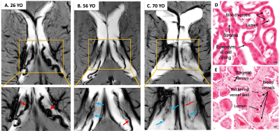 |
Age-related Vascular Changes in Choroid Plexus Evaluated Using
High-resolution USPIO-Enhanced 7T MRI
Zhe Sun1,2,
Chenyang Li1,2,
Marco Muccio1,
Li Jiang1,
and Yulin Ge1
1Department of Radiology, New York University Grossman School of Medicine, New York, NY, United States, 2Vilcek Institute of Graduate Biomedical Sciences, New York University Grossman School of Medicine, New York, NY, United States Keywords: Neurofluids, Aging The choroid plexus (ChP) is a highly vascularized structure that is important in CSF production and waste solute clearance. In this work, USPIO-enhanced 7T imaging was used to detect microvascular changes associated with normal aging. We showed improved susceptibility contrast of ChP vessels and surrounding stromal tissues on high-resolution USPIO-enhanced 2D T2*-weighted MRI. Both age-related sparser vasculature and distended stromal tissues were identified. ChP volume increases with age, whereas the difference between pre-/post-contrast quantitative susceptibility map (QSM) decreases with age (P<0.05); and vascular degenerative change may occur earlier than volume change. |
| 09:27 |
1183. |
Quantitative Flow Velocity in Cerebral Perforating Arteries with
7T MRI: the EUFIND study.
Stanley D.T. Pham1,
Jari T. van Vliet2,
Rick J van Tuijl2,
Geert Jan Biessels3,
Mauro Costagli4,
Mark A. van Buchem5,
Oliver Kraff6,
Arno Villringer7,
Mark E. Ladd8,
Jeroen C.W. Siero2,9,
Ludovic de Rochefort10,
and Jaco J.M. Zwanenburg2
1UMC Utrecht, Utrecht, Netherlands, 2Radiology, UMC Utrecht, Utrecht, Netherlands, 3Neurology, UMC Utrecht, Utrecht, Netherlands, 4IMAGO7 Research Foundation, Pisa, Italy, 5Neuroradiology, Leiden University Medical Center, Leiden, Netherlands, 6Radiology, University Hospital Essen, Essen, Germany, 7Max Planck Institute for Human Cognitive and Brain Sciences, Leipzig, Germany, 8German Cancer Research Center (DKFZ), Heidelberg, Germany, 9Spinoza Centre for Neuroimaging, Amsterdam, Netherlands, 10Centre for Magnetic Resonance in Biology and Medicine (UMR 7339), Marseille, France Keywords: Data Acquisition, High-Field MRI, Harmonization, two dimensional phase contrast We analyzed multi-center blood flow velocity measurements in perforating arteries from 7T MRI with the two-dimensional phase-contrast (2D-PC) sequence (eight sites, comprising three MRI vendors). Analysis was performed with the software tool Small vessEL MRI MArkers (SELMA). Inter-rater reliability of SELMA was excellent with overall intra-class coefficients for number of vessels (Ndetected), mean velocity (Vmean) and velocity pulsatility index (vPI) of at least 0.84 for 2D-PC data from all MRI vendors. Inter-vendor differences were larger than the intra-vendor differences (coefficients of variation: 0.62 vs. 0.39, 0.21 vs. 0.12 and 0.39 vs. 0.12 for Ndetected, Vmean and vPI, respectively). |
| 09:35 |
1184.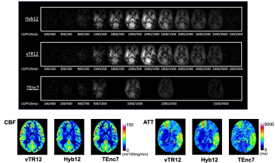 |
Hybrid multi-delay PCASL of time-encoded and variable-TR schemes
for the assessment of cerebral perfusion in Moyamoya disease
Osamu Togao1,
Makoto Obara2,
Koji Yamashita3,
Kazufumi Kikuchi4,
Tatsuhiro Wada5,
Chiaki Tokunaga5,
Ryoji Mikayama5,
Shota Ishida6,
Hiroshi Hamano2,
Lena Vaclavu7,
Matthias J.P. van Osch7,
Kim van de Ven8,
Marc Van Cauteren2,
and Kousei Ishigami4
1Department of Molecular Imaging & Diagnosis, Graduate School of Medical Sciences, Kyushu University, Fukuoka, Japan, 2Philips Japan, Tokyo, Japan, 3Departments of Radiology Informatics and Network, Graduate School of Medical Sciences, Kyushu University, Fukuoka, Japan, 4Department of Clinical Radiology, Graduate School of Medical Sciences, Kyushu University, Fukuoka, Japan, 5Division of Radiology, Department of Medical Technology, Graduate School of Medical Sciences, Kyushu University, Fukuoka, Japan, 6Department of Radiological Technology, Faculty of medical sciences, Kyoto College of Medical Science, Kyoto, Japan, 7C.J. Gorter MRI Center, Department of Radiology, Leiden University Medical Center, Leiden, Netherlands, 8Philips Healthcare, Best, Netherlands Keywords: Stroke, Perfusion, Arterial Spin Labeling We have developed a multi-delay PCASL acquisition with a hybrid scheme, combining time-encoded and variable-TR schemes. This hybrid scheme combines the advantage of high SNR obtained with the time-encoded scheme with the timing flexibility of the variable-TR scheme. We investigated the abilities for CBF (cerebral blood flow) and ATT (arterial transit time) quantification in Moyamoya disease. The hybrid scheme provided a higher temporal SNR than the other two schemes. Although slight differences in the CBF and ATT measurements were found between the hybrid and the other two schemes, the differences were acceptable, considering the strong correlations and excellent agreements. |
| 09:43 |
1185. |
Clinical feasibility of contrast-enhanced ASL for blood-brain
barrier water exchange rate measurements
Elizabeth Powell1,
Ben Dickie2,3,
Yolanda Ohene2,3,
Geoff JM Parker1,4,5,
and Laura M Parkes2,3 1Centre for Medical Image Computing, Medical Physics and Biomedical Engineering, University College London, London, United Kingdom, 2School of Health Sciences, Faculty of Biology, Medicine and Health, University of Manchester, Manchester, United Kingdom, 3Geoffrey Jefferson Brain Research Centre, Manchester Academic Health Science Centre, University of Manchester, Manchester, United Kingdom, 4Queen Square MS Centre, Institute of Neurology, University College London, London, United Kingdom, 5Bioxydyn Limited, Manchester, United Kingdom Keywords: Quantitative Imaging, Neurofluids, Blood-brain barrier water exchange Contrast-enhanced ASL (CE-ASL) has been proposed as a method for measuring blood-brain barrier water exchange, and is a technique that could be used in conjunction with DCE/DSC-MRI to provide complementary information on brain vasculature.
We demonstrate in this work the clinical feasibility and consistency of the CE-ASL technique in simulations and in healthy volunteers. Using simulations, we characterise the expected accuracy and precision of parameter estimates. We then evaluate the consistency of CE-ASL measurements across six healthy volunteers. |
| 09:51 |
1186.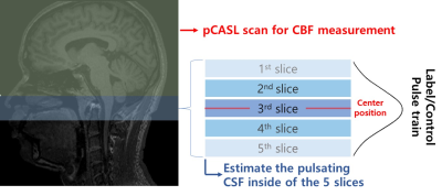 |
Estimation of CSF pulsations at the craniospinal junction using
pseudo-continuous arterial spin labeling
JaeGeun Im1,
JunHee Kim1,
and SungHong Park1
1KAIST, Daejeon, Korea, Republic of Keywords: Neurofluids, Neurofluids, CSF pulsation, fMRI, CBF, pCASL To measure CSF pulsation and CBF signals simultaneously with pCASL, we measured the influence of pCASL label pulse on the CSF signal at the craniospinal junction and compared the CSF signal differences and PC stroke volume to estimate CSF pulsation with pCASL. Our results showed that the variability of CSF signals at the nearby position from label pulses was negatively correlated with PC stroke volume. Based on this, the labeling pulse used for CBF tagging in pCASL also can be used for CSF pulsation estimation, and it will allow us to measure CBF and CSF pulsation simultaneously. |
| 09:59 |
1187.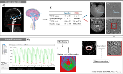 |
Effects of free breathing on cerebrospinal fluid hydrodynamics:
a study based on real-time phase-contrast MRI
Pan LIU1,2,
Kimi Piedad Owashi1,
Heimiri Monnier1,
Serge Metanbou3,
Cyrille Capel4,
and Olivier Balédent1,2
1CHIMERE UR 7516, Jules Verne University of Picardy, Amiens, France, 2Medical Image Processing Department, Amiens Picardy University Hospital, Amiens, France, 3Radiology Department, Amiens Picardy University Hospital, Amiens, France, 4Neurosurgery Department, Amiens Picardy University Hospital, Amiens, France Keywords: Neurofluids, Neurofluids, breathing effects, cerebrospinal fluid, real time phase contrast The effect of breathing on CSF is not well understood. Real-time phase-contrast MRI can quantify CSF flow continuously. A time-domain multi-parameter analysis method was developed to quantify the effect of free breathing on CSF. It was found that the cardiac period and the average stroke volume of CSF at second-to-third cervical vertebrae level and at the aqueduct were significantly increased during the expiratory phase, while the net flow of CSF was notably decreased. |
| 10:07 |
1188.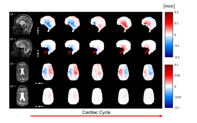 |
3D quantitative-amplified Magnetic Resonance Imaging (3D q-aMRI)
Itamar Terem1,
Nan Wang2,
Paul Condron3,
Kyan Younes4,
Javid Abderezaei5,
Berthy Feng 6,
Hari Kumar3,7,8,
Hillary Vossler9,
Mehmet Kurt5,
Katherine L. Bouman 6,
Elizabeth Mormino4,
Samantha Holdsworth3,10,
and Kawin Setsompop1,2
1Electrical Engineering, Stanford, Stanford, CA, United States, 2Radiology, Stanford, Stanford, CA, United States, 3Mātai Medical Research Institute, Tairāwhiti-Gisborne, New Zealand, 4Neurology & Neurological Sciences, Stanford, Stanford, CA, United States, 5Mechanical Engineering, University of Washington, Seattle, WA, United States, 6Computing and Mathematical Sciences (CMS), Cal Tech, Pasadena, CA, United States, 7Auckland Bioengineering Institute, University of Auckland, Auckland, New Zealand, 8General Electric Healthcare, Victoria, Australia, 9Neurology, Stanford, Stanford, CA, United States, 10Faculty of Medical & Health Sciences & Centre for Brain Research, University of Auckland, Auckland, New Zealand Keywords: Neurofluids, Data Processing, amplified MRI (aMRI), Alzheimer`s disease Amplified Magnetic Resonance Imaging (aMRI) is a pulsatile brain motion visualization method that delivers ‘videos’ with high contrast and temporal resolution. aMRI has been shown to be a promising tool in various neurological disorders. However, aMRI currently lacks the ability to quantify the sub-voxel motion field in physical units. Here, we introduce a quantitative-aMRI (q-aMRI) algorithm, which quantifies the sub-voxel motion of the 3D aMRI signal and validates its precision using phantom simulations with realistic noise. In-vivo experiments on healthy volunteers demonstrated repeatability of the measurements, and differences in brain motion were observed in subjects with positive/negative amyloid PET. |
 Back to Meeting Home
 Back to the Program-at-a-Glance
The International Society for Magnetic Resonance in Medicine is accredited by the Accreditation Council for Continuing Medical Education to provide continuing medical education for physicians.
 Back to Meeting Home
 Back to the Program-at-a-Glance
 View Presentation Video