Oral Session
Radiomics, Quantitative MRI & Miscellaneous Data Acquisition & Analysis
ISMRM & ISMRT Annual Meeting & Exhibition • 03-08 June 2023 • Toronto, ON, Canada

Oral Session
Radiomics, Quantitative MRI & Miscellaneous Data Acquisition & Analysis
16:00 |
0239.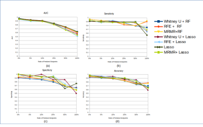 |
The Influence of Variability and Uncertainty in the Clinical Reference on MRI Radiomics Modelling and Performance
Cindy Xue1,2, Winnie CW Chu2, Jing Yuan1, Yihang Zhou1, Raymond WH Yung1, and Lo G Gladys3 1Research Department, Hong Kong Sanatorium and Hospital, Hong Kong, Hong Kong, 2Department of Imaging and Interventional Radiology, The Chinese University of Hong Kong, Hong Kong, Hong Kong, 3Department of Diagnostics and Interventional Radiology, Hong Kong Sanatorium and Hospital, Hong Kong, Hong Kong Keywords: Radiomics, Machine Learning/Artificial Intelligence, Data Modelling Radiomics uses quantitative analysis of medical imaging based on machine learning techniques and has shown its potentials of aiding personalized clinical decisions. A high standard of clinical reference (or ground truth, endpoint) is vital in radiomics feature selection and modeling, but is commonly overlooked, and assumed to be perfect. However, in reality, there are uncertainties and variability in these clinical references due to many factors. We aim to quantitatively assess the influence of clinical reference uncertainty and variability on MRI Radiomics modeling via endpoint annotation permutation with different levels. |
| 16:08 | 0240.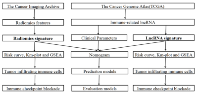 |
Prediction of immune status and overall survival of glioblastoma based on immune-related lncRNA signature and radiomics signature
Jixin Luan1, Chuanchen Zhang2, Di Zhang2, and Guolin Ma1
1China-Japan Friendship Hospital, Chinese Academy of Medical Sciences & Peking Union Medical College, Beijing, China, 2Department of Radiology, Liaocheng People's Hospital, Shandong First Medical University & Shandong Academy of Medical Sciences, Liaocheng, China Keywords: Radiomics, Brain, GBM In this study, we aimed to investigate the correlation between immune-related lncRNA signature and radiomics signature and immune cell infiltration and immune checkpoint blockade in glioblastoma multiform (GBM), and to develop a prognostic model to predict the overall survival of patients. We found that immune-related lncRNA signature and radiomics signature can better predict the immune status and overall survival of GBM patients, which can help clinical prognosis determination and immunotherapy selection. |
| 16:16 | 0241.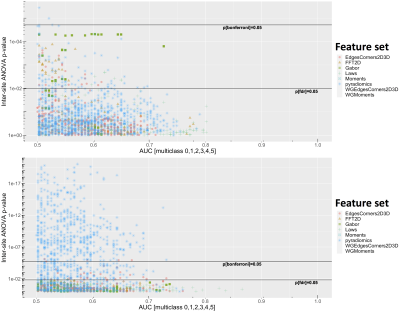 |
Multicenter reproducibility of hand-crafted radiomics and deep-learning based features for biparametric prostate MRI
Harri Merisaari1, Janne Verho2, Ileana Montoya Perez2,3, Otto Ettala4, Kari T Syvänen4, Pekka Taimen5, Aida Steiner2, Jani Saunavaara6, Ekaterina Saukko2, Peter Boström4, Hannu Aronen1, and Ivan Jambor1,2
1Department of Radiology, University of Turku, Raisio, Finland, 2Department of Radiology, Turku University Hospital, Turku, Finland, 3Department of Computing, University of Turku, Turku, Finland, 4Department of Urology, TYKS Turku University Hospital and University of Turku, Turku, Finland, 5Department of Pathology, TYKS Turku University Hospital, Turku, Finland, 6Department of Medical Physics, TYKS Turku University Hospital, Turku, Finland Keywords: Radiomics, Cancer, Inter-site reproducibility, Deep Learning, bi-parametric MRI In the current study, we aimed to explore reproducibility of various hand-crafted radiomics features, and deep learning autoencoder-based features within Gleason Grade Groups (GGG) using MULTI-IMPROD trial data. Differences between sites were evaluated with ANOVA test, corrected for GGG group, and multi-class AUC for GGG. We explored if systematic differences exist between the four centers taking part in the trial with conventionally used radiomic features. The results show differences between modalities, feature groups, and when intensity harmonization is applied for ADC. |
| 16:24 | 0242.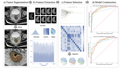 |
Automatic Rectal Tumor Segmentation and Extramural Venous Invasion Diagnosis based on Deep Learning and Radiomics Model
Jiyao Liu1, Rencheng Zheng1, Chengyan Wang2, Yigang Pei3, Yinghua Chu4, and He Wang1
1Institute of Science and Technology for Brain-Inspired Intelligence, Fudan University, Shanghai, China, 2Human Phenome Institute, Fudan University, Shanghai, China, 3Xiangya Hospital Central South University, Changsha, Hunan, China, 4Siemens Healthineers, Shanghai, China Keywords: Machine Learning/Artificial Intelligence, Segmentation This study developed an automatic diagnosis model for rectal cancer, which consists following steps: high-precision rectal tumor segmentation by Spatial Hybrid Network (SH-Net) and Adaboost Decision Tree based radiomics model to improve the diagnostic performance of extramural venous invasion (EMVI). The comparable diagnostic performance of the proposed model compared to the visual assessment by radiologists suggests the potential to help doctors with clinical diagnosis of EMVI.
|
| 16:32 | 0243.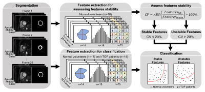 |
Impact of Radiomic Features Stability of Myocardial Motion on Classification of Repaired Tetralogy of Fallot Patients
Pao-Han Chiu1, Ming-Ting Wu2, Ken-Pen Weng3,4, Nai-Yu Pan5, Teng-Yi Huang5, and Hsu-Hsia Peng1
1Department of Biomedical Engineering and Environmental Sciences, National Tsing Hua University, Hsinchu, Taiwan, 2Department of Radiology, Kaohsiung Veterans General Hospital, Kaohsiung, Taiwan, 3Department of Pediatrics, Kaohsiung Veterans General Hospital, Kaohsiung, Taiwan, 4Department of Pediatrics, National Yang Ming Chiao Tung University, Taipei, Taiwan, 5Department of Electrical Engineering, National Taiwan University of Science and Technology, Taipei, Taiwan Keywords: Radiomics, Radiomics Myocardial motion influences the stability of radiomic features of cardiac magnetic resonance (CMR) cine images. We aimed to evaluate the impact of the stability of myocardial radiomic features on the classification performance of differentiation of repaired tetralogy of Fallot (rTOF) patients from normal volunteers. The stability of each radiomic feature during cardiac cycle was assessed by coefficient of variation (CV) of the feature value. 50 of 107 radiomic features (46.7%) in normal volunteers were stable features. The classification model established only with stable features presented the best classification performance (AUC=0.94) which was able to be improved by chi-square feature selection. |
16:40 |
0244.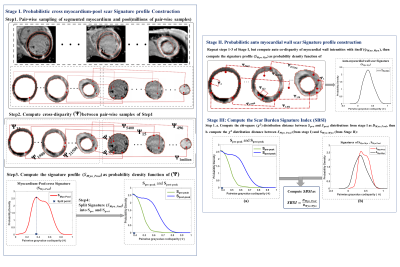 |
Novel LGE Myocardial Scar Burden Signatures: A Novel Concept for Comprehensive Scar Quantification in Myocardial Infarction
Mehri Mehrnia1 and Mohammed S.M. Elbaz1
1Northwestern University, Chicago, IL, United States Keywords: Radiomics, Cardiomyopathy, heart, cardiovascular, LGE, scar, Myocardial Infarction There remains no standard method for quantification of myocardial infarction (MI) from LGE images. Current methods lack reproducibility as they rely on manual or threshold-based scar segmentation. Notably, these methods only quantify scar volume/percentage but ignore the impact of myocardial LGE distribution pattern. Here, we propose a novel threshold-free concept that comprehensively quantifies scar burden in terms of both scar extent and the unique myocardial LGE distribution pattern: LGE Scar Burden Signatures. We demonstrated our technique’s strong correlation to scar extent, its independent association with serum biomarker of myocardial injury (Troponin), and reduced ejection fraction, independent of Scar percentage. |
| 16:48 | 0245.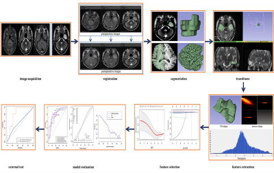 |
Multiparametric MRI radiomic model for diagnosing focal cortical dysplasia and laterality
Shiqi Chen1, Yawen Xiao1, Zhaotao Zhang1, Jiankun Dai2, Yifei Gui1, and Xinlan Xiao1
1Department of Radiology, The Second Affiliated Hospital of Nanchang University, Nanchang, China, 2GE Healthcare, MR Research China, Beijing, China Keywords: Radiomics, Epilepsy Focal cortical dysplasia (FCD) is the most common epileptogenic developmental malformation which remains challenging to diagnose. This study aimed to investigate the possibility of diagnosing FCD and laterality in epilepsy patients using the multiparametric MRI radiomics model. Radiomic features were extracted from the preoperative MRI images of 86 patients. Multivariable logistic regression was used to develop the diagnosis model. The performance of radiomic model was evaluated with general evaluation metrics and was compared with that of inexperienced radiologists.We concluded that radiomics features derived from the combined of T1WI, T2WI and FLAIR might help diagnosing FCD and laterality. |
| 16:56 | 0246.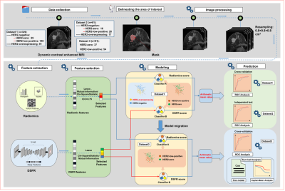 |
MRI-based deep learning radiomics can predict HER2 expression and disease-free survival in breast cancer
Wenjie Tang1, Yuan Guo1, Siyi Chen1, Bingsheng Huang2, Xiaotong Xie2, Mingyu Wang2, Yongzhou Xu3, Kuiming Jiang4, and Xinhua Wei1
1Guangzhou First People's Hospital, Guangzhou, China, 2Shenzhen University, Shenzhen, China, 3Philips Healthcare, Guangzhou, China, 4Guangdong Women and Children Hospital, Guangzhou, China Keywords: Radiomics, Breast, HER2 expressing; MRI; Deep learning; Prognosis To the best of our knowledge, our study is the first to non-invasively assess human epidermal growth factor receptor 2 (HER2) status, especially HER2-low-positive status in breast cancer. In this study, a deep learning radiomics (DLR) model based on contrast-enhanced MRI was constructed and showed high and stable performance in predicting HER2 status in both the training and validation cohorts, and the predicted status was an independently significant predictor of disease-free survival (DFS) in HER2-low-positive/HER2-zero breast cancers. The DLR model showed prospects as a computer-aided diagnostic tool to help more accurately identify HER2-low-positive breast cancers, thereby guiding patient treatment strategies. |
| 17:04 |
0247.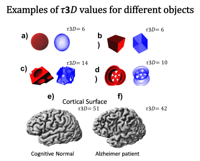 |
Quantifying geometrical properties of the Brain Surface in Alzheimer's Disease using 3D Tortuosity
Maria Julieta Mateos1, James J. Lah2, and Qiu Deqiang1 1Radiology and Imaging Sciences, Emory University School of Medicine, Atlanta, GA, United States, 2Department of Neurology, Emory University School of Medicine, Atlanta, GA, United States Keywords: Radiomics, Alzheimer's Disease Alzheimer's disease causes significant gray matter loss, which leads to changes in the brain surface's shape. In this work, we used the local gyrification index (LGI) and the three-dimensional tortuosity (𝜏3𝐷) to characterize the cortical morphology, and to determine if the obtained values were significantly different amog the Alzheimer's diagnosis. The subset of MRI studies was obtained from ADNI database. For the data analyzed, the results show that the 𝜏3𝐷 has a positive correlation with brain volume and can potentially be a biomarker for AD. |
| 17:12 | 0248.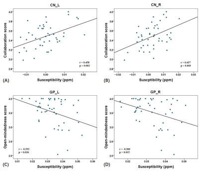 |
Associations of Brain Iron with Cognitive and Social Emotional Performance in Children using Quantitative Susceptibility Mapping
Gaiying Li1, Qifan Pang1, Mengying Chen1, Yang Song2, Qing Cai3, Kai Zhang3, Longnian Lin3, Yi Wang4, and Jianqi Li1,3
1School of Physics and Electronic Science, East China Normal University, Shanghai, China, 2MR Scientific Marketing, Siemens, Siemens Healthineers, Shanghai, China, 3Institute of Brain and Education Innovation, East China Normal University, Shanghai, China, 4Department of Radiology, Weill Medical College of Cornell University, New York, NY, United States Keywords: Data Analysis, Quantitative Susceptibility mapping Tissue iron play a critical role in cognitive functions. However, associations of basal ganglia iron concentration with cognition and emotion in children are less well understood. This study examined the correlation of susceptibility values in the bilateral basal ganglia nuclei with cognitive functions and social emotional capacity in children around the age of seven. The results highlighted that the inhibitory control, collaboration, open-mindedness showed significant association with susceptibility values in the basal ganglia. In conclusion, this QSM study indicated the potential for using brain iron content in the basal ganglia to assess cognitive and emotional performance during the children development. |
| 17:20 | 0249.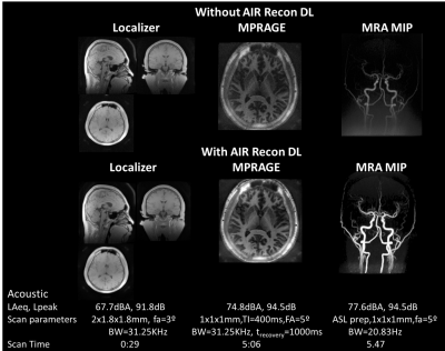 |
AI-enhanced comprehensive quiet neuroimaging
Ana Beatriz Solana1,2, Sagar Mandava3, Xinzeng Wang4, Marc Lebel5, David J Lythgoe2, Tobias C Wood2, Matthew Bowdler2, Steven CR Williams2, and Florian Wiesinger1,2
1GE Healthcare, Munich, Germany, 2King's College London, London, United Kingdom, 3GE Healthcare, Marietta, GA, United States, 4GE Healthcare, Houston, TX, United States, 5GE Healthcare, Calgary, AB, Canada Keywords: Pulse Sequence Design, MR Value, Neuro, quiet, multicontrast, fMRI, QSM Most of today’s MRI scans are very loud and can be challenging for patients of all ages, e.g. sleeping babies, anxious children or elderly with tinnitus. Existing quiet and/or acoustic noise reduced MRI scanning solutions are in their infancy, primarily because of their incomplete nature and associated trade-offs in terms of image quality and/or scan time. Here, we present a comprehensive quiet neuroimaging solution, including standard anatomical scans (localizer, T1w-MPRAGE, T2w-FSE, T2w-FLAIR, MRA, Diffusion), quantitative parameter mapping (ADC, T2*, QSM, PD, T1, T2) and functional BOLD-fMRI. Scan time and overall image quality was improved using DL-based image reconstruction framework. |
| 17:28 | 0250.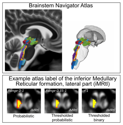 |
The Brainstem Navigator: a toolkit to investigate brainstem nuclei structure, function, and connectivity in living humans
Marta Bianciardi1,2, Simone Cauzzo1,3, Nicola Toschi1,4, Subhranil Koley1, Maria Guadalupe García-Gomar1,5, and Kavita Singh1 1Brainstem Imaging Laboratory, Department of Radiology, Athinoula A. Martinos Center for Biomedical Imaging, Massachusetts General Hospital and Harvard Medical School, Boston, MA, United States, 2Division of Sleep Medicine, Harvard University, Boston, MA, United States, 3Research Center “E. Piaggio”, School of Engineering, University of Pisa, Pisa, Italy, 4Medical Physics Section, Department of Biomedicine and Prevention, Faculty of Medicine, Tor Vergata University of Rome, Rome, Italy, 5Escuela Nacional de Estudios Superiores Unidad Juriquilla, Universidad Nacional Autónoma de México, Queretaro, Mexico Keywords: Software Tools, Brain, Brainstem atlas, segmentation, functional and structural connectome, 7 Tesla MRI, 3 Tesla MRI Brainstem nuclei are deep gray matter regions involved in vital functions such as arousal/sleep/motor/sensory/autonomic/nociceptive/limbic/sensory function. Due to their small size and limited MRI contrast, these nuclei are difficult to visualize in conventional imaging of living humans. We generated and released the Brainstem Navigator toolkit, which enables the automatic identification of brainstem nuclei location in conventional and advanced MRI of living humans. It includes in-vivo probabilistic atlas labels for 31 brainstem nuclei generated by multi-contrast 7 Tesla MRI. We also developed coregistration routines optimized for the brainstem in 3 Tesla and 7 Tesla MRI data in health and disease. |
| 17:36 | 0251.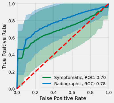 |
Association between Post-traumatic Osteoarthritis 10+ Years after ACLR and Quantitative MRI of Knee Cartilage, Menisci, and Thigh Muscles
William Zaylor1,2, Sameed Khan1,2,3, Richard Lartey1,2, Brendan L. Eck2,3, Mei Li1,2, Sibaji Gaj1,2, Jeehun Kim1,2, Carl S. Winalski1,2,3, Faysal Altahawi2,3, Morgan H. Jones4, Laura J. Huston5, Kevin D. Harkins6, Michael V. Knopp7, Christopher C. Kaeding8, Kurt P. Spindler2,9, and Xiaojuan Li1,2,3
1Biomedical Engineering, Cleveland Clinic, Cleveland, OH, United States, 2Program of Advanced Musculoskeletal Imaging (PAMI), Cleveland Clinic, Cleveland, OH, United States, 3Department of Diagnostic Radiology, Cleveland Clinic Imaging Institute, Cleveland, OH, United States, 4Department of Orthopaedic Surgery, Brigham and Women's Hospital, Boston, MA, United States, 5Department of Orthopaedics and Rehabilitation, Vanderbilt University, Nashville, TN, United States, 6Radiology and Radiological Sciences, Vanderbilt University, Nashville, TN, United States, 7Department of Radiology, The Ohio State University Wright Center of Innovation in Biomedical Imaging, Columbus, OH, United States, 8Department of Orthopaedic Surgery, The Ohio State University, Columbus, OH, United States, 9Department of Orthopaedic Surgery, Cleveland Clinic, Cleveland, OH, United States Keywords: Radiomics, Relaxometry The purpose of this study was to evaluate the associations between radiomic features in knee and thigh quantitative MRI with symptomatic and radiographic post-traumatic osteoarthritis (PTOA) after anterior cruciate ligament reconstruction (ACLR). Radiomic features of knee cartilage, menisci (extracted from T1ρ maps), and thigh muscles (extracted from anatomic images and fat fraction maps) were extracted from patient MRI 10+ years after ACLR, and features were selected for symptomatic or radiographic PTOA associations. Symptomatic and radiographic PTOA were associated with features from the medial cartilage compartments, and menisci; features from the thigh muscles were associated with symptomatic PTOA only. |
| 17:44 | 0252.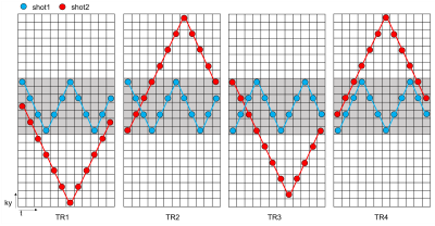 |
Ultrafast multi-parametric quantitative MRI using Multi-TR echo-planar time-resolved imaging (Multi-TR-EPTI)
Haoran Bai1, Yueqi Qiu1, Ke Dai1, Hao Chen1, and Zhiyong Zhang1 1School of Biomedical Engineering, Shanghai Jiao Tong University, Shanghai, China Keywords: Data Acquisition, Data Acquisition Echo planar time-resolved imaging (EPTI) is a recently developed multi-shot EPI method that provides time-resolved multi-echo images and providing multi-contrast images. Based on this basis, here we present Multi-TR-EPTI, a technique developed to achieve distortion-free and Multi-parametric quantitative MRI including T1, T2, T2*, and PD mapping. Multi-TR-EPTI exploits an optimized spatiotemporal CAIPI encoding in the k-TE-TR space.which can recover the k-TE-TR space within the same shot number as the original EPTI. A subspace reconstruction was employed to obtain hundreds of high-quality multi-contrast images at sub-millisecond temporal increments. Multi-TR-EPTI adopts different TRs to provide T1 contrast while improving scan efficiency. |
| 17:52 | 0253.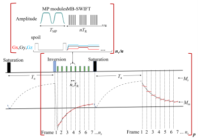 |
Alternating Look-Locker for 3D quantitative longitudinal and rotating frame relaxation mapping
Lin WU1, Chris Carchi2, Shalom Michaeli1, Silvia Mangia1, and Djaudat Idiyatullin1
1CMRR and Department of Radiology, University of Minnesota, Minneapolis, MN, United States, 2University of Minnesota, Minneapolis, MN, United States Keywords: High-Field MRI, Relaxometry, T1ρ A new method entitled alternating Look-Locker (aLL) for mapping of T1 and T1ρ is proposed. Magnetization preparation modules incorporating adiabatic full passage pulses were imbedded in a Look-Locker scheme that additionally alternates magnetization from +Z and -Z axes. MB-SWIFT was used as a readout. Analytical derivations and relevant simulations are presented. Phantom experiments and in vivo studies in the rat brain were conducted at 9.4 T. Results show that aLL allows more robust and faster acquisitions as compared to previously introduced steady–state technique, providing the possibility of simultaneous collection of T1 and T1ρ maps in one acquisition. |
 Back to Meeting Home
 Back to the Program-at-a-Glance
The International Society for Magnetic Resonance in Medicine is accredited by the Accreditation Council for Continuing Medical Education to provide continuing medical education for physicians.
 Back to Meeting Home
 Back to the Program-at-a-Glance
 View Presentation Video