Oral Session
Pulse Sequences & Encoding Methods
ISMRM & ISMRT Annual Meeting & Exhibition • 03-08 June 2023 • Toronto, ON, Canada

Oral Session
Pulse Sequences & Encoding Methods
| 13:30 |
0529.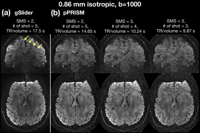 |
Pseudo Partition-encoded Simultaneous Multislab (pPRISM) for
Submillimeter Diffusion Imaging Without Navigator and
Slab-Boundary Artifacts
Wei-Tang Chang1,2,3
1Radiology, UNC at Chapel Hill, Chapel Hill, NC, United States, 2Biomedical Research Imaging Center, UNC at Chapel Hill, Chapel Hill, NC, United States, 3Biomedical Engineering, UNC at Chapel Hill, Chapel Hill, NC, United States Keywords: Pulse Sequence Design, Diffusion Tensor Imaging The ability to achieve submillimter isotropic resolution diffusion MR imaging (dMRI) is critically important to study fine-scale brain structures. While the multi-shot approaches, including SMSlab and gSlider-SMS, have been proposed to mitigate the inherently low SNR, the SMSlab sequences require additional navigators for phase estimation and both SMSlab and gSlider-SMS suffered from the slab-boundary artifacts. This study proposed two new concepts: PRISM encoding and 2) pseudo slab in order to mitigate the slab-boundary artifacts and shorten the scan time. Together, this study achieved the dMRI with 0.86 mm isotropic resolution with 16.3%-43.6% reduction of scan time compared to gSlider. |
13:38 |
0530. |
Multiphoton Simultaneous Multislice Imaging with CAIPIRINHA
Phase Encoding
Tanya Deniz Ipek1,
Victor Han1,
and Chunlei Liu1,2
1Electrical Engineering and Computer Sciences, University of California, Berkeley, Berkeley, CA, United States, 2Helen Wills Neuroscience Institute, University of California, Berkeley, Berkeley, CA, United States Keywords: RF Pulse Design & Fields, RF Pulse Design & Fields, Multiphoton, SAR Simultaneous multislice (SMS) imaging methods reduce scan time but increase specific absorption rate (SAR) because of additional radiofrequency (RF) pulses required for exciting multiple slices. Multiphoton excitation was recently proposed to significantly reduce SAR for SMS imaging. In this work, we further developed a phase-modulated multiphoton SMS excitation method that achieves CAIPIRINHA-type controlled aliasing, which improves image reconstruction quality significantly. Images obtained with our custom multiphoton CAIPIRINHA-SMS sequence have reduced aliasing artifacts and higher SNR compared to images obtained with a multiphoton SMS sequence without CAIPIRINHA. For a three-slice design, SAR is reduced by a factor of two. |
|
13:46 |
0531. |
SNR-efficient, motion-robust multi-echo SPGR with k-space
aliasing
Peter J Lally1,2,
Mark Chiew3,
Paul M Matthews1,4,
Karla L Miller5,
and Neal K Bangerter6
1Department of Brain Sciences, Imperial College London, London, United Kingdom, 2Centre for Care Research and Technology, UK Dementia Research Institute, London, United Kingdom, 3Medical Biophysics, University of Toronto, Toronto, ON, Canada, 4UK Dementia Research Institute at Imperial, London, United Kingdom, 5Wellcome Centre for Integrative Neuroimaging, University of Oxford, Oxford, United Kingdom, 6Department of Bioengineering, Imperial College London, London, United Kingdom Keywords: Data Acquisition, Pulse Sequence Design, SSFP, susceptibility The behaviour of magnetisation under RF spoiling is typically considered to be pseudorandom from TR to TR. Here we describe a model for the coherent underlying phase behaviour which we exploit in a new acquisition strategy. We propose this as an SNR-efficient and motion-robust alternative to multi-echo spoiled gradient echo acquisitions. |
|
13:54 |
0532. |
Multi-echo RF spatial phase encoding for gradient-free imaging
in a nonuniform B0-field at low-field
Kartiga Selvaganesan1,
Yonghyun Ha2,
Chenhao Sun2,
Anja Samardzija1,
Zhehong Zhang1,
Heng Sun1,
Gigi Galiana2,
and Todd Constable2
1Biomedical Engineering, Yale University, New Haven, CT, United States, 2Radiology and Biomedical Imaging, Yale University, New Haven, CT, United States Keywords: Pulse Sequence Design, Low-Field MRI The cost and complexity of MR scanners can be significantly reduced by eliminating conventional linear gradient coils, and instead using RF coils for spatial encoding. Here we have developed a novel multi-echo RF phase encoding pulse sequence that exploits the Bloch-Siegert shift for nonlinear spatial encoding in a nonuniform, field-cycling, low-field MR system. Phantoms of varying sizes were successfully imaged using this pulse sequence, demonstrating that this technique can be used to perform gradient-free imaging in an inhomogeneous B0-field at low-field. |
| 14:02 |
0533.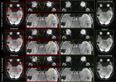 |
Semi-Randomized Trajectories to the Rescue: Reducing Artifacts
from Fluctuating Physiological Signals in 3D Steady-State MRI
Amir Seginer1 and
Rita Schmidt2
1Siemens Healthcare Ltd, Rosh Ha'ayin, Israel, 2Department of Brain Sciences, Weizmann Institute of Science, Rehovot, Israel Keywords: Data Acquisition, Artifacts, ultra-high field Rapid 3D steady-state sequences such as SWI are sensitive to semi-periodic physiological fluctuations (e.g., cardiac pulsation, breathing, and eye movement) resulting in repeating artifacts in the images. Randomization of the phase-encoding order reduces the above artifacts but results in apparent noise from slow global changes (like motion or eddy currents changes). We propose a new semi-randomized acquisition order that allows to set a cutoff frequency for artifact suppression; above which artifacts are suppressed, whereas artifacts from slower changes are unaffected. Simulations and SWI human brain scanning at 7T validate the method. |
|
14:10 |
0534.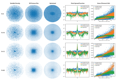 |
MR Sampling Patterns Learned with Variational Information
Maximization
Cagan Alkan1,
Shreyas Vasanawala2,
and John Pauly1
1Electrical Engineering, Stanford University, Stanford, CA, United States, 2Radiology, Stanford University, Stanford, CA, United States Keywords: Machine Learning/Artificial Intelligence, Data Acquisition, Data Sampling Variational information maximization allows joint optimization of MR data sampling and reconstruction and improves reconstruction quality upon the heuristically designed sampling patterns. Here, we analyze the learned sampling patterns with respect to changes in acceleration factor, measurement noise, anatomy, and coil sensitivities in order to provide some interpretation. We show that all of these factors contribute to the optimization result by impacting the sampling density, k-space coverage and point spread functions of the learned sampling patterns. |
14:18 |
0535.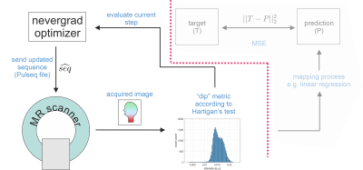 |
MR-double-zero in vivo: Model-free and live MRI contrast
optimization running a loop over a real scanner with a real
subject.
Sebastian Mueller1,2,
Felix Glang1,
Klaus Scheffler1,2,
and Moritz Zaiss1,3
1High-field Magnetic Resonance Center, Max Planck Institute for Biological Cybernetics, Tuebingen, Germany, 2Department for Biomedical Magnetic Resonance, University Hospital Tuebingen, Tuebingen, Germany, 3Institute of Neuroradiology, University Hospital Erlangen, Friedrich-Alexander University Erlangen-Nürnberg, Erlangen, Germany Keywords: Pulse Sequence Design, Pulse Sequence Design Recently, we proposed a framework for automatized, model-free MR sequence design for contrast generation. The concept was proven by optimizing the contrast-generating sequence using a real MRI scanner for scanning model solutions repeatedly until convergence to the target. In this work, we demonstrate for the first time that this live optimization loop can also be used directly on a real subject’s brain. As an example, GM/WM contrast for a GRE sequence was optimized fully automatically in a model-free and reference-free manner live in a healthy subject at a 3T MR scanner. |
|
14:26 |
0536.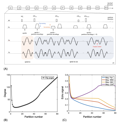 |
Variable-flip-angle 3D spiral-in-out TSE/SPACE using
echo-reordering and concomitant gradient compensation at 0.55 T
Zhixing Wang1,
Rajiv Ramasawmy2,
Ahsan Javed2,
John P. Mugler3,
Craig H. Meyer1,3,
and Adrienne E. Campbell-Washburn2 1Biomedical Engineering, University of Virginia, Charlottesville, VA, United States, 2Cardiovascular Branch, Division of Intramural Research, National Heart, Lung, and Blood Institute, National Institutes of Health, Bethesda, MD, United States, 3Radiology & Medical Imaging, University of Virginia, Charlottesville, VA, United States Keywords: Data Acquisition, Data Acquisition This study describes an alternative approach to Cartesian SPACE for 1 mm3 isotropic whole brain T2-weighted imaging on a high performance 0.55T scanner. In this technique, the Cartesian readouts were replaced by interleaved, rotated spiral-in-out trajectories, combined with a variable-flip-angle refocusing, echo-reordering, and concomitant gradient compensation. Parallel imaging (PI) and compressed sensing (CS) were utilized for further acceleration. Compared to 3D-Cartesian SPACE, this method can be leveraged to mitigate the lower SNR of 0.55T via the improved SNR efficiency of prolonged spiral trajectory sampling. |
| 14:34 |
0537.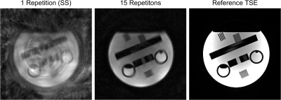 |
Spiral Fast Spin Echo MRI with Interleaved Diffusion
Sensitization for Simultaneous ADC and T2 Mapping
Lars Bielak1,2,
Jürgen Hennig1,
Thomas Lottner1,
and Michael Bock1,2
1Dept.of Radiology, Medical Physics, Medical Center University of Freiburg, Faculty of Medicine, University of Freiburg, Freiburg, Germany, 2German Cancer Consortium (DKTK), Partner Site Freiburg, Freiburg, Germany Keywords: Pulse Sequence Design, Multi-Contrast A sequence design is presented based on spiral fast spin echo acquisition with interleaved diffusion sensitization for simultaneous ADC and T2 mapping. With spiral readout trajectories only few echoes need to be acquired per (TE,b) pair allowing to accelerate the acquisition significantly. A phantom experiment with single echo per k-space is shown for T2 and ADC mapping. With a model-based reconstruction both parameter maps can be rapidly reconstructed. |
| 14:42 |
0538.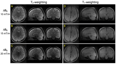 |
3D Fast Spin Echo using Frequency-Modulated RF Pulses for MRI in
Highly Inhomogeneous Magnetic Fields
Naoharu Kobayashi1 and
Michael Garwood1
1CMRR, Department of Radiology, University of Minnesota, Minneapolis, MN, United States Keywords: Data Acquisition, Pulse Sequence Design, Inhomogeneous field 3D Fast spin echo (FSE) with frequency-modulated (FM) pulses has been introduced for MRI in highly inhomogeneous fields. B1 dependent phase in FM pulse excitation and refocus was adjusted in the FM-FSE pulse sequence. Refocus flip angles in FM-FSE were determined with prospective extended phase graph (EPG). Proposed flip angle and phase in FM-FSE were validated in EPG simulation and experiments at 3T by introducing a linear inhomogeneous field. Finally, in vivo human brain imaging with T1- and T2-weighting was performed using the proposed 3D FM-FSE sequence. |
| 14:50 |
0539.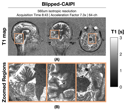 |
Rapid Mesoscale MP2RAGE Imaging at Ultra High Field with
Controlled Aliasing
Gabriel Varela-Mattatall1,2,
Wei-Ching Lo3,
Omer Oran4,
Jonathan Polimeni5,6,
Azma Mareyam5,7,
Borjan Gagoski8,9,
Ravi S. Menon1,2,
and Berkin Bilgic5,6
1Centre for Functional and Metabolic Mapping (CFMM), Robarts Research Institute, Western University, London, ON, Canada, 2Department of Medical Biophysics, Schulich School of Medicine and Dentistry, Western University, London, ON, Canada, 3Siemens Medical Solutions USA, Boston, MA, United States, 4Siemens Healthcare Limited, Oakville, ON, Canada, 5Athinoula A. Martinos Center for Biomedical Imaging, Massachusetts General Hospital & Harvard Medical School, Boston, MA, United States, 6Harvard-MIT Division of Health Sciences and Technology, Massachusetts Institute of Technology, Boston, MA, United States, 7Harvard-MIT Division of Health Sciences and Technology, Massachusetts Institute of Technology, Boston, Boston, MA, United States, 8Fetal-Neonatal Neuroimaging & Developmental Science Center, Boston Children’s Hospital, Boston, MA, United States, 9Department of Radiology, Harvard Medical School, Boston, MA, United States Keywords: Data Acquisition, New Trajectories & Spatial Encoding Methods MP2RAGE is considered the workhorse sequence at ultra-high field (UHF) as it provides high-contrast and bias-free structural imaging for both anatomical and segmentation purposes. However, high-resolution MP2RAGE imaging is hampered by lengthy acquisitions, which can be partially mitigated using parallel imaging, but this comes at the cost of g-factor and √R penalties on SNR; thus, limiting the usefulness of this sequence at the submillimeter scale. In this work, we show preliminary results on how wave encoding, and blipped-controlled aliasing could allow efficient MP2RAGE acquisitions at up to 560 um isotropic resolution at 7T. |
| 14:58 |
0540.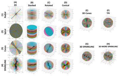 |
Benchmarking common and advanced non-Cartesian trajectories with
high acceleration and static off-resonance effects
Guillaume Daval-Frérot1,2,3,
Chaithya G R2,3,
Fernanda Ponce2,3,
Aurélien Massire1,
Boris Mailhe4,
Mariappan Nadar4,
Alexandre Vignaud2,
and Philippe Ciuciu2,3
1Siemens Healthineers, Saint-Denis, France, 2CEA, NeuroSpin, CNRS, Université Paris-Saclay, Gif-sur-Yvette, France, 3Inria, MIND, Palaiseau, France, 4Digital Technology & Innovation, Siemens Healthineers, Princeton, NJ, United States Keywords: Data Acquisition, Data Acquisition, Non-Cartesian; SPARKLING Non-Cartesian sampling patterns allow for highly accelerated MRI exams at the cost of exacerbated and more complex artifacts, each impacting image quality in unique ways. Patient-induced B0 field inhomogeneities can notably cause distortions and blurring, but some trajectories are by design more robust to them. We propose to retrospectively benchmark 16 different non-Cartesian sampling patterns with high acceleration factor (AF=20) and realistic off-resonance artifacts over 9 volumes acquired at 3T with 0.6 mm isotropic spatial resolution. SPARKLING and spiral-based trajectories achieve higher image quality scores, but only the former shows robustness to off-resonance effects through the MORE-SPARKLING extension. |
|
15:06 |
0541.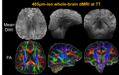 |
Mesoscale distortion-free in-vivo dMRI at 7T using ROtating-view
Motion-robust supEr Resolution EPTI (Romer-EPTI)
Zijing Dong1,2,
Jonathan R. Polimeni1,2,3,
Lawrence L. Wald1,2,3,
and Fuyixue Wang1,2
1Athinoula A. Martinos Center for Biomedical Imaging, MGH, Charlestown, MA, United States, 2Department of Radiology, Harvard Medical School, Charlestown, MA, United States, 3Harvard-MIT Health Sciences and Technology, MIT, Cambridge, MA, United States Keywords: Data Acquisition, Brain Pushing the spatial resolution of diffusion MRI to mesoscale is technically challenging due to the low SNR efficiency and severe image artifacts. Last year, we presented a novel ROtating-view Motion-robust supEr Resolution Echo Planar Time-resolved Imaging method (Romer-EPTI), providing significant gains (~3.5×) in SNR efficiency and high-quality images free from distortion with minimized motion artifacts. In this work, we further developed Romer-EPTI at 7T for in-vivo mesoscale dMRI. By exploiting the higher signal of 7T and the short-TE, high-SNR, distortion-free acquisition provided by Romer-EPTI, mesoscale whole-brain dMRI at an unprecedented 485-μm isotropic resolution was acquired. |
| 15:14 |
0542.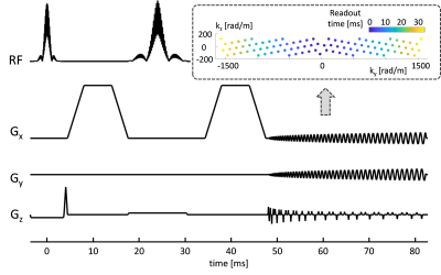 |
Multiband T-Hex spirals for rapid and SNR-efficient
diffusion-weighted MRI
Maria Engel1,
Lars Müller1,2,
André Döring1,
and Derek Jones1
1Cardiff University Brain Research Imaging Centre (CUBRIC), Cardiff University, Cardiff, United Kingdom, 2Leeds Institute of Cardiovascular and Metabolic Medicine, University of Leeds, Leeds, United Kingdom Keywords: New Trajectories & Spatial Encoding Methods, New Trajectories & Spatial Encoding Methods Diffusion weighted imaging (DWI) for advanced modelling of tissue microstructure is notoriously short in SNR and requires long scan times. In this work we boost the SNR of DWI by combining highly efficient T-Hex sampling with multiband imaging and spiral readout schemes. This allows for an unprecedented SNR efficiency and holds promise for advanced microstructural scans especially in clinical populations. |
| 15:22 |
0543.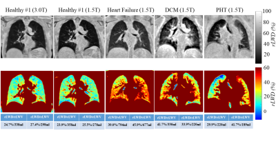 |
Cartesian Ultrashort Echo Time (CUTE) Imaging of the Lung
Parenchyma
Richard Thompson1,
Christopher Keen1,
Hefin Jones2,
Richard Coulden2,
Peter Seres1,
and Justin Grenier1
1Biomedical Engineering, University of Alberta, Edmonton, AB, Canada, 2Radiology & Diagnostic Imaging, University of Alberta, Edmonton, AB, Canada Keywords: Pulse Sequence Design, Lung, UTE, Heart Failure, Cartesian The Cartesian UTE (CUTE) sequence provides high quality three-dimensional imaging of the lung parenchyma (TE = 140 us) without the requirement for k-space gridding in a short breath-hold. Phantom validation and comparison to an existing UTE sequence (Spiral VIBE) is provided in 64 healthy subjects and in clinical examples. |
 Back to Meeting Home
 Back to the Program-at-a-Glance
The International Society for Magnetic Resonance in Medicine is accredited by the Accreditation Council for Continuing Medical Education to provide continuing medical education for physicians.
 Back to Meeting Home
 Back to the Program-at-a-Glance
 View Presentation Video