Oral Session
Advances in Image Reconstruction
ISMRM & ISMRT Annual Meeting & Exhibition • 03-08 June 2023 • Toronto, ON, Canada

13:30 |
0859. |
K2S Challenge: From Undersampled K-Space to Automatic
Segmentation
Aniket Tolpadi1,
Upasana Bharadwaj1,
Kenneth Gao1,
Rupsa Bhattacharjee1,
Felix Gassert1,
Johanna Luitjens1,
Jan Nikolas Morshuis2,3,
Paul Fischer2,
Matthias Hein2,
Christian F. Baumgartner2,
Artem Razumov4,
Dmitry Dylov4,
Quintin van Lohuizen5,
Stefan Fransen5,
Xiaoxia Zhang6,
Radhika Tibrewaka6,
Hector Lise de Moura6,
Kangning Liu6,
Marcelo Zibetti6,
Ravinder Regatte6,
Sharmila Majumdar1,
and Valentina Pedoia1
1Radiology and Biomedical Imaging, UCSF, San Francisco, CA, United States, 2Cluster of Excellence Machine Learning, University of Tübingen, Tübingen, Germany, 3International Max Planck Research School for Intelligent Systems, Tübingen, Germany, 4Skolkovo Institute of Science and Technology, Moscow, Russian Federation, 5Department of Radiology, University Medical Center Groningen, Groningen, Netherlands, 6Center for Advanced Imaging Innovation and Research, New York University Grossman School of Medicine, New York, NY, United States Keywords: Image Reconstruction, MSK Image reconstruction and downstream tasks have typically been treated independently by the image processing community, but we hypothesized performing them end-to-end could facilitate further optimization. To these ends, UCSF organized the K2S challenge, where challenge participants were tasked with segmenting bone and cartilage from 8X undersampled knee MRI acquisitions. Top challenge submissions produced high-quality segmentations maintaining fidelity to ground truth, but strong reconstruction performance proved not to be required for accurate tissue segmentation, and there was no correlation between reconstruction and segmentation performance. This challenge showed reconstruction algorithms can be optimized for downstream tasks in an end-to-end fashion. |
| 13:38 |
0860.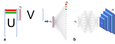 |
A Generative Subspace Model for High-dimensional MR Imaging
Xi Peng1 and
Fan Lam2 1Mayo Clinic, Rochester, MN, United States, 2University of Illinois Urbana-Champaign, Urbana, IL, United States Keywords: Sparse & Low-Rank Models, Quantitative Imaging Subspace or low-rank models have been demonstrated useful in producing efficient representations and reconstructions for high dimensional imaging problems. In this study, we propose an unsupervised learning-based approach by generalizing the subspace model using a deep generative network. The generative subspace model can then be incorporated into the physics-based reconstruction formalism. The network parameters can be self-trained by minimizing the cost function with the flexibility to integrate with conventional constraints. We demonstrated the effectiveness of the proposed method over standard linear subspace and deep image prior models using in vivo T2 mapping dataset. |
|
13:46 |
0861.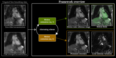 |
Time-resolved cardiac imaging and motion analysis using a
multi-scale dynamics decomposition
Thomas E. Olausson1,
Casper Beijst2,
Alessandro Sbrizzi1,
Cornelis A.T. van den Berg1,
and Niek R.F. Huttinga1 1Computational Imaging Group for MR therapy & Diagnostics, Department of Radiotherapy, University Medical Center Utrecht, Utrecht, Netherlands, Utrecht, Netherlands, 2UMC Utrecht Cancer Center, Department of Radiotherapy, University Medical Center Utrecht, Utrecht, Netherlands Keywords: Sparse & Low-Rank Models, Cardiovascular, Motion estimation; Motion correction; Low-Rank & Sparse;Time-resolved imaging In this work we present a time-resolved (ungated, free-breathing) dynamic cardiac MRI method which disentangles and jointly estimates motion-fields and time-varying contrast images. Different sources of intensity variations are represented with L+S decompositions and motion-fields, that can naturally provide the information required for downstream tasks such as perfusion (contrast dynamics) and myocardial strain analysis (tissue dynamics). Results indicate the feasibility of disentangling the different sources of temporal intensity variations such as respiratory motion or contrast enhancement. Moreover, no breath holds nor ECG triggering/sorting are required since the time-resolved framework can simultaneously resolve the many overlapping dynamics from a continuous data-stream. |
|
13:54 |
0862.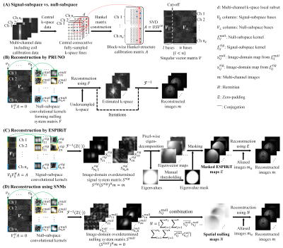 |
Robust Image Reconstruction using Multi-channel Spatial Nulling
Maps: An Alternative to ESPIRiT?
Jiahao Hu1,2,3,
Yi Zheyuan1,2,3,
Yujiao Zhao1,2,
Junhao Zhang1,2,
Linfang Xiao1,2,
Christopher Man1,2,
Vick Lau1,2,
Alex T. L. Leong1,2,
Fei Chen3,
and Ed X. Wu1,2 1Laboratory of Biomedical Imaging and Signal Processing, The University of Hong Kong, Hong Kong, Hong Kong, 2Department of Electrical and Electronic Engineering, The University of Hong Kong, Hong Kong, Hong Kong, 3Department of Electrical and Electronic Engineering, Southern University of Science and Technology, Shenzhen, China Keywords: Signal Representations, Parallel Imaging We develop a novel parallel imaging reconstruction method by extracting null-subspace bases of calibration data/matrix to calculate image-domain spatial nulling maps that contain both coil sensitivity and finite image support information. Images are reconstructed by solving a nulling system formed by multi-channel spatial nulling maps without any masking-related procedure (i.e., in existing SENSE/ESPIRiT for minimizing noise propagation). We demonstrate this method with 2D brain, knee and cardiac data under various conditions, yielding results highly comparable to ESPIRiT with optimal manual masking. Our proposed hybrid-domain reconstruction method is efficient, and more robust than existing ESPIRiT for parallel imaging in practice. |
| 14:02 |
0863.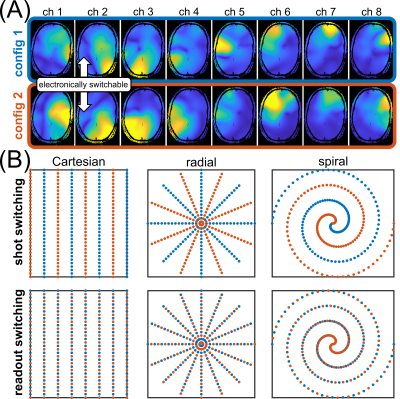 |
Investigations on non-Cartesian parallel imaging with
time-varying receive sensitivities
Felix Glang1,
Praveen Iyyappan Valsala1,
Anton V. Nikulin1,2,
Nikolai Avdievich1,
Theodor Steffen1,
and Klaus Scheffler1,2
1High-field Magnetic Resonance Center, Max Planck Institute for Biological Cybernetics, Tübingen, Germany, 2Department of Biomedical Magnetic Resonance, Eberhard Karls University Tübingen, Tübingen, Germany Keywords: Parallel Imaging, High-Field MRI Non-Cartesian trajectories have several advantageous properties, including favorable parallel imaging performance. Recently, a novel concept of improving parallel imaging by electronically modulated time-varying receive sensitivities has been introduced. In the present work, we investigate if these two concepts can be combined, i.e., how dynamic sensitivity modulation impacts non-Cartesian parallel imaging reconstruction. To that end, numerical experiments are performed based on data from the novel reconfigurable coil array. We find improvement in convergence, reconstruction error and noise amplification due to rapid sensitivity modulation for radial, spiral, and Cartesian trajectories, implying the potential of this method for advanced encoding and reconstruction schemes. |
14:10 |
0864.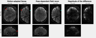 |
Pose-dependent field reconstruction enabled by prospective
motion navigation and randomized sampling
Malte Riedel1,
Thomas Ulrich2,
and Klaas Pruessmann2
1ETH Zurich, Zurich, Switzerland, 2Institute for Biomedical Engineering, ETH Zurich and University of Zurich, Zurich, Switzerland Keywords: Image Reconstruction, Brain Head motion is accompanied by a multitude of second order motion effects like changing coil sensitivity maps, background field inhomogeneities, and susceptibility-induced fields. While scan geometries and shims can be corrected in real-time, the scanner has no means to counteract changes of the coil sensitivity maps or the susceptibility-induced fields requiring data-driven retrospective motion correction algorithms for these issues. Randomized sampling can further be exploited to improve the problem conditioning of the parameter estimations on temporal sub-segments of the scan. In this work, we evaluate the combination prospective motion navigation with randomized sampling in a pose-dependent field correction algorithm. |
|
14:18 |
0865.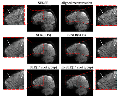 |
Motion Compensated Structured Low-rank Reconstruction for Robust
3D Multi-shot EPI fMRI
Xi Chen1,
Wenchuan Wu1,
and Mark chiew1,2,3
1Wellcome Centre for Integrative Neuroimaging, FMRIB, Nuffield Department of Clinical Neurosciences, University of Oxford, Oxford, United Kingdom, 2Department of Medical Biophysics, University of Toronto, Toronto, ON, Canada, 3Physical Sciences, Sunnybrook Research Institute, Toronto, ON, Canada Keywords: Sparse & Low-Rank Models, Motion Correction, fMRI; 3D EPI Structured low-rank (SLR) reconstruction has been successfully used in 3D multi-shot EPI for fMRI to improve its robustness to inter-shot phase variations. This work proposed a motion compensated structured low-rank (mcSLR) reconstruction, which further improves the robustness of 3D multi-shot EPI by joint modelling of both inter-shot motion and phase variations. |
| 14:26 |
0866.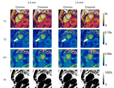 |
Alternating Low-Rank Tensor Reconstruction for Improved
Multi-Dimensional MRI with MR Multitasking
Tianle Cao1,2,
Yibin Xie1,
Debiao Li1,
and Anthony G. Christodoulou1
1Cedars Sinai Medical Center, Los Angeles, CA, United States, 2University of California, Los Angeles, Los Angeles, CA, United States Keywords: Sparse & Low-Rank Models, Sparse & Low-Rank Models Low-rank tensor modelling is promising for multi-dimensional MR imaging. In this work, we developed a new low-rank tensor reconstruction approach using alternating minimization of spatial and temporal bases from the whole k-t space data instead of from split subsets of data. The approach was evaluated for 2D motion-resolved myocardial T1/T2/T2*/fat-fraction mapping and could potentially be used for imporving reconstruction quality and/or further reducing scan time. |
| 14:34 |
0867.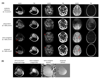 |
Navigator-free water/fat separation for multi-shot
diffusion-weighted EPI using structured low-rank reconstruction
Yiming Dong1,
Kirsten Koolstra2,
Ziyu Li3,
Matthias J.P. van Osch1,
and Peter Börnert1,4
1C.J. Gorter MRI Center, Department of Radiology, LUMC, Leiden, Netherlands, 2Philips, Best, Netherlands, 3Wellcome Centre for Integrative Neuroimaging, FMRIB, Nuffield Department of Clinical Neurosciences, University of Oxford, Oxford, United Kingdom, 4Philips Research, Hamburg, Germany Keywords: Image Reconstruction, Diffusion/other diffusion imaging techniques Multi-shot EPI readout-approaches provide high spatial resolution at reduced geometric distortions and improved SNR in diffusion weighted imaging (DWI). As a specific challenge, physiological motion induces shot-to-shot phase variations and needs specific handling, e.g., using additionally measured phase navigators, data-driven phase estimation and/or low-rank regularizations. Furthermore, good fat-suppression is also needed in DWI, making the use of chemical-shift encoding interesting. In this work, a structured low-rank-based water/fat separation pipeline is proposed to jointly estimate water/fat images while correcting motion-induced phase variations with improved time efficiency. In-vivo examples from different anatomies demonstrate improved water/fat separation compared to conventional approaches. |
| 14:42 |
0868. |
Robust Quantitative Susceptibility Mapping via Approximate
Message Passing with Parameter Estimation
Shuai Huang1,
James J. Lah1,
Jason W. Allen1,
and Deqiang Qiu1
1Emory University, Atlanta, GA, United States Keywords: Image Reconstruction, Quantitative Susceptibility mapping We propose a robust Bayesian approach with built-in parameter estimation for quantitative susceptibility mapping (QSM). From a Bayesian perspective, wavelet coefficients of the susceptibility map are modeled by Laplace distribution. Noise is modeled by a two-component Gaussian-mixture distribution, where the second component is reserved to model the noise outliers. The susceptibility map and distribution parameters are jointly recovered using approximate message passing (AMP). The proposed approach achieves better performance in challenging cases of brain hemorrhage and calcification. It automatically estimates the parameters, which avoids subjective bias from the usual visual-tuning step of in vivo reconstruction. |
| 14:50 |
0869.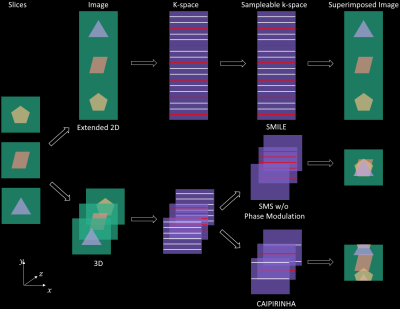 |
Accelerated Simultaneous Multislice Imaging via Linear Phase
Modulated Extended Field of View (SMILE)
Shen Zhao1,
Junyu Wang1,
and Michael Salerno1
1Cardiovascular Medicine, Stanford University, Stanford, CA, United States Keywords: Image Reconstruction, Image Reconstruction, SMS Slice leakage is a significant issue in simultaneous multislice (SMS) imaging. In this work, we extend POMP and develop a new efficient accelerated SMS acquisition technique: Simultaneous Multislice Imaging via Linear phase modulated Extended field of view (SMILE). SMILE transforms the SMS problem into a 2D imaging task and enables direct implementation of 2D non-SMS-only reconstruction algorithms. SMILE increases the sampling degree of freedom by a factor of the number of slices and could theoretically avoid the significant "slice-leakage" issue. |
|
14:58 |
0870.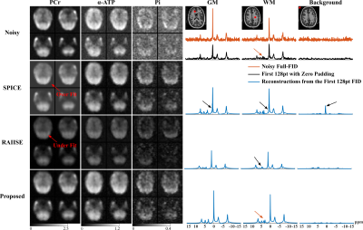 |
Fast MRSI Reconstruction Combining Linear and Nonlinear Manifold
Models
Yahang Li1,2,
Zepeng Wang 1,2,
Aaron Anderson 2,3,
Ruiyang Zhao 2,4,
Paul Arnold 2,3,
Graham Huesmann 2,3,5,
and Fan Lam 1,2,4
1Department of Bioengineering, University of Illinois Urbana-Champaign, Urbana, IL, United States, 2Beckman Institute for Advanced Science and Technology, Urbaba, IL, United States, 3Neuroscience Institute, Carle Foundation Hospital, Urbaba, IL, United States, 4Department of Electrical and Computer Engineering, University of Illinois Urbana-Champaign, Urbana, IL, United States, 5School of Molecular and Cellular Biology, University of Illinois Urbana-Champaign, Urbana, IL, United States Keywords: Image Reconstruction, Spectroscopy A computationally efficient MRSI reconstruction method is presented. The proposed problem formulation integrates a subspace model of the high-dimensional spatiotemporal function (SPICE) and a network-based learned projector on to a low-dimensional manifold of generic spectroscopic signals. The subspace representation allows for more flexible spatiotemporal sampling designs than using nonlinear manifold constraint alone, while the manifold constraint effectively regularizes the subspace fitting, especially at higher orders. An efficient algorithm is designed to solve the optimization problem. The benefits of the proposed synergy have been demonstrated using simulations as well as experimental 31P and 1H-MRSI data. |
|
15:06 |
0871.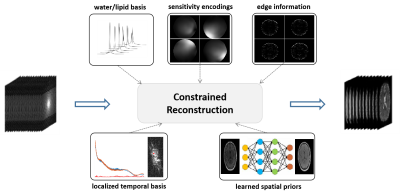 |
Improved Reconstruction for High-Resolution QSM from Highly
Sparse Unsuppressed Water Signals of 1H-MRSI Scans
Ziwen Ke1,
Yibo Zhao2,3,
Yudu Li2,4,
Rong Guo2,5,
Zhi-Pei Liang2,3,
and Yao Li1
1School of Biomedical Engineering, Shanghai Jiao Tong University, Shanghai, China, 2Beckman Institute for Advanced Science and Technology, University of Illinois at Urbana-Champaign, Urbana, IL, United States, 3Department of Electrical and Computer Engineering, University of Illinois at Urbana-Champaign, Urbana, IL, United States, 4National Center for Supercomputing Applications, University of Illinois at Urbana-Champaign, Urbana, IL, United States, 5Siemens Medical Solutions USA, Urbana, IL, United States Keywords: Image Reconstruction, Quantitative Susceptibility mapping The SPICE technique has demonstrated a unique capability of simultaneous QSM/MRSI. To achieve fast high-resolution QSM, highly sparse sampling of (k,t)-space is used in SPICE data acquisition, which poses a significant challenge in image reconstruction. In this work, we solved this problem using a subspace-assisted parallel imaging technique with learned image priors. The proposed method has been validated using experimental data, producing high-quality QSM maps from the unsuppressed water signals of 1H-MRSI scans. |
| 15:14 |
0872. |
A Time-Saving View-Sharing Interleaved Block-Segmented
Diffusion-Tensor Imaging (VS iblocks-DTI)
Liyuan Liang1,2,
Mei-Lan Chu3,
Nan-Kuei Chen4,5,
and Hing-Chiu Chang1,2
1Department of Biomedical Engineering, The Chinese University of Hong Kong, Shatin, Hong Kong, 2Multi-Scale Medical Robotics Center, Shatin, Hong Kong, 3Graduate Institute of Biomedical Electronics and Bioinformatics, National Taiwan University, Taipei, Taiwan, 4Department of Biomedical Engineering, University of Arizona, Tucson, AZ, United States, 5Brain Imaging and Analysis Center, Duke University Medical Center, Durham, NC, United States Keywords: Image Reconstruction, Diffusion Tensor Imaging Recently, a novel self-navigated multi-shot EPI data acquisition scheme called iblocks-EPI was proposed to achieve high-resolution DWI images with further reduction of distortion compared to the conventional interleaved EPI. However, the total acquisition time of iblocks-EPI is several times longer than the conventional interleaved EPI because it requires multiple patch patterns for covering a complete k space. Consequently, it is time-consuming and impractical for routine acquisition of DTI data. In this work we propose a view-sharing iblocks-DTI scheme which can substantially reduce the scan time for iblocks-DTI acquisition while providing accurate DTI tensor calculation. |
|
15:22 |
0873. |
Interactive Real-Time MRI with BART
Philip Schaten1 and
Martin Uecker1
1Institute of Biomedical Imaging, Graz University of Technology, Graz, Austria Keywords: Image Reconstruction, Software Tools The open-source software tool BART provides various state-of-the-art image reconstruction algorithms for MRI. We add an interface for streaming image and k-space data to BART, which should facilitate the application of modern image reconstruction to real-time MRI. Our implementation matches the requirements of interventional MRI in terms of frame rate and latency.Furthermore, we present an intuitive way of interactive slice positioning for MR-guided Interventions using a game controller featuring an inertial measurement unit. |
 Back to Meeting Home
 Back to the Program-at-a-Glance
The International Society for Magnetic Resonance in Medicine is accredited by the Accreditation Council for Continuing Medical Education to provide continuing medical education for physicians.
 Back to Meeting Home
 Back to the Program-at-a-Glance
 View Presentation Video