Oral Session
Signal Modeling & Representation
ISMRM & ISMRT Annual Meeting & Exhibition • 03-08 June 2023 • Toronto, ON, Canada

| 08:15 |
0759.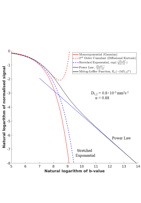 |
Quasi-Diffusion: A Model Of Signal Attenuation In Diffusion
Magnetic Resonance Imaging Of Brain Tissue
Thomas R Barrick1,
Carson Ingo2,3,
and Franklyn A Howe1
1Department of Neurosciences, St George's, University of London, London, United Kingdom, 2Department of Neurology, Northwestern University, Chicago, IL, United States, 3Department of Physical Therapy, Human Movement Sciences, Northwestern University, Chicago, IL, United States Keywords: Signal Modeling, Diffusion/other diffusion imaging techniques We show the quasi-diffusion model describes the dMRI signal from low b-value (stretched exponential) to high b-value (power law) regimes via a single, parsimonious function of two independent parameters. We identify new tissue contrast via a signal inflection point indicating the transition to the localisation regime. Quasi-Diffusion Imaging (QDI) parameter estimates converge to stable values as maximum b-value is increased, suggesting quasi-diffusion is a valid model for brain tissue dMRI signal decay. Accuracy of QDI parameters computed from 4 b-values of a 12 b-value acquisition indicate QDI measures may be derived from data acquired in clinically feasible times. |
| 08:23 |
0760.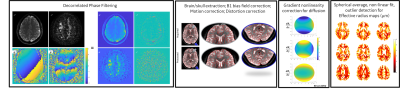 |
Axonal Diameter Mapping using High Performance Gradients:
Feasibility study and Repeatability of Estimates
Nastaren Abad1,
Afis Ajala1,
Chitresh Bhushan1,
Ante Zhu1,
Luca Marinelli1,
Eric Fiveland1,
Seung-Kyun Lee1,
J Kevin DeMarco2,3,
Robert Shih2,3,
Maureen Hood2,3,
Gail Kohls2,
H. Doug Morris3,
Kimbra Kenney3,
Vincent Ho3,
and Thomas K.F Foo1,3 1GE Research, Niskayuna, NY, United States, 2Walter Reed National Military Medical Center, Bethesda, MD, United States, 3Uniformed Services University of the Health Sciences, Bethesda, MD, United States Keywords: Signal Modeling, Diffusion/other diffusion imaging techniques High performance gradients allow for the exploration of an expanded diffusion parameter space, that simplifies biophysical model at ultra-high b=7-30 ms/μm2. The choice of b-encoding space can suppress contributions from extra-axonal water signal while utilization of high performance gradient systems allows for maintaining short echo times (TE<63 ms) with adequate SNR. In this study, the feasibility and reproducibility of mapping effective axonal diameter distributions in the in-vivo brain was assessed by making use of a test-retest paradigm. Whole brain white-matter and parcel based reproducibility and sensitivity were evaluated for this promising biomarker. |
| 08:31 |
0761.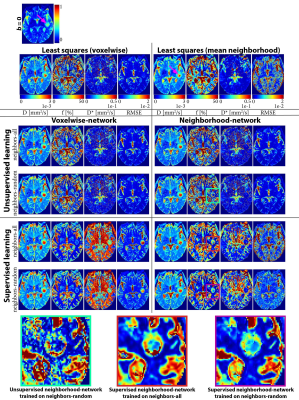 |
Synthetic data shows the potential of unsupervised and
supervised learning for incorporating spatial information in
IVIM fitting
Misha Pieter Thijs Kaandorp1,2,
Peter Thomas While1,2,
and Frank Zijlstra1,2
1Department of Radiology and Nuclear Medicine, St. Olav's University Hospital, Trondheim, Norway, 2Department of Circulation and Medical Imaging, NTNU – Norwegian University of Science and Technology, Trondheim, Norway Keywords: Signal Modeling, Diffusion/other diffusion imaging techniques DWI data is spatially homogeneous, yet microstructures are irregular, where neighboring voxels do not always share information. Therefore, we need to utilize neighboring correlations only when they are present. In simulations, we show that by training on synthetic data with all plausible combinations of neighboring correlations, the accuracy of supervised deep learning IVIM model fitting can be improved. Conversely, unsupervised learning did not benefit from incorporating spatial information. In in-vivo data from a glioma patient, supervised training on this synthetic data improved the performance of IVIM fitting by effectively denoising the DWI data while preserving edge-like structures. |
| 08:39 |
0762.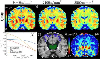 |
Characterization of T2-dependent microstructural spectra using
combined relaxation-diffusion MRI
Lipeng Ning1,2,
Carl-Fredrik Westin1,2,
and Yogesh Rathi1,2
1Brigham and Women's Hospital, Boston, MA, United States, 2Harvard Medical School, Boston, MA, United States Keywords: Signal Representations, Diffusion/other diffusion imaging techniques We proposed a method for joint modeling and analysis for diffusion MRI data with multiple TEs and examined the performance of using in vivo data acquired from a clinical scanner. The contribution of this work includes the characterization of b-value-dependent T2 relaxation rates, comparing prediction results using subsampled TEs, and characterization of microstructural spectral and long and short TEs using extrapolated signals. The results show that the proposed method can provide novel features to characterize T2-dependent microstructure using rdMRI acquired from clinical scanners. |
| 08:47 |
0763.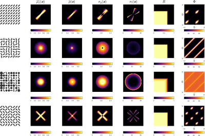 |
Propagator-sensitive diffusion measurements hear the percussion
ensemble
Evren Özarslan1,
Deneb Boito1,
and Alfredo Ordinola1
1Department of Biomedical Engineering, Linköping University, Linköping, Sweden Keywords: Signal Modeling, Diffusion/other diffusion imaging techniques In 1966, Kac asked the question ‘Can one hear the shape of a drum?’ which refers to linking the density of states function to the shape of the pores. We illustrate how the density of eigenstates as well as the pore shape can be obtained through diffusion measurements performed via a recently introduced arrangement of long and narrow gradient pulses. In the presence of dispersity, maps derived from the signal provide exquisite details about the ensemble of pore shapes within the voxel. |
08:55 |
0764.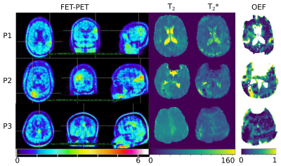 |
Simultaneous T2/T2* quantification for oxygen extraction
fraction estimation using GE-SE EPIK: A preliminary study in
brain tumor patients
Fabian Küppers1,2,
Seong Dae Yun1,
Philipp Lohmann1,
Christian Filss1,3,
Gabriele Stoffels1,
Karl-Josef Langen1,3,
and Nadim Jon Shah1,4,5,6
1Institute of Neuroscience and Medicine - 4, Forschungszentrum Juelich GmbH, Jülich, Germany, 2RWTH Aachen University, Aachen, Germany, 3Department of Nuclear Medicine, RWTH Aachen University Hospital, Aachen, Germany, 4JARA-BRAIN - Translational Medicine, Aachen, Germany, 5Institute of Neuroscience and Medicine - 11, Forschungszentrum Juelich GmbH, Jülich, Germany, 6Department of Neurology, RWTH Aachen University Hospital, Aachen, Germany Keywords: Multi-Contrast, Oxygenation, Oxygen Extraction Fraction The versatile information provided by simultaneous multi-contrast GE/SE acquisitions keeps interest in this area high. This work extends the previously published 10-echo GE-SE EPIK to investigate brain tumor patients. Within this scope, a single-slice 12-second acquisition provides quantification of T2 and T2* with application to oxygen extraction fraction (OEF) information. Data from four tumor patients were acquired, revealing increased T2/T2* values and increased OEF in regions with variable amino acid uptake in O-(2-[18F]fluoroethyl)-L-tyrosine (FET) PET. |
| 09:03 |
0765.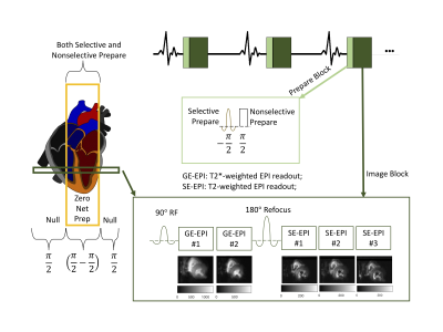 |
Effects of SNR on T2 and T2* estimates from a cardiac
gradient-echo spin-echo (GESE) echo planar sequence
Qi Huang1,
Jason Mendes1,
Ganesh Adluru2,
and Edward DiBella2 1University of Utah, Salt Lake City, UT, United States, 2UCAIR, salt lake city, UT, United States Keywords: Signal Modeling, Multi-Contrast, Gradient-echo spin-echo (GESE); Echo Planar; The bias and precision of T2 and T2* estimates for a five-echo GESE model were studied using CRLB and Monte Carlo simulations. Different SNR cases were implemented by varying the standard deviation of added noise or the T2/T2* ratio. An in-vivo study with high SNRs for all EPI readouts was performed to validate the simulation. |
09:11 |
0766.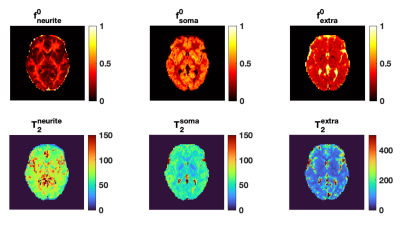 |
Multi-TE SANDI: Quantifying compartmental T2 relaxation times in
the grey matter
Ting Gong1,
Chantal MW Tax2,3,
Matteo Mancini4,
Derek K Jones4,
Hui Zhang1,
and Marco Palombo4,5
1Centre for Medical Image Computing & Department of Computer Science, University College London, London, United Kingdom, 2Cardiff University Brain Research Imaging Centre, School of Physics and Astronomy, Cardiff University, Cardiff, United Kingdom, 3University Medical Center Utrecht, Utrecht University, Utrecht, Netherlands, 4Cardiff University Brain Research Imaging Centre, School of Psychology, Cardiff University, Cardiff, United Kingdom, 5School of Computer Science and Informatics, Cardiff University, Cardiff, United Kingdom Keywords: Signal Modeling, Diffusion/other diffusion imaging techniques This study models the echo time (TE) dependence of the apparent soma and neurite signal fractions derived from the soma and neurite density imaging (SANDI) and quantifies the apparent compartmental T2 relaxation times in the grey matter (GM). The SANDI model divides the intra-cellular space into intra-neurite and intra-soma compartments to account for contribution of water diffusion in cell bodies. By collecting multi-TE SANDI datasets, we provide the first ever estimates of apparent intra-neurite and intra-soma T2 relaxation times in the human brain and characterise their distribution in cortical and subcortical GM regions. |
| 09:19 |
0767.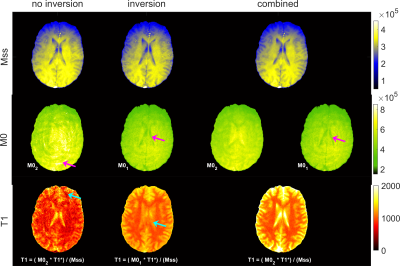 |
No Wait Inversion – a novel model for T1 mapping from inversion
recovery measurements without the waiting times
Anne Slawig1,2,
Juliana Bibiano2,
and Herbert Köstler2
1University Clinic and Outpatient Clinic for Radiology, University Hospital Halle (Saale), Halle (Saale), Germany, 2Department of Diagnostic and Interventional Radiology, University Hospital Würzburg, Würzburg, Germany Keywords: Signal Modeling, Relaxometry, T1 mapping, inversion recovery, model based reconstruction This study proposes a combined fit of an inversion-prepared and non-prepared measurement for robust fast T1-mapping. Therefore, 24 slices in the brain were acquired with and without a global inversion pulse before each slice and no waiting time was heeded in between. Results of exponential model reconstructions for inversion measurement only, non-prepared measurement only and combined fitting of both measurements were compared towards their performance for T1-mapping. Robust and accurate T1-maps were achieved by the proposed combined model even in cases of imperfect inversion. It can thus eliminate long waiting times in between inversions or the necessity of perfect inversion pulses. |
| 09:27 |
0768.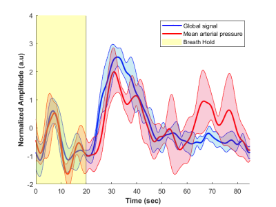 |
A practical methodology to study the relationship between
arterial blood pressure fluctuations and the BOLD signal
Rémi Dagenais1 and
Georgios D. Mitsis2
1Biological and Biomedical Engineering, McGill University, Montréal, QC, Canada, 2Bioengineering, McGill University, Montréal, QC, Canada Keywords: Data Acquisition, fMRI, Physiological denoising, ABP regulation The relationship between arterial blood pressure (ABP) fluctuations and the BOLD-fMRI signal is mostly unknown due to the limited availability of reliable MR-compatible ABP devices. Here, we propose a combined experimental and mathematical modeling methodology that uses two calibration recordings outside the scanner to estimate the ABP during the MR scan. Significant correlations between BOLD and ABP were observed in all tested subjects, which also revealed that the global BOLD signal is intrinsically correlated with the underlying ABP fluctuations. Our method is expected to improve our understanding of ABP regulation in the brain and allow for improved physiological fMRI denoising. |
| 09:35 |
0769.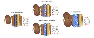 |
Biophysical model to correct for blood volume confounders of T2*
mapping based renal MR oximetry
Thomas Gladytz1,
Kathleen Cantow2,
Jason Michael Millward1,3,
Sonia Waiczies1,3,
Erdmann Seeliger2,
and Thoralf Niendorf1,3
1Berlin Ultrahigh Field Facility (B.U.F.F.), Max Delbrück Center for Molecular Medicine in the Helmholtz Association, Berlin, Germany, 2Institute of Translational Physiology, Charité - Universitätsmedizin Berlin, Berlin, Germany, 3Experimental and Clinical Research Center, a joint cooperation between the Charité Medical Faculty and the Max Delbrück Center for Molecular Medicine in the Helmholtz Association, Berlin, Germany Keywords: Oxygenation, Quantitative Imaging Quantitative MRI of renal hypoxia, an early key feature in acute kidney injury, is a valuable tool for diagnostics and pathophysiological studies. En route to MR oximetry we propose a biophysical model that uses the kidney size as an additional MRI derivable information to correct for blood volume confounders of oxygenation sensitive T2* maps. The model is demonstrated in a preclinical study involving three different vascular occlusions highlighting the confounding effect of blood volume changes and its correction. |
| 09:43 |
0770.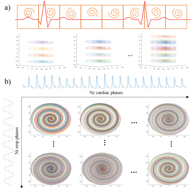 |
Free-breathing 3D Stack-of-Spiral Cardiac Quantitative
Susceptibility Mapping for Noninvasive Measurement of Cardiac
Chamber Oxygenation
Jiahao Li1,2,
Pablo Villar-Calle3,
Jinwei Zhang1,2,
Chao Li2,4,
Thanh D. Nguyen2,
Yi Wang1,2,
Jiwon Kim3,
Jonathan W. Weinsaft3,
and Pascal Spincemaille2
1Biomedical Engineering, Cornell University, Ithaca, NY, United States, 2Radiology, Weill Cornell Medicine, New York, NY, United States, 3Medicine, Weill Cornell Medicine, New York, NY, United States, 4Applied and Engineering Physics, Cornell University, Ithaca, NY, United States Keywords: Oxygenation, Motion Correction A free-breathing 3D stack-of-spiral data acquisition was developed for motion-free cardiac quantitative susceptibility mapping, to tackle the challenge where even a short breath-hold is difficult for patients to perform. ECG and respiratory bellow signal were recorded for retrospective motion binning. A 5D dataset incorporating additional cardiac and respiratory phase dimensions were reconstructed with joint spatiotemporal regularization to generate a motion-free cardiac QSM. In healthy volunteers, this method was compared with a motion-averaged reconstruction and with a separate breath-hold spiral cardiac QSM using Cartesian navigator QSM as reference. Equivalent right-to-left heart chamber differential blood oxygenation was observed among all method studied. |
| 09:51 |
0771.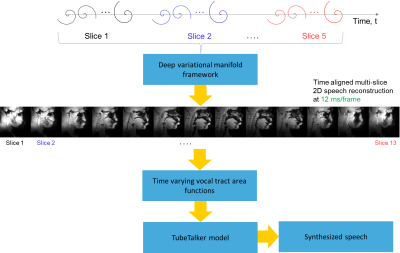 |
Synthesizing speech through a tube talker model informed by
dynamic MRI-derived vocal tract area functions
Rushdi Zahid Rusho1,
Brad H. Story2,
David Meyer3,
Mathews Jacob4,
and Sajan Goud Lingala1,5
1Roy J. Carver Department of Biomedical Engineering, The University of Iowa, Iowa City, IA, United States, 2Speech, Language and Hearing Sciences, University of Arizona, Tucson, AZ, United States, 3Janette Ogg Voice Research Center, Shenandoah University, Winchester, VA, United States, 4Electrical and Computer Engineering, The University of Iowa, Iowa City, IA, United States, 5Department of Radiology, The University of Iowa, Iowa City, IA, United States Keywords: Signal Modeling, Head & Neck/ENT The vocal tract encompasses the airspace from the glottis (the space between the vocal folds) to the external lips. This irregular tube filters the glottal sound source, and is modulated by many structures (e.g. the tongue, lips, and velum) to produce speech sounds (e.g. vowels and consonants). In this work, we determine the preliminary feasibility of integrating vocal tract area functions derived from a recently proposed accelerated pseudo-3D dynamic speech MRI scheme to a parametric tube talker model to synthesize speech. |
| 09:59 |
0772.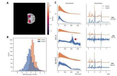 |
Two-voxel MRS with B0 correction using a model-based
reconstruction
Adam Berrington1 and
Olivier Mougin1 1Sir Peter Mansfield Imaging Centre, School of Physics and Astronomy, University of Nottingham, Nottingham, United Kingdom Keywords: Signal Modeling, Spectroscopy Performing simultaneous MRS from two-regions is often desirable in spectroscopy studies. However, achieving an optimal B0 shim in two regions is difficult, leading to poor data quality. We investigate a model-based reconstruction for two-voxel MRS, incorporating B0 information, to reconstruct data with reduced T2* decay. We test this approach using least squares and regularization approaches for Hadamard encoding in simulations and phantom. Despite a noise penalty for B0-correction (2-4)-fold, FIDs from two voxels better matched un-broadened spectra under conditions of sufficient SNR. Regularization also produced time-domain reconstructions with less noise than a least-squares approach. |
| 10:07 |
0773.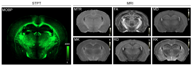 |
Enhanced Myelin Mapping based on Histology and MRI from the Same
Subjects
Zifei Liang1,
Choong Heon Lee1,
Jennifer A. Minteer2,
Yongsoo Kim2,
and Jiangyang Zhang1
1Radiology, NYU Langone health, new york, NY, United States, 2Penn State University, Hershey, PA, United States Keywords: Signal Representations, Brain, MR-histology To infer cellular-level information from MR signals with high sensitivity and specificity is a challenging task. We previously demonstrated the feasibility of mapping myelin in the mouse brain based on multi-contrast MRI using deep learning, but the results were based on limited histological data and MRI data from separate cohorts. In this study, we acquired serial 2-photon and MRI data from the same mice and trained a neural network for mapping myelin. Our results demonstrated enhanced sensitivity and specificity compared to conventional MRI myelin markers, our previous network, and polynomial fitting. |
 Back to Meeting Home
 Back to the Program-at-a-Glance
The International Society for Magnetic Resonance in Medicine is accredited by the Accreditation Council for Continuing Medical Education to provide continuing medical education for physicians.
 Back to Meeting Home
 Back to the Program-at-a-Glance
 View Presentation Video