Oral Session
Keeping you Abreast: Advances in Breast MRI
ISMRM & ISMRT Annual Meeting & Exhibition • 03-08 June 2023 • Toronto, ON, Canada

Oral Session
Keeping you Abreast: Advances in Breast MRI
| 13:45 |
0184.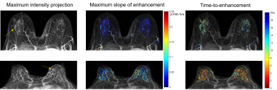 |
Screening abbreviated breast MRI with ultrafast imaging:
prospective study results
Federico Pineda1,
Zhen Ren2,
Rabia Safi2,
Elle Hill2,
Kirti Kulkarki2,
Hiroyuki Abe2,
and Gregory Karczmar2 1Radiology, University of Pittsburgh, Pittsburgh, PA, United States, 2University of Chicago, Chicago, IL, United States Keywords: Breast, Breast, Abbreviated Breast MRI We report on the results of a prospective screening trial using abbreviated breast MRI with ultrafast dynamic contrast-enhanced imaging (3.5 seconds per time point) to screen for breast cancer in 166 women at any risk of developing breast cancer. The cancer detection rate was 24.1 per 1000 women while maintaining a specificity of 97.6%. The biopsy recommendation and abnormal interpretation rates were lower than in other published trials that did not include ultrafast imaging in their abbreviated protocol. |
| 13:53 |
0185.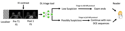 |
Personalized Breast MRI Scanning Using Deep Learning
Sarah Eskreis-Winkler1,
Arka Bhowmik1,
Christopher Comstock1,
Elizabeth Sutton1,
Vardan Sevilimedu1,
Sunitha Thakur1,
and Katja Pinker1 1MSK, New York, NY, United States Keywords: Breast, Breast, Artificial Intelligence Contrast-enhanced breast MRI exams typically last more than 20 minutes even though the dynamic contrast-enhanced sequences alone would be sufficient for interpretation in over 95% of cases. Thus, we present a personalized on-the-fly MRI protocoling paradigm, where a deep learning algorithm uses images acquired during the first few minutes of the exam to triage patients to an abbreviated versus full MRI protocol. We conduct a retrospective reader study to show that this personalized scanning paradigm decreases scan time and cost while maintaining diagnostic performance. |
14:01 |
0186.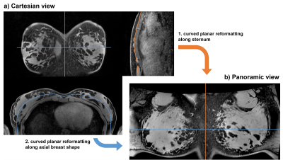 |
A panoramic view for supine breast MRI
Lena Nohava1,
Michael Obermann1,
and Elmar Laistler1
1High Field MR Center, Center for Medical Physics and Biomedical Engineering, Medical University of Vienna, Vienna, Austria Keywords: Breast, Visualization A wearable coil worn like a vest (“BraCoil”) has recently been shown to improve signal-to-noise ratio by a factor of up to three in supine breast MRI. However, the visualization of supine breast images is inefficient when using Cartesian views due to the bent shape of the breast along the chest wall. We introduce a panoramic view similar to panoramic dental X-ray to make reading of supine breast MRI more efficient by reducing the number of slices by a factor of 2-4, depending on breast size. |
| 14:09 |
0187.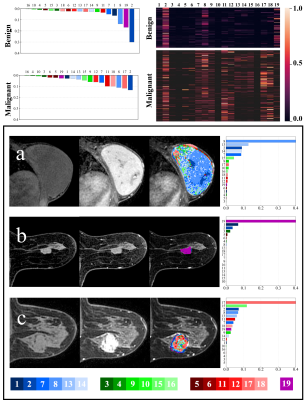 |
A voxel-wise composition ratio of DCE-MRI time-intensity curve
profiles allows for visualizing and quantifying hemodynamic
heterogeneity
Bingyu Yao1,2,
Zhou Liu3,
Yumin Chen2,
Jie Wen3,
Meng Wang3,
Ya Ren3,
Dong Liang1,
Xin Liu1,
Hairong Zheng1,
Dehong Luo3,
and Na Zhang1
1Paul C. Lauterbur Research Center for Biomedical Imaging, Shenzhen Institute of Advanced Technology, Chinese Academy of Sciences, Shenzhen, China, 2College of Computer and Information Engineering, Xiamen University of Technology, Xiamen, China, 3Department of Radiology, National Cancer Center/National Clinical Research Center for Cancer/Cancer Hospital & Shenzhen Hospital, Chinese Academy of Medical Sciences and Peking Union Medical College, Shenzhen, China Keywords: Breast, Cancer We propose a novel model-free and data-driven approach, i.e., voxel-wise composition ratio on 19 dynamic contrast-enhanced MRI (DCE-MRI) time-intensity curve (TIC) profiles (Type-19) to visualize and quantify spatial hemodynamic heterogeneity. The proposed quantitative method for breast tumor was evaluated and compared with the two existing methods (qualitative and semi-quantitative methods) in 4 different clinical applications. In distinguishing malignancy on breast cancer lesions and predicting tumor proliferation status, we found that the machine learning model based on the Type-19 feature outperformed other two models in the validation set. |
| 14:17 |
0188.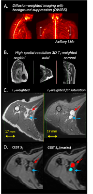 |
In vivo CEST-Dixon MRI in axillary lymph nodes with and without
carcinoma: potential for noninvasive determination of lymph node
metastasis
Rachelle Crescenzi1,2,
Paula M.C. Donahue3,4,
R. Sky Jones5,
Chelsea Lee6,
Maria Garza1,5,
Niral J Patel6,
Ingrid Meszoely7,
and Manus J Donahue5,8
1Radiology and Radiological Sciences, Vanderbilt University Medical Center, Nashville, TN, United States, 2Biomedical Engineering, Vanderbilt University, Nashville, TN, United States, 3Physical Medicine and Rehabilitation, Vanderbilt University Medical Center, Nashville, TN, United States, 4Dayani Center for Health and Wellness, Vanderbilt University Medical Center, Nashville, TN, United States, 5Department of Neurology, Vanderbilt University Medical Center, Nashville, TN, United States, 6Division of Pediatric Neurology, Vanderbilt University Medical Center, Nashville, TN, United States, 7Department of Surgical Oncology, Vanderbilt University Medical Center, Nashville, TN, United States, 8Department of Psychiatry and Behavioral Sciences, Vanderbilt University Medical Center, Nashville, TN, United States Keywords: Breast, CEST & MT, cancer, lymph node, metastasis, breast cancer The overall goal of this work is to apply a CEST-Dixon MRI approach in the axillary lymph nodes (LNs) of women with breast cancer to test fundamental hypotheses about biochemical LN profiles with carcinoma. Mean z-spectra and corresponding significant differences in cohorts with metastatic vs. benign LNs were observed in regions of known chemical exchange for the nuclear Overhauser effect (PTR=0.087 vs. 0.051, p=0.04), hydroxyl (PTR=0.272 vs. 0.212, p=0.003), and amine (PTR=0.081 vs. 0.033, p=0.003) protons. CEST-Dixon MRI of LNs may have relevance for pre-surgical breast cancer staging and for guiding LN sparing resections to reduce risk for lymphedema. |
|
14:25 |
0189.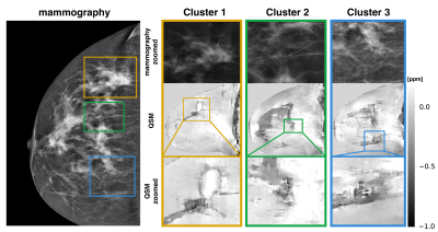 |
High spatial resolution quantitative susceptibility mapping
using in-phase echoes enables the depiction of breast
microcalcifications
Christof Boehm1,
Alexander Komenda1,
Kilian Weiss2,
Jonathan K. Stelter1,
Tabea Borde1,
Jakob Meineke3,
Marcus R. Makowski1,
Eva M. Fallenberg1,
and Dimitrios C. Karampinos1
1Department of Diagnostic and Interventional Radiology, School of Medicine, Klinikum rechts der Isar, Technical University of Munich, Munich, Germany, 2Philips GmbH Market DACH, Hamburg, Germany, 3Philips Research, Hamburg, Germany Keywords: Breast, Susceptibility Breast microcalcifications (MCs) can be the only sign of carcinoma and other precursor lesions of breast cancer in x-ray mammography. Mammography is routinely used for population-based breast cancer screening. However, due to the ionizing nature of x-ray radiation, the use of an MR-based technique would be desirable for repeated examinations especially in screening targeted sub-cohorts at high-risk or younger women. In this work, we present initial results on the visualization of MCs using MR with an optimized high-resolution GRE-scan and quantitative susceptibility mapping. The proposed methodology allows for the robust visualization of MC clusters in MR for the first time. |
| 14:33 |
0190. |
T2 and ADC correlated mapping of breast cancer lesions: A
spatiotemporal encoding 3T MRI analysis
Martins Otikovs1,
Noam Nissan2,
Edna Furman-Haran1,
Debbie Anaby2,
Ravit Agassi3,
Miri Sklair-Levy2,4,
and Lucio Frydman1
1Weizmann Institute of Science, Rehovot, Israel, 2Sheba Medical Center, Ramat Gan, Israel, 3Ben Gurion University Hospital, Beer Sheva, Israel, 4Sackler School of Medicine, Tel Aviv, Israel Keywords: Breast, Cancer The possibility to improve the MRI contrast on cancerous breast lesions was explored by correlating quantitative T2 and ADC values. To this end, T2 maps of patients were measured at more than one b-value and ADC maps at more than one TE value –in all cases using spatiotemporal encoding (SPEN) MRI. SPEN delivered quality, artifact-free TE-weighted DW images, from which ADC and T2 maps could be correlated despite relatively long acquisitions and heavy signal weighting. Data suggest there may be potential value in combining T2 and ADC mappings, as these low-correlated variables can provide complementary information about breast lesions. |
| 14:41 |
0191.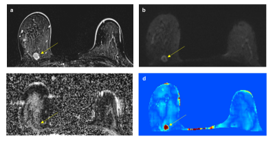 |
MR conductivity imaging to decrease false-positive biopsy caused
by breast ultrasound: comparison with DWI and abbreviated MRI
Soo-Yeon Kim1,
Jun-Hyung Kim 2,
Nariya Cho1,
and Dong-Hyun Kim2
1Radiology, Seoul National University Hospital, Seoul, Korea, Republic of, 2Electrical and Electronic Engineering, Yonsei university, Seoul, Korea, Republic of Keywords: Breast, Electromagnetic Tissue Properties We evaluated the role of MRI as a problem-solving tool to reduce false-positive biopsy caused by breast ultrasound. Seventy nine participants underwent MRI prior to ultrasound-guided core needle biopsy. The MRI protocol consisted of T2-weighted imaging for conductivity map reconstruction, diffusion-weighted imaging, and abbreviated contrast-enhanced MRI. The conductivity, ADC, and BI-RADS criterion lowered false-positive biopsy by 23%, 38%, and 43%, respectively, while being able to detect all cancers. Conductivity imaging showed the potential but showed lower performance than diffusion-weighted imaging and abbreviated MRI in reducing unnecessary biopsies caused by breast ultrasound. |
| 14:49 |
0192.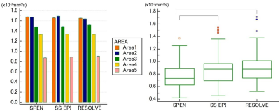 |
High-resolution DWI in the breast by Spatiotemporal encoding
(SPEN): clinical utility in comparison with SS-EPI and RESOLVE
Rena Nakayama1,
Mami Iima2,3,
Masako Kataoka2,
Maya Honda2,4,
Yuta Urushibata5,
Martins Otikovs6,
Noam Nissan7,
Lucio Frydman6,
Rie Ota2,8,
Aika Okazawa2,
Kazuki Tsuji1,
Masakazu Toi9,
and Yuji Nakamoto2
1Kyoto University Faculty of Medicine, Kyoto, Japan, 2Diagnostic Imaging and Nuclear Medicine, Kyoto University Graduate School of Medicine, Kyoto, Japan, 3Institute for Advancement of Clinical and Translational Science (iACT), Kyoto University Hospital, Kyoto, Japan, 4Diagnostic Radiology, Kansai Electric Power Hospital, Osaka, Japan, 5Siemens Healthcare K.K., Tokyo, Japan, 6Chemical and Biological Physics, Weizmann Institute of Science, Rehovot, Israel, 7Radiology, Sheba-Medical-Center, Ramat-Gan, Israel, 8Tenri Hospital, Nara, Japan, 9Breast Surgery, Kyoto University Hospital, Kyoto, Japan Keywords: Breast, Breast This study investigated the breast lesion conspicuity and ADC reliability for 3 different DWI protocols; SPEN, SS-EPI, and RESOLVE. The in-plane resolution for SPEN and RESOLVE was 1x1mm2, and SS-EPI was 2x2mm2. SPEN showed a higher score for lesion conspicuity than SS-EPI, and a lower score than RESOLVE. ADC values in breast lesions were significantly lower in SPEN than others, presumably due to the choice of different b values, the sequences having different T1/T2 weightings, different robustness vs motions, the presence of unsuppressed fat, or different viewer systems used to analyze the data, which will need further investigation. |
| 14:57 |
0193.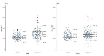 |
Automated breast tumor segmentation in DWI using multi-modality
image registration: a feasibility study using multi-center data
Nu N Le1,
David C Newitt1,
Wen Li1,
Deep Hathi1,
Jiachao Liang1,
Lisa J Wilmes1,
Jessica E Gibbs1,
Natsuko Onishi1,
Bonnie N Joe1,
John Kornak2,
Patrick J Bolan3,
Savannah C Partridge4,
and Nola M Hylton1
1Radiology, University of California, San Francisco, San Francisco, CA, United States, 2Epidemiology & Biostatistics, University of California, San Francisco, San Francisco, CA, United States, 3Radiology, University of Minnesota, Minneapolis, MN, United States, 4Radiology, University of Washington, Seattle, WA, United States Keywords: Breast, Cancer Diffusion-weighted imaging (DWI) in MRI plays an important role in diagnostic applications. Automated tumor segmentation is an important yet challenging step for quantitative breast imaging analysis. While methods have been developed for dynamic contrast enhanced (DCE) MRI, automatic segmentation in breast DWI-MRI is still underdeveloped. The purpose of this study is to develop methods to transfer functional tumor volume (FTV) analysis ROI from DCE-MRI image space to DWI-MRI image space using image registration and extract ADC statistics in regions corresponding to those of DCE segmentations for prediction of pathologic treatment response (pCR). |
| 15:05 |
0194.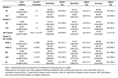 |
Assessment of Breast Lesions by the Kaiser Score for
Differential Diagnosis on MRI: The Added Value of ADC and
Machine Learning Modeling
Zhong-Wei Chen1,
You-Fan Zhao1,
Hui-Ru Liu1,
Jie-Jie Zhou1,
Hai-Wei Miao1,
Shu-Xin Ye1,
Yun He1,
Xin-Miao Liu1,
Min-Ying Su2,3,
and Mei-Hao Wang1
1the First Affiliated Hospital of Wenzhou Medical University, Wenzhou, China, 2University of California, Irvine, CA, United States, 3Kaohsiung Medical University, Kaohsiung, Taiwan Keywords: Breast, Breast How ADC could be combined with Kaiser score (KS) for the diagnosis of breast cancer was emerged as an interesting research area. We modified KS to KS+ based on the dichotomized ADC >1.4×10⁻³ mm2/s, and integrated KS and the continuous ADC values to build machine learning (ML) models for assessment. The diagnostic specificity of KS+ was higher than that of KS with a slightly degraded sensitivity. The AUCs of them were not significantly different. When the KS and the continuous ADC values were used to train ML models, the performance could be further improved while maintaining at a high sensitivity. |
| 15:13 |
0195.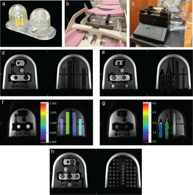 |
Multisite Intravoxel Incoherent Motion Repeatability and
Reproducibility in a Breast Diffusion Phantom at 3T
Dibash Basukala1,
Artem Mikheev1,
Nima Gilani1,
Thomas Benkert2,
Linda Moy1,
Katja Pinker-Domenig3,
Sunitha B. Thakur3,
and Eric E. Sigmund1
1Radiology, NYU Langone Health, New York, NY, United States, 2MR Application Predevelopment, Siemens Healthcare GmbH, Erlangen, Germany, 3Radiology, Memorial Sloan Kettering Cancer Center, New York, NY, United States Keywords: Breast, Phantoms, IVIM, Reproducibility Monoexponential apparent diffusion coefficient (ADC) and biexponential intravoxel incoherent motion (IVIM) analysis of diffusion-weighted imaging (DWI) is helpful in the characterization of breast tumors. Toward this goal, a novel breast phantom containing tubes of different polyvinylpyrrolidone (PVP) concentrations, water, fat, and sponge flow chambers was utilized. This work tests this breast phantom at two sites employing different vendor MRI scanners to estimate the ADC and IVIM parameters. The results are reproducible within sites, and show progress towards reproducibility across sites and vendors, and can be used in the future in multicenter clinical trials for breast cancer characterization, prediction and prognosis. |
| 15:21 |
0196.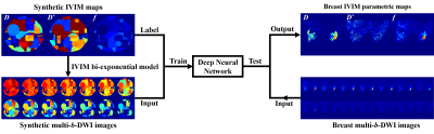 |
Deep learning improves breast IVIM estimation in better benign
and malignant lesion differentiation
Shuhao Shi1,
Lu Wang1,
Jianfeng Bao2,
Zhigang Wu3,
Congbo Cai1,
Zhong Chen1,
Jiazheng Wang3,
and Shuhui Cai1
1Department of Electronic Science, Xiamen University, Xiamen, China, 2Department of Radiology, First Affiliated Hospital of Zhengzhou University, Zhengzhou, China, 3MSC Clinical & Technical Solutions, Philips Healthcare, Shenzhen, China Keywords: Breast, Cancer Intravoxel incoherent motion (IVIM) with multiple b-values, as an advanced diffusion model, provides accurate identification of breast cancer. However, the IVIM-derived parameters vary greatly depending on different fitting methods, especially for parameters D* and f. In this study, we proposed a method for high-quality breast IVIM reconstruction based on deep neural network. Data analysis shows that our proposed method improves the visual quality of breast IVIM parametric maps with better benign and malignant breast lesion differentiation ability compared to the traditional least-square fitting method. |
| 15:29 |
0197.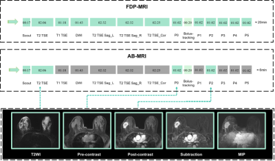 |
The performance of evaluation in neoadjuvant chemotherapy for
breast cancer on abbreviated breast MRI: comparison with full
diagnostic protocol
Wenjie Tang1,
Yuan Guo1,
Siyi Chen1,
Xueli Li1,
Yongzhou Xu2,
and Xinhua Wei1
1Guangzhou First People's Hospital, Guangzhou, China, 2Philips Healthcare, Guangzhou, China Keywords: Breast, Cancer, Breast neoplasms, Neoadjuvant chemotherapy, Radiologists To compare the diagnostic efficacy of abbreviated breast MRI (AB-MRI) and regular full diagnostic protocol MRI (FDP-MRI) in the assessment of response to neoadjuvant chemotherapy (NAC) in breast cancer, this study retrospectively analyzed the performance of AB-MRI and FDP-MRI in breast cancer patients undergoing NAC. We found that, for experienced radiologists, compared with FDP-MRI, AB-MRI showed similar sensitivity, specificity, and accuracy in the evaluation of pCR and similar accuracy in the measurement of residual tumor size for breast cancer. |
| 15:37 |
0198.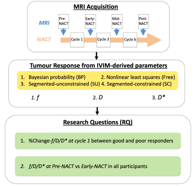 |
Intravoxel incoherent motion improves diffusion-weighted imaging
in the detection of response to neoadjuvant chemotherapy in
breast cancer
Sai Man Cheung1,
Wing Shan Wu1,
Nicholas Senn1,
Ravi Sharma2,
Trevor McGoldrick2,
Tanja Gagliardi1,3,
Ehab Husain4,
Yazan Masannat5,
and Jiabao He1,6
1Institute of Medical Sciences, University of Aberdeen, Aberdeen, United Kingdom, 2Department of Oncology, Aberdeen Royal Infirmary, Aberdeen, United Kingdom, 3Department of Radiology, Royal Marsden Hospital, London, United Kingdom, 4Department of Pathology, Aberdeen Royal Infirmary, Aberdeen, United Kingdom, 5Breast Unit, Aberdeen Royal Infirmary, Aberdeen, United Kingdom, 6Translational and Clinical Research Institute, Newcastle University, Aberdeen, United Kingdom Keywords: Breast, MR-Guided Interventions Breast cancer is a major and expanding health challenge, and neoadjuvant chemotherapy (NACT) is increasingly prescribed to facilitate breast surgery. However, response to NACT is highly inconsistent, imposing an ongoing demand for improved imaging methods for early response identification. Tissue perfusion, a sensitive marker of cancer metabolism, can be derived from intravoxel incoherent motion (IVIM) model, and recent Bayesian algorithm yields increased sensitivity and precision in pancreatic cancer. We therefore hypothesise that IVIM model powered by Bayesian algorithm is able to detect early treatment-induced changes in tumour perfusion and diffusion, with the potential to impact patient care pathway. |
 Back to Meeting Home
 Back to the Program-at-a-Glance
The International Society for Magnetic Resonance in Medicine is accredited by the Accreditation Council for Continuing Medical Education to provide continuing medical education for physicians.
 Back to Meeting Home
 Back to the Program-at-a-Glance
 View Presentation Video