Oral Session
Reproduction & Reproductive Pathologies
ISMRM & ISMRT Annual Meeting & Exhibition • 03-08 June 2023 • Toronto, ON, Canada

Oral Session
Reproduction & Reproductive Pathologies
08:15 |
0436.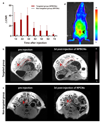 |
Endometriosis targeted MRI imaging using bevacizumab-modified
nanoparticles aiming at vascular endothelial growth factor
Qi Zhang1,
Caixia Fu2,
Qing Li3,
and Yajie Li4
1Department of Radiology, Huashan hospital, Fudan University, Shanghai, China, 2MR Application Development, Siemens Shenzhen Magnetic Resonance Ltd., Shenzhen, China, 3MR Collaborations, Siemens Healthineers Digital Technology (Shanghai) Co., Ltd., Shanghai, China, 4Fudan University, Shanghai, China Keywords: Uterus, Molecular Imaging In vivo, NPBCNs generated strong signal enhancement in endometriosis lesion in rat on T1-weighted images via MRI. |
08:23 |
0437.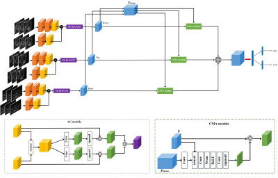 |
Using deep learning to identify LNM and LVSI of endometrial
cancer from conventional MRI: a preliminary two-center study
Yida Wang1,
He Zhang2,
Xiance Zhao3,
and Guang Yang1
1Shanghai Key Laboratory of Magnetic Resonance, East China Normal University, Shanghai, China, 2Department of Radiology, Obstetrics and Gynecology Hospital, Fudan University, Shanghai, China, 3Philips Healthcare, Shanghai, China Keywords: Uterus, Cancer We developed a multi-task deep learning model using multi-parametric MRI to simultaneously predict lymphatic nodes metastasis (LNM) and lymphatic vascular space invasion (LVSI) in patients with endometrial cancer. Cross-modality attention mechanism was integrated with the model to learn the within and cross modality-specific features which could enhance the performance of network. In this study, we also treated endometrial cancer regions as the anatomical prior knowledge to capture the discriminative information from the whole MR images. The results showed the proposed model predicted LNM and LVSI with a high accuracy in both internal and external test datasets. |
| 08:31 |
0438.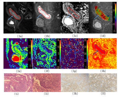 |
APT weighted imaging combined with IVIM to evaluate Her-2 gene
expression in endometrial cancer
Ma Changjun1,
Tian Shifeng1,
Song Qingling1,
Chen Lihua1,
Wang Nan2,
Lin Liangjie3,
Wang Jiazheng4,
and Liu Ailian1
1Department of Radiology,, The First Affiliated Hospital of Dalian Medical University, Dalian, China, China, 2Department of Radiology, The First Affiliated Hospital of Dalian Medical University, Dalian, China, China, 3Clinical & Technical Support, Philips Healthcare, Beijing, China, China, 4Clinical & Technical Support, Philips Healthcare, Shanghai, China, China Keywords: Pelvis, Quantitative Imaging it is necessary to find a method for non-invasive monitoring of Her-2 status. MRI with characteristics of multi-direction, multi-parameter, multi-function, high soft-tissue resolution, and non-invasiveness, has become the preferred method for evaluation of uterine lesions. |
| 08:39 |
0439.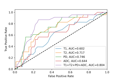 |
Radiomics of Multiparametric MRI in Tumor Grading of Endometrial
Cancer
Yiang Wang1,
Mengge He1,
Peng Cao1,
Chien-Yuan Lin2,
Weiyin Liu2,
Chia-Wei Lee2,
and Elaine Y.P. Lee1
1Department of Diagnostic Radiology, The University of Hong Kong, Hong Kong, Hong Kong, 2GE Healthcare, Taipei, Taiwan Keywords: Uterus, Cancer, Endometrial Cancer; Tumor Grade Random forest models were constructed to predict tumor grade (grade 1-2 vs. grade 3) of endometrial cancer based on radiomics features extracted from quantitative T1, T2, proton density maps generated by synthetic MRI and apparent diffusion coefficient maps generated by diffusion-weighted imaging. The classification model based on features extracted from all the quantitative maps achieved the highest area under the curve of 0.804 compared to models constructed based on single quantitative map. |
|
08:47 |
0440.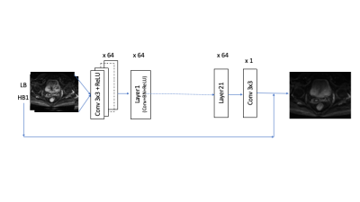 |
Accelerated multi-shot diffusion MRI using deep learning
denoising
Or Alus1,
Maria El Homsi2,
Lee Rodriguez2,
Yousef Mazaheri1,2,
Youngwook Kee1,
Iva Petkovska2,
and Ricardo Otazo1,2 1Department of Medical Physics, Memorial Sloan Kettering Cancer Center, New York, NY, United States, 2Department of Radiology, Memorial Sloan Kettering Cancer Center, New York, NY, United States Keywords: Pelvis, Diffusion/other diffusion imaging techniques, image reconstruction Multi-shot EPI is commonly used to compensate for geometric distortions and increase spatial resolution in body diffusion MRI, with a price tag of longer scan times. This work presents an alternative technique to k-space undersampling to accelerate the acquisition, which is based on reducing the number of repetitions at high b-value and denoising the resulting images using a convolutional neural network. The proposed deep learning denoising technique is demonstrated to accelerate the acquisition of multi-shot diffusion MRI acquisition of patients with rectal cancer and reduce the scan time beyond the duration of a single-shot diffusion MRI acquisition. |
| 08:55 |
0441.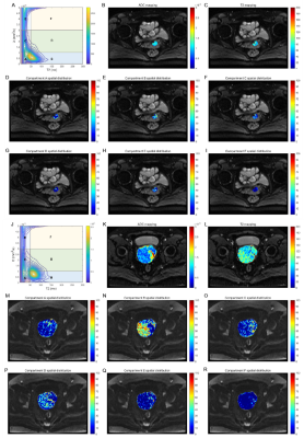 |
The value of diffusion-relaxation correlation spectrum imaging
in differential diagnosis of early and advanced squamous
cervical carcinoma
Yihe Gao1,
Ke Xue2,
Yongming Dai2,
and Jing Ye1
1Northern Jiangsu People's Hospital, Yangzhou, Jiangsu Province, China, 2MR Collaboration, Central Research Institute, United Imaging Healthcare, Shanghai, China, Shanghai, China Keywords: Uterus, Microstructure, multi dimension Accurately staging squamous cell carcinoma (SCC) is of vital importance for determining the treatment plan, prognosis and outcome. Diffusion-relaxation correlation spectrum imaging (DR-CSI) can jointly encode diffusion and relaxation information and resolve information of tissue compartments and heterogeneity at a sub-voxel level. In this study we investigated the feasibility of DR-CSI in characterizing tissue microenvironment and staging SCC. The DR-CSI technique can non-invasively provide information on microscopic tissue compartments within voxels, reflecting intra-tissue heterogeneity. The diagnostic performance of DR-CSI in identifying early and advanced SCC was significantly superior to conventional ADC and T2. |
| 09:03 |
0442.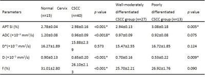 |
Identifying pathological differentiation of cervical squamous
cell carcinoma with APTw and IVIM
Zhonghong Xin1,
Junqiang Lei1,
Jianhong Peng2,
Xiande Lu2,
Jiang Nan2,
Yaping Zhang2,
Xiaohui Wang2,
Jun Zhu2,
and Jianxiu Lian3 1Radiology, the First Hospital of Lanzhou University, Lanzhou, China, 2the First Hospital of Lanzhou University, Lanzhou, China, 3Philips Healthcare, Beijing, China Keywords: Uterus, Diffusion/other diffusion imaging techniques, amide proton transfer weighted imaging,cervical cancer,squamous cell carcinoma of the cervix The pathological differentiation of cervical squamous cell carcinoma (CSCC) determines the therapy method and prognosis. APTw, DWI and IVIM sequences were performed for predicting pathological differentiation. 27 patients with well-moderately differentiation, 13 patients with poorly differentiation and 15 healthy volunteers were enrolled. APT SI, ADC, D*, D and f values were calculated for comparing among different groups. Results showed parameters except D* differed significantly between CSCC and normal. There were statistically significant differences in AUC of APT SI, D and f between well-moderately group and poorly differentiated group. APTw and IVIM can be used to identify pathological differentiation of CSCC. |
| 09:11 |
0443.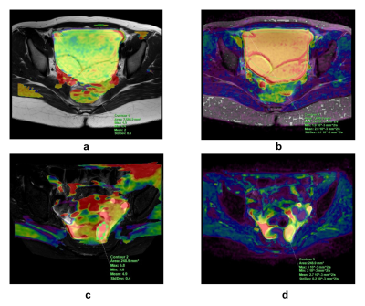 |
Can 3D APTw Imaging improve the differential diagnosis ability
of traditional MRI in Ovarian Cystadenoma and
Cystadenocarcinoma?
Yibei Yu1,
Xiaolei Song2,
Lixue Wang1,
Dandan Zheng3,
and Zhuozhao Zheng1
1Radiology department, Beijing Tsinghua Changgung Hospital, School of Clinical Medicine, Tsinghua University, Beijing, China, 2Center for Biomedical Imaging Research, School of Medicine, Tsinghua University, Beijing, China, 3Clinical& Technical Support, Philips Healthcare, Beijing, China Keywords: Pelvis, CEST & MT The therapies for ovarian cystadenoma and cystadenocarcinoma are different, thus the differential diagnosis of ovarian lesions is critical. The diagnostic efficacy increased with the use of DWI and DCE-MRI, but the differential diagnosis of cystic ovarian lesions is still a challenge. This study evaluates the diagnostic efficacy of 3D APTw MRI in the differentiation between cystadenoma and cystadenocarcinoma based on the analysis of the cystic regions. The results showed that APTw SIs had better diagnostic performance than ADC values and may be used as a noninvasive tool for differential diagnosis and therapy guidance. |
| 09:19 |
0444.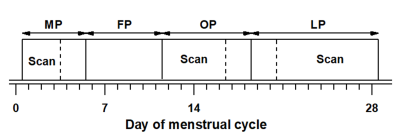 |
Microcirculatory changes in the uterus of women during the
menstrual cycle - based on intravoxel incoherent motion MRI
Yajie Li1,2,
Qi Zhang2,
Caixia Fu3,
Qing Li4,
and Robert Grimm5
1Shanghai Institute of Medical Imaging, Shanghai, China, 2Department of Radiology, Huashan Hospital, Shanghai, China, 3MR Application Development, Siemens Shenzhen Magnetic Resonance Ltd., Shenzhen, China, 4MR Collaborations, Siemens Healthineers Digital Technology (Shanghai) Co., Ltd., Shanghai, China, 5MR Application Predevelopment, Siemens Healthcare GmbH, Erlangen, Germany Keywords: Uterus, Diffusion/other diffusion imaging techniques, Intravoxel incoherent motion, Microcirculation, Perfusion Purpose: To assess the feasibility of intravoxel incoherent motion MRI on detecting the microcirculatory changes in the uterus of women during the menstrual cycle. Methods: Volunteers underwent MRI scans by using IVIM. The changes of D value, D* and f value of three layers of uterine structure during menstrual cycle were analyzed quantitatively. Results: During menstrual cycle, the D values of junctional zone were significantly lower than in myometrium. Meanwhile, the f values of the three zones structure of uterus were significantly different. Conclusion: IVIM can be used for noninvasive and quantitative evaluation of uterine blood microcirculation changes. |
| 09:27 |
0445.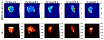 |
Mapping Placenta Structure and Function with Low-Field MRI
Paddy J. Slator1,
Jana Hutter 2,3,
Raphael Tomi Tricot2,3,4,
Jordina Aviles Verdera2,3,
Joseph V. Hajnal2,3,
and Daniel C. Alexander1
1Centre for Medical Image Computing and Department of Computer Science, University College London, London, United Kingdom, 2Centre for the Developing Brain, School of Biomedicial Engineering and Imaging Sciences, King's College London, London, United Kingdom, 3Biomedical Engineering Department, School of Biomedical Engineering and Imaging Sciences, King's College London, London, United Kingdom, 4MR Research Collaborations, Siemens Healthcare Limited, Frimley, United Kingdom Keywords: Placenta, Low-Field MRI Placental MRI is emerging as a promising adjunct to ultrasound during pregnancy. Low field MRI is appealing for multiple reasons and can support the widespread roll out of placental MRI. Here we provide a proof-of-concept that a quantitative placental imaging technique that has been demonstrated at high-field (1.5T / 3T) – combined T2*-diffusion MRI – is also viable at low field (0.55T). We highlight similarities and differences in low-field maps compared to the current state-of-the-art high-field maps and highlight key areas for future work to realise the potential of low-field placental quantitative MRI during pregnancy. |
| 09:35 |
0446.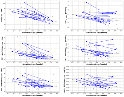 |
Assessing within-subject rates of change of placental magnetic
resonance imaging diffusion metrics
Daniel Cromb1,2,
Paddy J Slator3,
Anthony Price1,2,
Miguel De La Fuente1,
Alexia Egloff-Collado1,
Mary Rutherford1,4,
Serena J Counsell1,2,
and Jana J Hutter1,2
1Centre for the Developing Brain, School of Biomedical Engineering and Imaging Sciences, King's College London, London, United Kingdom, 2Biomedical Engineering Department, School of Biomedical Engineering and Imaging Sciences, King's College London, London, United Kingdom, 3Centre for Medical Image Computing, Department of Computer Science, University College London, London, United Kingdom, 4MRC Centre for Neurodevelopmental Disorders, King's College London, London, United Kingdom Keywords: Placenta, Placenta This study provides unique data on the evolution of quantitative multi-modal placenta MRl measures over gestation, including, crucially, within-subject results. A multi-compartmental T2*-IVIM model was employed, suited to the complex physiology of the human placenta, and matched to the deployed multi-parametric acquisition technique. |
| 09:43 |
0447.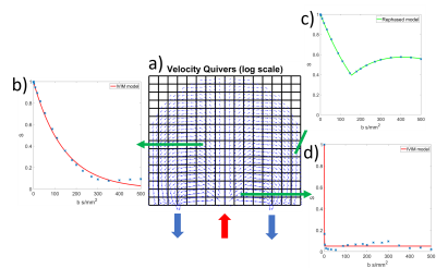 |
The effects of maternal flow on placental DWI data
George Hutchinson1,
Adam Blakey2,
Neele Dellschaft1,
Nia Jones3,
Reuben O'Dea2,
Lopa Leach4,
Matthew Hubbard2,
Paul Houston2,
and Penny Gowland1
1The Sir Peter Mansfield Imaging Centre, The University of Nottingham, Nottingham, United Kingdom, 2Mathematical Sciences, The University of Nottingham, Nottingham, United Kingdom, 3Medicine, The University of Nottingham, Nottingham, United Kingdom, 4Life Sciences, The University of Nottingham, Nottingham, United Kingdom Keywords: Placenta, Diffusion/other diffusion imaging techniques Placental diffusion imaging data is assumed to be driven by a combination of slow diffusive processes, as well as faster incoherent terms. How these faster incoherent terms combine within a voxel is not obvious, and here we investigate this further by comparing a mathematical simulation of maternal flow through a single placentone to data collected in utero. We observe maternal flows can cause IVIM like effects, with slow exponential decays, but also regions of fast IVIM similar to those observed in the placenta, as well as 'rebounding' of signal. |
| 09:51 |
0448. |
Inspecting placental microstructure in pregnancies affected by
fetal congenital heart disease
Daniel Cromb1,2,
Paddy J Slator3,
Anthony Price1,2,
Megan Hall1,2,
Daniel Alexander3,
Joseph V Hajnal1,2,
Mary Rutherford1,4,
Jana Hutter1,2,
and Serena J Counsell1,2
1Centre for the Developing Brain, School of Biomedical Engineering and Imaging Sciences, King's College London, London, United Kingdom, 2Biomedical Engineering Department, School of Biomedical Engineering and Imaging Sciences, King's College London, London, United Kingdom, 3Centre for Medical Image Computing, Department of Computer Science, University College London, London, United Kingdom, 4MRC Centre for Neurodevelopmental Disorders, King's College London, London, United Kingdom Keywords: Placenta, Placenta, Diffusion, Microstructure Congenital heart disease (CHD) is common and associated with abnormal placental development. We used novel diffusion MRI acquisition and analysis techniques to inspect placental microstructure in pregnancies affected by congenital heart disease (CHD), highlighting the potential value of this approach for in-utero identification of how placental development may deviate from normal in CHD. |
|
09:59 |
0449.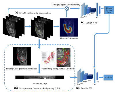 |
A deep learning pipeline using priori knowledge for automatic
evaluation of placenta accreta spectrum disorders with MRI
Haijie Wang1,
Yida Wang1,
Chenglong Wang1,
He Zhang2,
Hao Zhu3,
Yuanyuan Lu4,
Yang Song5,
and Guang Yang1
1Shanghai key lab of magnetic resonance, East China Normal University, Shanghai, China, 2Department of Radiology, Obstetrics and Gynecology Hospital of Fudan University, Shanghai, China, 3Department of Obstetrics, Obstetrics and Gynecology Hospital of Fudan University, Shanghai, China, 4Department of Radiology, Shanghai First Maternity and Infant Health Hospital, Shanghai, China, 5MR Scientific Marketing, Siemens Healthcare, Shanghai, China Keywords: Placenta, Placenta Placenta accreta spectrum (PAS) is a pathologic condition of placentation associated with significant maternal morbidity and mortality. We enrolled 540 patients from two institutions to build an automatic pipeline for early diagnosis of PAS based on T2W images. An nnU-Net model was trained for automatic segmentation of the placenta, then an image stripe was created, in which utero-placental borderline (UPB) was straightened and centered. The UPB image was fed into a DenseNet-based network together with placental position for PAS diagnosis. The pipeline achieved good performance with AUCs of 0.860 and 0.897 in internal and external test cohorts, respectively. |
|
10:07 |
0450.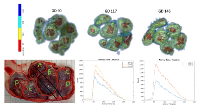 |
Cotyledon-Specific Flow Evaluation of Rhesus Macaque Placental
Injury using Ferumoxytol Dynamic Contrast Enhanced MRI
Ruiming Chen1,
Daniel Seiter1,
Logan T. Keding2,3,
Jessica Vazquez2,3,
Kathleen Antony4,
Heather A. Simmons2,3,
Kevin M. Johnson1,5,6,
Aleksandar K. Stanic4,
Ruo-Yu Liu1,
Dinesh Shah4,
Thaddus G. Golos2,3,
and Oliver Wieben1,5,6
1Medical Physics, University of Wisconsin - Madison, Madison, WI, United States, 2Wisconsin National Primate Research Center, University of Wisconsin - Madison, Madison, WI, United States, 3Comparative Biosciences, University of Wisconsin - Madison, Madison, WI, United States, 4Obstetrics & Gynecology, University of Wisconsin - Madison, Madison, WI, United States, 5Radiology, University of Wisconsin - Madison, Madison, WI, United States, 6Biomedical Engineering, University of Wisconsin - Madison, Madison, WI, United States Keywords: Placenta, DSC & DCE Perfusion Placental blood flow is a marker that reflects the health of the utero-placental vasculature. Dynamic contrast-enhanced (DCE) MRI is more robust than arterial spin labeling and can be utilized in animal models to provide cotyledon-specific blood flow and blood volume measurements in vivo throughout gestation. This study investigated the distribution of blood flow to the placental at a cotyledon level in pregnant rhesus monkeys before and following the injection of Tisseel (a fibrin sealant) and MCP1 (an inflammatory response inducer) to assess the predictabilities of flow for placental injury. |
 Back to Meeting Home
 Back to the Program-at-a-Glance
The International Society for Magnetic Resonance in Medicine is accredited by the Accreditation Council for Continuing Medical Education to provide continuing medical education for physicians.
 Back to Meeting Home
 Back to the Program-at-a-Glance
 View Presentation Video