Oral Session
Liver: Diffuse Disease & Quantification
ISMRM & ISMRT Annual Meeting & Exhibition • 03-08 June 2023 • Toronto, ON, Canada

Oral Session
Liver: Diffuse Disease & Quantification
08:15 |
0054.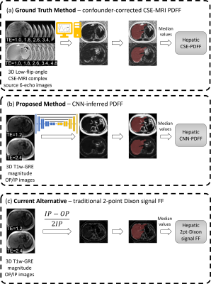 |
Deep learning improves accuracy of proton-density fat fraction
estimation from In-phase and out-of-phase T1-weighted MRI
Kang Wang1,
Guilherme Mourna Cunha2,
Kyle Hasenstab3,
Walter C Henderson4,
Michael S Middleton4,
Rohit Loomba5,
Shelley A Cole6,
Albert Hsiao4,
and Claude B Sirlin7 1Radiology, Stanford, Palo Alto, CA, United States, 2Radiology, University of Washington Medicine, Seattle, WA, United States, 3Department of Mathematics and Statistics, San Diego State University, San Diego, CA, United States, 4Radiology, UC San Diego, La Jolla, CA, United States, 5Department of Hepatology, UC San Diego, La Jolla, CA, United States, 6Texas Biomedical Research Institute, San Antonio, TX, United States, 7UC San Diego, La Jolla, CA, United States Keywords: Liver, Quantitative Imaging, Fat, Data analysis Proton density fat fraction (PDFF) is an established quantitative-imaging-biomarker for hepatic -fat quantification, but typically requires specialized confounder-corrected chemical-shift-encoded (CSE) magnetic-resonance-imaging (MRI) pulse sequences. We developed and assessed the feasibility of deep learning to infer hepatic PDFF maps from conventional T1-weighted-in-and-opposed-phase (T1w-IOP) MRI. Using PDFF maps reconstructed from CSE-MRI as reference, we trained a convolutional-neural-network (CNN) to infer voxel-wise PDFF maps from T1w-IOP MRI. The CNN was evaluated using both internal and external test datasets. Participant-level median CNN-inferred-PDFF were compared with reference CSE-MRI using linear regression, intraclass correlation, and Bland-Altman analysis. Median CNN-inferred PDFF agreed closely with reference CSE-MRI PDFF. |
|
08:23 |
0055.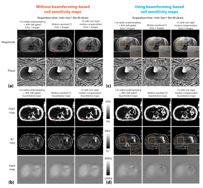 |
Motion-Resolved Self-Gated Free-Breathing 3D Liver PDFF and R2*
Mapping using Phase-Preserving Beamforming and Non-Rigid Motion
Compensation
Shu-Fu Shih1,2,
Sevgi Gokce Kafali1,2,
Kara L. Calkins3,
and Holden Wu1,2
1Radiological Sciences, David Geffen School of Medicine, University of California Los Angeles, Los Angeles, CA, United States, 2Bioengineering, University of California Los Angeles, Los Angeles, CA, United States, 3Pediatrics, David Geffen School of Medicine, University of California Los Angeles, Los Angeles, CA, United States Keywords: Liver, Fat 3D self-navigated multi-echo stack-of-radial Dixon sequence has been used to quantify fat and R2* with free-breathing acquisitions. To compensate motion, motion-resolved compressed sensing (CS) uses self-navigation for data binning, and applies sparsity constraint along the dimension of motion states. However, this approach does not explicitly model non-rigid motion in the liver. In addition to artifacts caused by respiratory motion, hardware imperfection such as gradient nonlinearity can lead to artifacts and affect the image quality. In this work, use a phase-preserving beamforming-based coil sensitivity estimation method and non-rigid motion compensation in a CS model to improve free-breathing PDFF and R2* quantification. |
|
08:31 |
0056.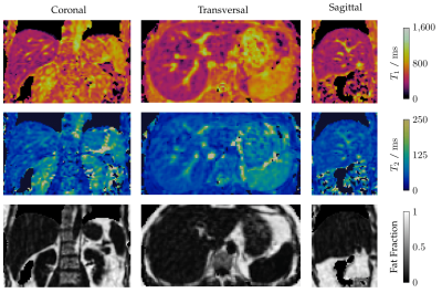 |
Respiratory-motion-corrected simultaneous 3D T1, T2, and
fat-fraction mapping at 0.55T, for comprehensive
characterization of liver tissue
Donovan Tripp1,
Karl P Kunze1,2,
Michael Crabb1,
Radhouene Neji1,2,
Claudia Prieto1,3,
and René Botnar1,3,4
1School of Biomedical Engineering and Imaging Sciences, King's College London, London, United Kingdom, 2MR Research Collaborations, Siemens Healthcare Limited, Camberley, United Kingdom, 3School of Engineering, Pontificia Universidad Católica de Chile, Santiago, Chile, 4Institute for Biological and Medical Engineering, Pontificia Universidad Católica de Chile, Santiago, Chile Keywords: Liver, Low-Field MRI Through T1, T2, and fat fraction mapping, MRI is a formidable tool for the comprehensive assessment of liver health, but has been held back by slow, 2D, single-parameter mapping sequences. We propose a novel respiratory-motion-corrected framework to acquire all three of these maps from a single 6.5-minute scan, on the latest generation of 0.55T scanner. The resulting maps are 3D, with 3 mm isotropic resolution, and co-registered, making them suitable for multiparametric assessment of tissue health. Liver joint T1, T2, and fat fraction maps are in good agreement with reference values and first in-vivo scans provide promising image quality. |
| 08:39 |
0057.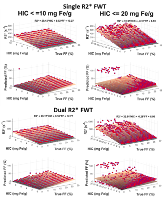 |
Comparing the accuracy of single- and dual-R2* fat-water models
for assessing hepatic steatosis and iron overload using Monte
Carlo simulations
Utsav Shrestha1,
Juan Pablo Esparza1,
Sanjaya Satapathy2,
Jason Vanatta3,
and Aaryani Tipirneni-Sajja1,4
1The University of Memphis, Memphis, TN, United States, 2North Shore University Hospital/Northwell Health, Manhasset, NY, United States, 3University of Tennessee Health Science Center, Memphis, TN, United States, 4St. Jude Children’s Research Hospital, Memphis, TN, United States Keywords: Liver, Liver Hepatic iron concentration (HIC) and fat fraction (FF) is assessed by chemical-shift encoded multi-spectral fat-water model that incorporates single- or dual-R2* correction. In this study, we designed a realistic virtual liver model simulating the combined presence of hepatic steatosis and iron overload using histology data and synthesized MRI signal in the virtual model using Monte-Carlo simulation to compare the accuracy of the R2* models to quantify FF in the presence of iron. Our results show that single-R2* is consistent than dual-R2* in estimating FF for HIC<=10 mg Fe/g after which both R2* models show higher error. |
| 08:47 |
0058.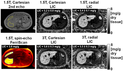 |
Free-Breathing Liver Iron Quantification Using 3D
Stack-of-Radial GRE Dixon MRI at Two Field Strengths: A
Preliminary Study
Xiaodong Zhong1,
Stephan A.R. Kannengiesser2,
Pedro Itriago Leon3,
Vibhas Deshpande4,
and Takeshi Yokoo5
1MR R&D Collaborations, Siemens Medical Solutions USA, Inc., Los Angeles, CA, United States, 2MR Application Predevelopment, Siemens Healthcare GmbH, Erlangen, Germany, 3MR R&D Collaborations, Siemens Medical Solutions USA, Inc., Dallas, CA, United States, 4MR R&D Collaborations, Siemens Medical Solutions USA, Inc., Austin, CA, United States, 5Department of Radiology and Advanced Imaging Research Center, University of Texas Southwestern Medical Center, Dallas, TX, United States Keywords: Liver, Quantitative Imaging, R2*, Iron, LIC, free-breathing This study compared free-breathing stack-of-radial and breath-hold Cartesian multi-echo GRE MRI for liver iron quantification at 1.5T and 3T to the reference standard R2-based liver iron concentration (LIC) at 1.5T (FerriScan), in patients at risk for liver iron overload. Agreement was observed for liver iron concentration measured by free-breathing 3D stack-of-radial GRE MRI compared the reference standard R2-based LIC, as well as breath-hold 3D Cartesian GRE MRI. With further validation in larger number of iron-overloaded subjects, 3D stack-of-radial GRE MRI may allow free-breathing R2* and LIC mapping at both 1.5T and 3T. |
| 08:55 |
0059.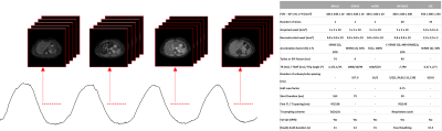 |
Free-breathing Simultaneous T1, T2, and T2* Mapping of the
Whole-Liver with Multi-Inversion Spin and Gradient Echo in Under
a Minute
Mary Kate Manhard1,2,
Anandh Kilpattu Ramaniharan1,
Jean A Tkach1,2,
Andrew T Trout1,2,
Jonathan R Dillman1,2,
and Amol Pednekar1,2
1Radiology, Cincinnati Children's Hospital Medical Center, Cincinnati, OH, United States, 2Radiology, University of Cincinnati College of Medicine, Cincinnati, OH, United States Keywords: Liver, Quantitative Imaging Quantitative parametric mapping is an increasingly important tool for non-invasive assessment of liver disease. Conventional parametric mapping techniques require multiple breath-held acquisitions providing limited coverage. A free-breathing multi-inversion spin and gradient echo (MI-SAGE) technique is demonstrated to simultaneously estimate T1, T2, and T2* over the whole-liver in a single scan of less than 1 minute. In 11 research participants, hepatic T1, T2, and T2* estimates obtained using the free-breathing MI-SAGE technique were comparable to those obtained using modified Lock Locker, multiple gradient and spin echo, and multiple gradient echo sequences respectively and exhibited good to excellent (ICC>0.77) repeatability and reproducibility. |
| 09:03 |
0060.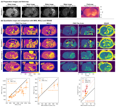 |
Free-breathing Isotropic resolution self-Navigated
B1-insensitive whOle liver simultaneous T1 and T2 mapping (FINO)
Jonathan K. Stelter1,
Kilian Weiss2,
Elizabeth Huaroc Moquillaza1,
Felix N. Harder1,
Marcus R. Makowski1,
Rickmer F. Braren1,
and Dimitrios C. Karampinos1
1Department of Diagnostic and Interventional Radiology, Technical University of Munich, Munich, Germany, 2Philips Healthcare, Hamburg, Germany Keywords: Liver, Relaxometry Volumetric T1 and T2 relaxation mapping is of interest in the characterization of diffuse and oncological liver diseases. Current methods for free-breathing whole-liver relaxation mapping have primarily been optimized to quantify a water-specific T1, are restricted to large voxel sizes in the feet-head-direction, and are not compatible with self-navigation. The present work proposes a novel methodology for Free-breathing Isotropic resolution self-Navigated B1-insensitive whOle-liver simultaneous water-specific T1 and T2 mapping (FINO). Phantom results illustrate the accuracy of the method and in vivo results demonstrate the feasibility of whole-liver water-specific T1 and T2 mapping at an isotropic resolution of 3mm in 6min. |
| 09:11 |
0061.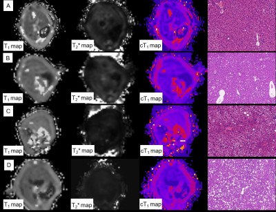 |
Assessment of Inflammation and Spatial Heterogeneity for
Nonalcoholic Fatty Liver Disease in Mice Based on Corrected T1
Lujie Li1,
Kan Deng2,
Yinhong Zhang1,
and Shi-ting Feng1
1the First Affiliated Hospital of Sun Yet-sen University, Guangzhou, China, 2Philips Healthcare, Guangzhou, China Keywords: Liver, Quantitative Imaging Corrected T1 (cT1) is potential to be a novel imaging biomarker, which was based on T1 mapping corrected for iron content. The results of this study showed the high correlation between cT1 value and NAFLD activity, without correlation with fibrosis. There was high accuracy of cT1 to identify normal liver and liver with NAFLD, as well as mild and severe NAFLD. The severer inflammation, the higher heterogeneity of cT1 mapping. Thus, cT1 may be used for evaluating liver inflammation of patient with NAFLD, especially for those without severe fibrosis, and may evaluate heterogeneity further. |
| 09:19 |
0062.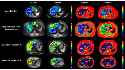 |
Longitudinal MR Elastography and Proton Density Fat Fraction
Measurements in Patients with Alcoholic and Nonalcoholic Fatty
Liver Disease
Jiahui Li1,
Zheng Zhu1,
Kevin J. Glaser1,
Yi Sui1,
Douglas A. Simonetto2,
Alina M Allen2,
Armando Manduca1,
Sudhakar K. Venkatesh1,
Vijay H. Shah2,
Richard L. Ehman1,
and Meng Yin1
1Radiology, Mayo Clinic, Rochester, MN, United States, 2Gastroenterology and Hepatology, Mayo Clinic, Rochester, MN, United States Keywords: Liver, Liver To evaluate MR imaging biomarkers in monitoring chronic liver diseases, we analyzed longitudinal MRE-assessed liver stiffness (LS) and proton density fat fraction (PDFF) measurements in 46 patients with alcoholic liver disease (ALD) or nonalcoholic fatty liver disease (NAFLD). Results demonstrated that yearly LS change rate was significantly associated with current LS and PDFF change rate in NAFLD, although with low correlation (R2=0.30, p=0.01). ALD patients had bidirectional changes in LS/PDFF change rate but not significant correlation (R2=0.20, p=0.10). We conclude that longitudinal MRE-LS and MRI-PDFF provide independent and complementary assessments of disease status and dynamic change in ALD and NAFLD. |
| 09:27 |
0063. |
Evaluation of liver fibrosis using histogram analysis with
diffusion kurtosis imaging and stretched-exponential diffusion
model
Fengxian Fan1,
Yanli Jiang1,
Jie Zou1,
Pin Yang1,
and Jing Zhang1
1Department of Magnetic Resonance, Lanzhou University Second Hospital, Lanzhou, China Keywords: Liver, Diffusion/other diffusion imaging techniques, histogram analysis; stretched exponential model; diffusion kurtosis imaging This study aims to evaluate the potential role of histogram analysis of diffusion kurtosis imaging (DKI) and stretched exponential model (SEM) for preoperative prediction of liver fibrosis and inflammation. Histogram metrics were extracted from mean diffusion (MD), mean kurtosis (MK), distributed diffusion coefficient (DDC) and intravoxel heterogeneity index (α). The histogram metrics of MD, DDC and α demonstrate significant correlation with inflammation, the histogram metrics of MD and DDC were significantly different between S0-1 and ≥S2. Histogram analysis of DKI and SEM may be potentially useful for evaluating heterogeneity of liver. |
| 09:35 |
0064. |
Comparing and combining MRI-ECV, DKI, and DWI for the staging of
liver fibrosis in chronic hepatitis B Patients: A
multiparametric model
Zhou Wei1,
Yupin Liu*1,
Xiaohuan Li1,
Zhiyuan Chen1,
Robert Grimm2,
and Yunzhu Wu3
1Department of Radiology, The Second Affiliated Hospital of Guangzhou University of Chinese Medicine, Guangdong Provincial Hospital of Traditional Chinese Medicine, Guangzhou, China, 2MR Application Predevelopment, Siemens Healthcare GmbH, Erlangen, Germany, 3MR Scientific Marketing, SIEMENS Healthineers Ltd., Shanghai, China Keywords: Liver, Liver Liver fibrosis (LF) is a dynamic, reversible process which can result in liver failure. Diagnosis and monitoring of LF are clinically important. This study was to evaluate the diagnostic capability of the model based on multiparametric MRI in identifying significant LF in patients with chronic hepatitis B (CHB). The predictive model based on multiparametric MRI further could improve diagnostic accuracy and serve for identifying significant LF in CHB patients, also provide a reliable basis for clinical diagnosis and treatment. |
| 09:43 |
0065. |
Non-invasive evaluation of anti-integrin αvβ6 treatment of liver
fibrosis by collagen-targeted MRI
Yingying Ning1,
Iris Y. Zhou1,
Nicholas J Rotile1,
Avery Boice1,
Johanna Schaub2,
Scott Turner2,
and Peter Caravan1
1Athinoula A. Martinos Center for Biomedical Imaging, Institute for Innovation in Imaging, Department of Radiology, Massachusetts General Hospital and Harvard Medical School, Charlestown, MA, United States, 2Pliant Therapeutics, Inc., South San Francisco, CA, United States Keywords: Liver, Liver, liver fibrogenesis A collagen-binding molecular MR probe CM-101 was employed to non-invasively assess liver fibrosis and the anti-fibrotic effects of a novel αvβ6/αvβ1 integrin antagonist treatment in bile duct-ligated rats. Reduction of fibrosis was detected by collagen imaging and correlated with morphometric assessment of fibrosis from histology. CM-101 enhanced MR is a useful tool to monitor drug treatment response in chronic liver disease. |
09:51 |
0066.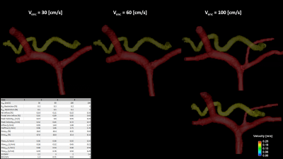 |
Anatomically Accurate In vitro Model of the Liver for 4D Flow
MRI Validation
James Rice1,2,
Thekla Oechtering2,3,
Kevin M Johnson2,4,
Oliver Wieben2,4,
Scott B Reeder2,4,5,6,
and Alejandro Roldan-Alzate1,2,6
1Mechanical Engineering, University of Wisconsin-Madison, Madison, WI, United States, 2Radiology, University of Wisconsin-Madison, Madison, WI, United States, 3Radiology, Universität zu Lübeck, Lübeck, Germany, 4Medical Physics, University of Wisconsin-Madison, Madison, WI, United States, 5Emergency Medicine, University of Wisconsin-Madison, Madison, WI, United States, 6Biomedical Engineering, University of Wisconsin-Madison, Madison, WI, United States Keywords: Liver, Modelling 4D flow MRI can be used to assess hemodynamics in the hepatic circulation system. This study presents a framework for in vitro hemodynamic evaluation of hepatic flow. One patient-specific in vitro model was created representing an anatomically realistic portal vein and hepatic artery and connected to a physiologic flow loop. The model was imaged with 4D flow MRI to assess hemodynamics. Altering hepatic artery and left gastric vein resistance changed flow proportions and velocities in the portal vein, highlighting the benefit of using in vitro modeling for systematic evaluation of 4D flow MRI in the liver. |
|
09:59 |
0067.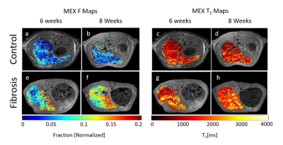 |
Quantitative Magnetization EXchange (MEX) MRI Measurement of
Liver Fibrosis Model in Rodents
Ella Wilczynski1,
Efrat Sasson2,
Uzi Eliav2,
Gil Navon2,
and Uri Nevo1,3 1Biomedical Engineering, Tel Aviv University, Tel Aviv, Israel, 2School of Chemistry, Tel Aviv University, Tel Aviv, Israel, 3Sagol School of Neuroscience, Tel Aviv University, Tel Aviv, Israel Keywords: Liver, Preclinical, MEX Quantitative MRI can elucidate the complex microstructural changes in liver disease. The MEX method estimates macromolecular fraction, such as collagen, and can potentially aid in this task. Rats and mice were given CCl4 to induce liver fibrosis and canned using a 7T scanner with MEX sequence (selective suppression and magnetization exchange) and macromolecular fraction (F) and T1 were extracted. Histology quantitative evaluation of collagen and inflammation scoring was conducted. F-values of fibrotic rats and mice were significantly increased and correlated with quantitative collagen percentage. T1 was significantly elevated with higher inflammation scores. |
| 10:07 |
0068.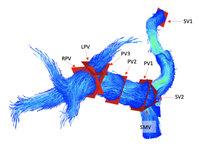 |
4D Flow MRI Reference Values of the Portal Venous System
Andrew Pan Huang1,2,
Grant S Roberts3,
Alejandro Roldán-Alzate4,5,
Oliver Wieben3,
Scott B Reeder2,3,5,6,7,
and Thekla Oechtering2,8 1Department of Surgery, Section of Vascular Surgery, University of Michigan, Ann Arbor, MI, United States, 2Department of Radiology, University of Wisconsin, Madison, WI, United States, 3Department of Medical Physics, University of Wisconsin, Madison, WI, United States, 4Department of Mechanical Engineering, University of Wisconsin, Madison, WI, United States, 5Department of Biomedical Engineering, University of Wisconsin, Madison, WI, United States, 6Department of Medicine, University of Wisconsin, Madison, WI, United States, 7Department of Emergency Medicine, University of Wisconsin, Madison, WI, United States, 8Department of Radiology and Nuclear Medicine, Universität zu Lübeck, Lübeck, Germany Keywords: Liver, Vessels 4D flow MRI can aid in diagnosing pathologies associated with hemodynamic changes in the portal vein, such as portal hypertension. Normal reference ranges for portal venous flow are lacking but essential for future clinical implementation. We report reference values based on radial 4D flow MRI measurements of mean flow, velocity, and effective diameter in the portal venous vasculature in 44 healthy subjects (59% female, 18-74 years). Faint helical and linear flow are physiological flow patterns observed in the portal vein. This study provides normative values for emerging clinical applications of 4D flow MRI currently under development. Keywords: Liver, Velocity&Flow, Vessels |
 Back to Meeting Home
 Back to the Program-at-a-Glance
The International Society for Magnetic Resonance in Medicine is accredited by the Accreditation Council for Continuing Medical Education to provide continuing medical education for physicians.
 Back to Meeting Home
 Back to the Program-at-a-Glance
 View Presentation Video