Oral Session
Pancreas, Fat & Gut
ISMRM & ISMRT Annual Meeting & Exhibition • 03-08 June 2023 • Toronto, ON, Canada

| 13:30 |
0504.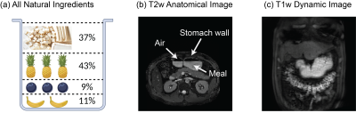 |
Mapping human gastric motility using contrast-enhanced MRI with
a natural test meal
Xiaokai Wang1,
Jiayue Cao1,
Kuan Han1,
Minkyu Choi1,
Ana Cecilia Saavedra Bazan1,
Jon-Fredrik Nielsen1,
Douglas C. Noll1,
and Zhongming Liu1
1University of Michigan, Ann Arbor, MI, United States Keywords: Digestive, Digestive MRI with oral contrast agents has been increasingly used to image the gastrointestinal tract in animals and humans. However, it has not been established for broad clinical applications. This study describes a set of natural ingredients abundant in manganese ions to enable rapid and contrast-enhanced MRI of the human gastrointestinal tract. A pipeline of advanced analyses reveals the structure of the stomach, dynamic transit of the food inside the stomach, and muscle activity of the stomach wall. Results offer quantitative biomarkers and mechanistic insights for gastric emptying, accommodation, and motility in humans, and merit future studies of gastrointestinal functional disorders. |
| 13:38 |
0505.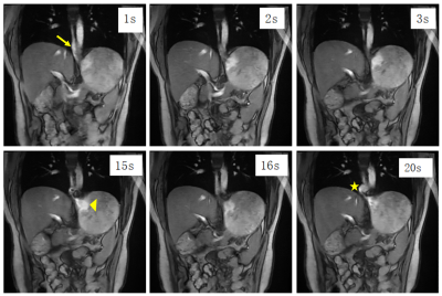 |
Real-time MRI using deep learning in gastroesophageal reflux
disease: a feasibility study
Qing Liu1,
Qi Liu2,
Jing Li1,
Eric Z Chen3,
Zhongqi Zhang2,
Xiao Chen3,
Shanhui Sun3,
Jian Xu2,
and Haoran Sun4
1Radiology, Beichen Hospital, Tianjin, China, 2UIH America, Inc., Houston, TX, United States, 3United Imaging Intelligence, Cambridge, MA, United States, 4Radiology, Tianjin Medical University General Hospital, Tianjin, China Keywords: Digestive, Digestive A real-time MRI technique is developed using deep-learning reconstruction. Its feasibility is evaluated in both healthy subjects and patients with gastroesophageal reflux diseases. |
| 13:46 |
0506.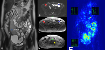 |
Characterization of small bowel strictures in Crohn’s disease
with multiparametric MRI
Emre Altinmakas1,
Himanshu Sharma2,
Octavia Bane1,3,
Ghadi Abboud1,
Alex Menys4,
Yansheng Hao5,
Jean-Frederic Colombel6,
Noam Harpaz5,
and Bachir Taouli1,3
1Department of Diagnostic, Molecular and Interventional Radiology, Icahn School of Medicine at Mount Sinai, New York, NY, United States, 2Icahn School of Medicine at Mount Sinai, NY, NY, United States, 3BioMedical Engineering and Imaging Institute, Icahn School of Medicine at Mount Sinai, New York, NY, United States, 4Motilent Ltd, London, United Kingdom, 5Department of Pathology, Icahn School of Medicine at Mount Sinai, NY, NY, United States, 6Department of Gastroenterology, Icahn School of Medicine at Mount Sinai, New York, NY, United States Keywords: Digestive, Quantitative Imaging, Crohn's disease; stricture; mpMRI; Small bowel stricture (SBS) is the one of the most common complications of Crohn’s Disease (CD) and it usually requires surgical management. Imaging diagnosis and characterization of SBS is essential for proper patient management, however this may be challenging with conventional MRI techniques. In current study, we investigate the utility of advance MRI techniques namely multiparametric MRI (mpMRI) including diffusion, perfusion, and motility in the characterization and tissue composition in CD-related SBS. Our preliminary results suggest that mpMRI parameters are promising tools to characterize SBS and may be helpful in the stratification of patients for medical treatment vs surgery. |
| 13:54 |
0507.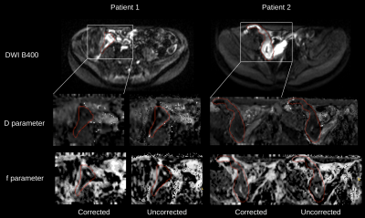 |
Slice Level 3D Motion Tracking For Motion Corrected IVIM DW-MRI
In Crohn’s Disease
SERGE DIDENKO VASYLECHKO1,
LINA LU1,
CEMRE ARIYUREK1,
JEANNETTE PEREZ-ROSSELLO1,
MICHAEL CALLAHAN1,
ONUR AFACAN1,
and SILA KURUGOL1
1RADIOLOGY, BOSTON CHILDREN'S HOSPITAL, HARVARD MEDICAL SCHOOL, BOSTON, MA, United States Keywords: Digestive, Motion Correction Diffusion-weighted MRI is increasingly used for detection and characterization of Crohn’s disease. However, unavoidable respiratory motion and bowel motility reduces accuracy and precision of quantitative parameter fitting, which hinders clinical applicability DW-MRI. We use a 3D slice-to-volume registration approach that sequentially tracks rigid motion parameters for each slice and regularises the parameters with a Kalman filter in the order of acquisition of each slice. We assess the quality of images and estimated parameter maps and the precision of IVIM parameters in the areas of disease using the proposed motion correction technique, and compare them with results from the uncorrected data. |
| 14:02 |
0508.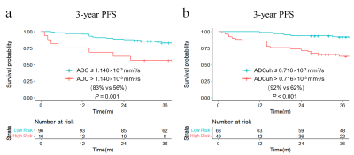 |
Ultra-High b-Value DWI in Predicting Progression risk of Locally
Advanced Rectal Cancer: A Comparative Study with Routine DWI
Guangwen Zhang1,
Jinsong Zhang1,
Ziliang Xu1,
Xiaocheng Wei2,
and Jialiang Ren3 1Department of Radiology, Xijing Hospital, Xi’an, China, 2Department of MR Research, GE Healthcare China,, Beijing, China, 3Department of Pharmaceuticals Diagnostics, GE Healthcare China, Beijing, China Keywords: Cancer, Diffusion/other diffusion imaging techniques The prognosis prediction of locally advanced rectal cancer (LARC) was important to individualized treatment, we investigated the performance of ultra-high b-value DWI (UHBV-DWI) in progression risk prediction of LARC and compare with routine DWI. It was found that ADCuh derived from UHBV-DWI performed better than ADC based on routine DWI in predicting prognosis of LARC. The model based on combination of ADCuh, TNM-stage and extramural venous invasion (EMVI) could help to indicate progression risk before treatment. |
| 14:10 |
0509.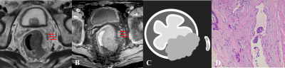 |
Synthetic phase-sensitive inversion-recovery imaging for
assessing extramural venous invasion in patients with rectal
cancer
Ge yuxi1,
Dai Jiankun2,
Hu Shudong1,
and Jia Yanlong3
1Affiliated Hospital of Jiangnan University, Wuxi, China, 2GE Healthcare, Shanghai, China, 3Department of Radiology,Xiangyang Central Hospital, Affiliated Hospital of Hubei University of Arts and Science, Xiangyang, China Keywords: Pelvis, Quantitative Imaging, synthetic MR, rectal cancer, vessels This study aimed to investigate the feasibility of synthetic phase-sensitive inversion recovery (SyPSIR) vessel for extramural venous invasion (EMVI) detection in patients with rectal cancer. One hundred and six histologically confirmed rectal cancer patients (35 EMVI+ and 71 EMVI−) were evaluated. Compared with using T2WI alone, the area under the receiver operating characteristic curve using the combination of T2WI and SyPSIR was increased from 0.86 to 0.96 and from 0.65 to 0.88 for the senior and junior radiologist, respectively. Therefore, SyPSIR can provide additional information to improve EMVI diagnostic efficiency of in rectal cancer. |
| 14:18 |
0510.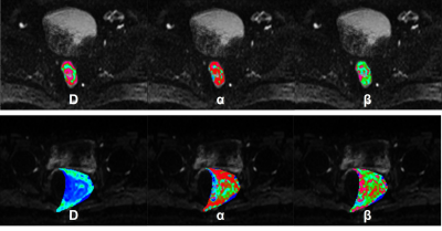 |
Preoperatively Grading Rectal Cancer with the Continue Time
Random Walk DWI model.
Zhijun Geng1,
Shaolei Li2,
Yunfei Zhang2,
Yongming Dai2,
and Chuanmiao Xie1
1Sun Yat-sen University Cancer Center, Guangzhou, China, 2MR Collaboration, Central Research Institute, United Imaging Healthcare, Shanghai, China Keywords: Pelvis, Diffusion/other diffusion imaging techniques This study evaluates a new pre-operatively method to grade rectal cancer based on Continue Time Random Walk (CTRW) model. The method is to calculate three parameters of the CTRW model by fitting the model with DWI signal and a series of b-values and to differentiate low- and high-grade tumors by fitting a logistic regression with different combinations of parameters. An additional k-mean clustering analysis is performed to evaluate how differentiable the low and high-grade groups are in the CTRW parameters’ phase space. Our study shows that CTRW model has the potential to accurately grade rectal cancer. |
| 14:26 |
0511.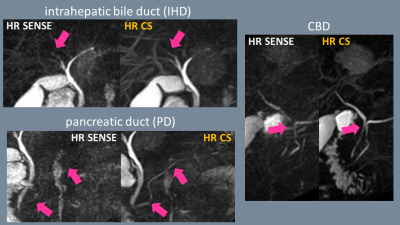 |
High-resolution single-breath-hold 3D MRCP using accelerated 3D
Gradient and Spin-Echo (GraSE) with Compressed SENSE
Takumi Ogawa1,
Michinobu Nagao2,
Yasuhiro Goto1,
Masami Yoneyama3,
Johannes M Peeters4,
Isao Shiina1,
Yutaka Hamatani1,
Kazuo Kodaira1,
Mana Kato1,
and Shuji Sakai2
1Department of Radiological Services, Tokyo Women's Medical University, Tokyo, Japan, 2Department of Diagnostic image & Nuclear Medicine, Tokyo Women's Medical University, Tokyo, Japan, 3Philips Japan, Tokyo, Japan, 4Philips Healthcare, Best, Netherlands Keywords: Digestive, Biliary Breath-hold MRCP has gained more attention in routine clinical MRI, but its limited scan time during the breath-hold period often results in poor signal-to-noise ratio (SNR) and spatial-resolution. Despite being a single breath-hold method, 3D gradient and spin-echo (GraSE) sequence has been reported to provide high image quality. In this study, accelerated GraSE sequence combined with Compressed SENSE has been developed to obtain high-resolution MRCP images with a single breath-hold. |
| 14:34 |
0512.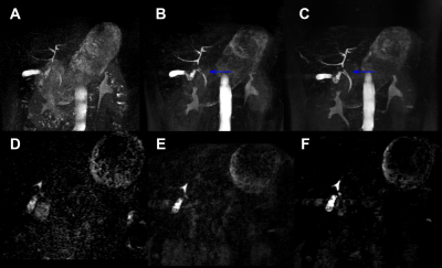 |
Accelerated 3D MR cholangiopancreatography using a deep
learning-based reconstruction in patients with cholelithiasis
Yu Zhang1,
Chunchao Xia1,
Zhenlin Li1,
and Xiaoyong Zhang2
1West China Hospital of Sichuan University, Chengdu, China, 2Clinical Science, Philips Healthcare, Chengdu, China Keywords: Liver, Biliary In this work, we aimed to compare image quality and lesion detectability in patients suspected with gallstones among single breath-hold three-dimensional magnetic resonance cholangiopancreatography (MRCP) with gradient and spin-echo (GRASE) technique, with compressed sensing (CS) and with deep learning (DL) technique. DL MRCP showed the best image quality and better lesion conspicuity and lesion edge sharpness. |
| 14:42 |
0513.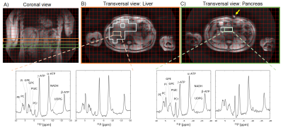 |
Comparison of 31P MRS in human pancreas and liver at 7 Tesla
Leonard W.F. Seelen1,2,
Lieke van den Wildenberg1,
Ayhan Gursan1,
W.J. Mark Gosselink1,
Wybe J.M. van der Kemp1,
Firdaus A.A. Mohamed Hoesein3,
Nadia Haj Mohammad4,
I. Quintus Molenaar2,
Hjalmar C. van Santvoort2,
Dennis W.J. Klomp1,
and Jeanine J. Prompers1
1Center for Image Sciences, University Medical Center Utrecht, Utrecht, Netherlands, 2Dept. of Surgery, UMC Utrecht Cancer Center and St Antonius Hospital Nieuwegein: Regional Academic Cancer Center Utrecht, Utrecht University, Utrecht, Netherlands, 3Dept. of Radiology, University Medical Center Utrecht, Utrecht University, Utrecht, Netherlands, 4Dept. of Medical Oncology, UMC Utrecht Cancer Center, Regional Academic Cancer Center Utrecht, Utrecht University, Utrecht, Netherlands Keywords: Pancreas, Non-Proton 31P MRS could be valuable for the assessment of treatment response in pancreatic cancer. Using a 31P whole-body transmit coil in combination with a 16-channel receive array at 7T, we could obtain 31P MRSI data covering both the whole liver and the deeply lying pancreas. The aim of this study was to compare 31P metabolite levels in the pancreas and liver in healthy volunteers. The PME/PDE ratio, which has been used as a tumor marker, was 2.1-fold higher in the pancreas compared to the liver. This will be important to take into account when applying 31P MRS in pancreatic tumors. |
| 14:50 |
0514.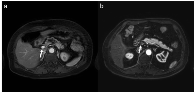 |
Free-Breathing Dynamic Pancreatic MRI Using Stack-of-Stars
Radial Sampling Algorithm and Compressed SENSE
Yoshifumi Noda1,
Nobuyuki Kawai1,
Tetsuro Kaga1,
Kimihiro Kajita2,
Yu Ueda3,
Masatoshi Honda3,
Fuminori Hyodo1,4,
Hiroki Kato1,
and Masayuki Matsuo1
1Department of Radiology, Gifu University, Gifu, Japan, 2Department of Radiology Services, Gifu University Hospital, Gifu, Japan, 3Philips Japan, Tokyo, Japan, 4Institute for Advanced Study, Gifu University, Gifu, Japan Keywords: Pancreas, Pancreas Dynamic contrast-enhanced imaging is an essential examination in pancreatic MRI. However, non-diagnosable image quality due to motion artifacts and inappropriate scan timing for pancreatic phase are common problems. Recently, free-breathing sequence (4D FreeBreathing) has been introduced and it can provide diagnosable image quality even in free-breathing. In this study, we evaluated the feasibility of 4D FreeBreathing in pancreatic MRI. Our results showed that 4D FreeBreathing could provide diagnosable image quality and appropriate pancreatic phase scanning could be achieved in all examinations. |
| 14:58 |
0515.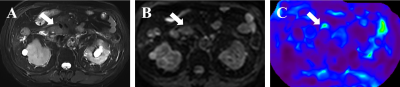 |
Magnetic resonance elastography-derived stiffness: potential
biomarker for differentiation of benign and malignant pancreatic
masses
Dingxia Liu1,
Jiejun Chen1,
Yunfei Zhang2,
Yongming Dai2,
and Xiuzhong Yao1
1Shanghai Institute of Medical Imaging, Shanghai, China, 2MR Collaboration, Central Research Institute, United Imaging Healthcare, Shanghai, China Keywords: Pancreas, Cancer Magnetic resonance elastography (MRE) is a noninvasive technique capable of quantifying tissue mechanical properties (stiffness) in vivo. Given its specialty, it is supposed to be potential for identifying malignant tumors that are characterized by a marked desmoplastic reaction and build-up of fibrotic tissues in cancer cells and tumor microenvironment. However, the field has been largely restricted to studies in pancreatic MRE. This study sought to determine the diagnostic performance of MRE for pancreatic solid masses, compared with diffusion-weighted imaging (DWI) and serum CA19-9, to establish a threshold for differentiating between pancreatic ductal adenocarcinoma (PDAC) and benign tumors in pancreas. |
| 15:06 |
0516.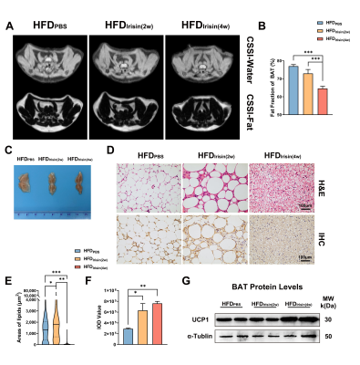 |
In vivo Quantitative evaluation of MR Imaging: Irisin Activates
Brown Adipose Tissue and Improves Systemic Metabolic Disorders
in Obese Mice
Jingyue Dai1,
Yufei Zhao1,
and Xingui Peng1
1Jiangsu Key Laboratory of Molecular and Functional Imaging, Department of Radiology, Southeast University, Zhongda Hospital, NANJING, China Keywords: Endocrine, Metabolism, Brown Adipose Tissue Irisin is an exercise-induced myokine which can induce white adipose tissue browning; however, its impact on BAT remains unclear. Here, we employed MR chemical shift-selective imaging to quantify fat fraction, and to verify these MR-derived data by that from laboratory analysis to investigate the effects of irisin on BAT and systemic metabolism in obese mice. Irisin reduced the fat fraction and increased the UCP1 protein expression of BAT. Besides, irisin helped obese mice to loss weigh and improve systemic metabolic disorders. These results suggests that irisin can reactivate brown adipose tissue and improve systemic metabolic disorders in obese mice. |
| 15:14 |
0517.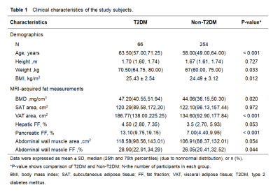 |
Relationship between muscle fat,vertebral density and ectopic
fat deposition in patients with T2DM using IDEAL-IQ
qi an1,
qinhe zhang1,
ailian liu1,
liangjie lin2,
and lizhi xie3
1Department of Radiology, the First Affiliated Hospital of Dalian Medical University, Dalian, China, 2Philips health care, Dalian, China, 3GE health care, Dalian, China Keywords: Endocrine, Diabetes This study was carried out to evaluate the correlations between abdominal fat, vertebral density and ectopic fat deposition patients with T2DM using IDEAL-IQ.We found that SAT area,VAT area,Pancreatic FF and Abdominal wall muscle FF was positively correlated with BMD in patients with T2DM. |
| 15:22 |
0518.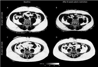 |
Correlations between adipose tissue volume changes and its
baseline MR-characteristics in people with obesity undergoing a
caloric restriction
Daniela Junker1,
Mingming Wu1,
Selina Rupp1,
Jessie Han1,
Stella Näbauer1,
Anna Reik2,
Meike Wiechert2,
Arun Somasundaram1,
Marcus R. Makowski1,
Hans Hauner2,
Christina Holzapfel2,
and Dimitrios C. Karampinos1
1Department of Diagnostic and Interventional Radiology, School of Medicine, Technical University of Munich, Munich, Germany, 2Institute for Nutritional Medicine, School of Medicine, Technical University of Munich, Munich, Germany Keywords: Endocrine, Fat Results of weight loss interventions differ individually. MRI-based quantification and characterization of adipose tissue (AT) offers methods to identify possible AT-phenotypes that facilitate AT loss. The purpose of this analysis was to evaluate how, in people with obesity undergoing an 8-week formula-based weight loss intervention, the relative AT and lipid volume loss of the subcutaneous and visceral AT depot correlate to the proton density fat fraction (PDFF) and the total volume of AT as well as the volume of lipids in each depot at the beginning of the diet. |
 Back to Meeting Home
 Back to the Program-at-a-Glance
The International Society for Magnetic Resonance in Medicine is accredited by the Accreditation Council for Continuing Medical Education to provide continuing medical education for physicians.
 Back to Meeting Home
 Back to the Program-at-a-Glance
 View Presentation Video