Oral Session
Lung: From Breathing to Blood Flow
ISMRM & ISMRT Annual Meeting & Exhibition • 03-08 June 2023 • Toronto, ON, Canada

Oral Session
Lung: From Breathing to Blood Flow
| 16:00 |
1398.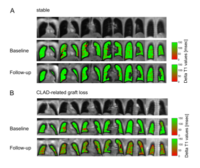 |
UTILITY OF OXYGEN-ENHANCED LUNG MRI IN LONG TERM POST-LUNG
TRANSPLANT PATIENT CARE
Milan Speth1,2,
Till Frederik Kaireit1,2,
Marcel Gutberlet1,2,
Filip Klimeš1,2,
Lea Behrendt1,2,
Andreas Voskrebenzev1,2,
Frank Wacker1,2,
Tobias Welte2,3,
Jens Gottlieb2,3,
and Jens Vogel-Claussen1,2
1Department of Diagnostic and Interventional Radiology, Hannover Medical School, Hannover, Germany, 2Biomedical Research in Endstage and Obstructive Lung Disease (BREATH), German Center for Lung Research (DZL), Hannover, Germany, 3Department of Respiratory Medicine, Hannover Medical School, Hannover, Germany Keywords: Lung, Transplantation, Graft loss Aim of this prospective single-center surveillance study was to assess the ability of oxygen-enhanced MRI to predict future chronic lung allograft dysfunction (CLAD) related transplant loss. Baseline MRI scans were acquired 6-12 months and follow-up MRI 2.5 years after double lung transplantation. T1 mapping was carried out with patients breathing room air and 100% oxygen, Delta T1 maps were calculated. Median, quartile coefficient of dispersion and ventilated volume parameters were correlated with graft loss and compared with same day lung function testing. Oxygen-enhanced MRI predicted future CLAD-related transplant loss 6-12 months post transplantation and, when evaluating %change, at follow-up MRI. |
| 16:08 |
1399.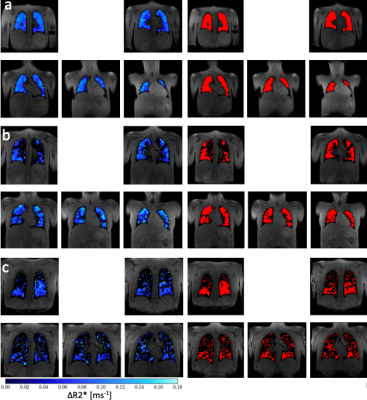 |
Double-echo Oxygen Enhanced MRI at 1.5 T correlates with
clinical lung function in CF patients
Marta Tibiletti1,
Christopher Short2,3,
Josephine H Naish1,4,
Mary Abkir2,3,
Thomas Semple2,3,5,
Simon Padley2,3,
Jane C. Davies2,6,
and Geoff JM Parker1,7
1Bioxydyn Ltd, Manchester, United Kingdom, 2National Heart & Lung Institute, Imperial College London, London, United Kingdom, 3Royal Brompton Hospital, Guy's & St Thomas’ Trust, London, United Kingdom, 4MCMR, Manchester University NHS Foundation Trust, Wythenshawe, United Kingdom, 5Centre for Paediatric and Child Health, Imperial College London, London, United Kingdom, 6Royal Brompton Hospital, , Guy's & St Thomas’ Trust, London, United Kingdom, 7Centre for Medical Image Computing, Department of Medical Physics and Biomedical Engineering, University College London, London, United Kingdom Keywords: Lung, Oxygenation This study presents the first results from a population with cystic fibrosis (CF) of a novel, easy-to-implement, multi-slice, free breathing OE-MRI acquisition method based on measuring R2* changes in the lung with varying level of oxygen delivered to the patients. The oxygen ventilated volume fraction obtained presents a very good correlation with lung clearance index (LCI) obtained by multiple breath N2 washout. LCI is a marker of overall lung ventilation inhomogeneity, which has been shown to be a sensitive marker of lung disease severity in CF, particularly in the early stage of the disease when spirometry outcomes are normal. |
| 16:16 |
1400.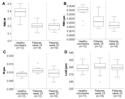 |
Impaired xenon gas transfer observed one year after
hospitalisation due to COVID-19 in patients with signs of
interstitial lung disease.
Laura Saunders1,
Guilhem Collier1,
Ho-Fung Chan1,
Paul Hughes1,
Laurie Smith1,
Neil Stewart1,
Jonathan Brooke2,
James Watson3,
James Meiring3,
Zoë Gabriel3,
Thomas Newman3,
Megan Plowright3,
Phillip Wade3,
James Eaden3,
Jody Bray1,
Helen Marshall1,
David Capener1,
Leanne Armstrong1,
Jennifer Rodgers1,
Martin Brook1,
Alberto Biancardi1,
Madhwesha Rao1,
Graham Norquay1,
Oliver Rodgers1,
Ryan Munroe1,
James Ball1,
Neil Stewart1,
Gisli Jenkins4,
James Grist5,
Kher Lik Ng6,
Ling-pei Ho5,
Fergus Gleeson5,
Ian Hall7,
Thomas Meersmann7,
Galina Pavlovskaya7,
Arthur Harrison7,
Jonathan Brooke7,
Joseph Jacob8,
Andrew Swift1,
Smitha Rajaram3,
Gary Mills1,
Lisa Watson3,
Paul Collini1,
Rod Lawson3,
A A Roger Thompson1,
and Jim Wild1
1The University of Sheffield, Sheffield, United Kingdom, 2University of Nottingham, Nottingham, United Kingdom, 3Sheffield Teaching Hospitals, Sheffield, United Kingdom, 4Imperial College London, London, United Kingdom, 5University of Oxford, Oxford, United Kingdom, 6Oxford NHS Foundation Trust, Oxford, United Kingdom, 7Nottingham University, Nottingham, United Kingdom, 8University College London, London, United Kingdom Keywords: Lung, COVID-19 Patients with signs of interstitial lung disease at 12 weeks after hospitalisation due to COVID-19 underwent 1H and 129Xe MRI. 129Xe MRI showed impaired xenon gas transfer (RBC:M and RBC:gas) at 24 and 52 weeks after hospital admission, with no longitudinal change between 24 and 52 weeks observed in 129Xe MRI metrics or PFT transfer factor. Xenon MRI metrics correlated significantly with PFT transfer factor at 24 weeks (RBC:M, RBC:gas, LmD) and 52 weeks (RBC:M, RBC:gas, LmD). |
| 16:24 |
1401.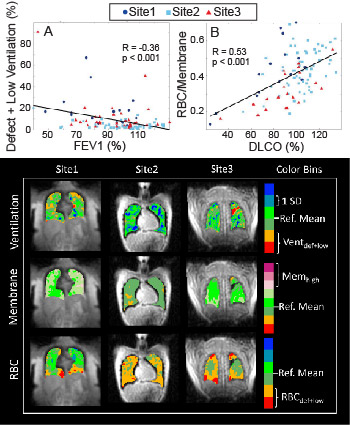 |
Retrospective analysis of Hyperpolarized 129Xe MRI Gas Exchange
Images in Healthy and Post-COVID-19 Individuals acquired at
Three Sites
Peter Niedbalski1,
David Mummy2,
Haoran Dai2,
Aryil Bechtel3,
Alexandra Schmidt4,
Bradie Frizzell1,
Sakib Kabir2,
Jonathon Leipsic4,5,
Janice Leung4,6,
Bastiaan Driehuys2,
Loretta Que7,
Mario Castro1,
Don Sin4,6,
and Rachel Eddy4,6 1Pulmonary, Critical Care, and Sleep Medicine, University of Kansas Medical Center, Kansas City, KS, United States, 2Department of Radiology, Duke University, Durham, NC, United States, 3Department of Medical Physics, Duke University, Durham, NC, United States, 4Centre for Heart Lung Innovation, St. Paul's Hospital, Vancouver, BC, Canada, 5Department of Radiology, University of British Columbia, Vancouver, BC, Canada, 6Division of Respiratory Medicine, University of British Columbia, Vancouver, BC, Canada, 7Division of Pulmonary Medicine, Duke University, Durham, NC, United States Keywords: Lung, COVID-19 Gas exchange hyperpolarized 129Xe MRI (Xe-MRI) is increasingly being considered as an outcome measure in multi-site clinical trials, but there is limited evidence of between-site comparability. In this study, we analyzed 121 gas exchange Xe-MRI images in healthy and post-acute COVID-19 participants independently acquired at three sites. In healthy volunteers, quantitative Xe-MRI measures are indistinguishable across sites. In post-acute COVID-19, cross-site differences in Xe-MRI measures are evident but appear to be driven by differences in patient population. Moreover, Xe-MRI measures across sites correlate strongly with pulmonary function testing. These results support the feasibility of multi-site trials using gas exchange Xe-MRI. |
| 16:32 |
1402.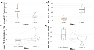 |
Improved 129Xe MRI of cardiopulmonary oscillations in patients
with chronic thromboembolic pulmonary hypertension
Junlan Lu1,
Elianna Bier2,
Suphachart Leewiwatwong2,
David Mummy3,
Sakib Kabir3,
Fawaz Alanezi4,
Sudarshan Rajagopal4,
Scott Haile Robertson5,
Peter J Niedbalski6,
and Bastiaan Driehuys3
1Medical Physics, Duke University, Durham, NC, United States, 2Biomedical Engineering, Duke University, Durham, NC, United States, 3Radiology, Duke University, Durham, NC, United States, 4Cardiology, Duke University, Durham, NC, United States, 5Clinical Imaging Physics Group, Duke University, Durham, NC, United States, 6Pulmonary, Critical Care, and Sleep Medicine, University of Kansas Medical Center, Kansas City, KS, United States Keywords: Lung, Hyperpolarized MR (Gas), CTEPH, keyhole reconstruction Dynamic spectroscopy of hyperpolarized 129Xe in red blood cells exhibits cardiopulmonary oscillations that can be used to detect both pre- and post-capillary pulmonary hypertension (PH). However, this whole-lung measurement cannot resolve spatially heterogeneous variations in oscillations. This limitation can be addressed by using keyhole reconstruction approaches to spatially resolve the oscillations. Here we demonstrate several extensions of this technique that make both the reconstruction and analysis more robust. We have used this to establish updated healthy reference distributions and demonstrate the utility of spatially resolved mapping in a cohort of patients with chronic thromboembolic pulmonary hypertension pre- and post-thromboendarterectomy. |
16:40 |
1403.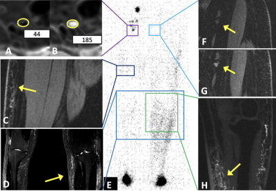 |
Iron Oxide Nanoparticle MR Lymphangiography (ION-MRL): A
quantitative technique for assessing the peripheral and central
lymphatic systems.
Robert Carson Sibley1,
Shreyas Vasanawala1,
and Andreas M Loening1 1Radiology, Stanford, Stanford, CA, United States Keywords: Vessels, Quantitative Imaging, lymphangiography A lack of clinical quantitative lymphatic metrics slows progress for innovation in treatment of lymphatic disease. MR lymphangiography provides high resolution anatomic analysis of the lymphatic system necessary for guiding new and emerging therapeutics but has not been explored as a quantitative metric. We coupled MR lymphangiography with iron oxide nanoparticles as the contrast agent and demonstrate the time to visualization of the thoracic duct is decreased in patients with bilateral lymphedema. Additionally, the change in R2* of inguinal lymph nodes after transpedal contrast injection quantifies lymphatic transit in patients with lymphedema. |
| 16:48 |
1404.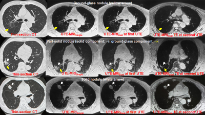 |
UTE-MRI with Single- and Dual-Echo Methods vs. CT:
Differentiation Capability of Non- or Minimally Invasive
Adenocarcinomas from Other Cancers
Yoshiharu Ohno1,2,
Masao Yui3,
Kaori Yamamoto3,
Masato Ikedo3,
Yuka Oshima4,
Nayu Hamabuchi4,
Satomu Hanamatsu4,
Hiroyuki Nagata2,
Takahiro Ueda1,
Hirotaka Ikeda1,
Daisuke Takenaka1,5,
Takeshi Yoshikawa1,5,
Akiyoshi Iwase6,
Yoshiyuki Ozawa1,
and Hiroshi Toyama1
1Radiology, Fujita Health University School of Medicine, Toyoake, Japan, 2Joint Research Laboratory of Advanced Medical Imaging, Fujita Health University School of Medicine, Toyoake, Japan, 3Canon Medical Systems Corporation, Otawara, Japan, 4Fujita Health University School of Medicine, Toyoake, Japan, 5Diagnostic Radiology, Hyogo Cancer Center, Akashi, Japan, 6Fujita Health University Hospital, Toyoake, Japan Keywords: Lung, Cancer We hypothesized that pulmonary MRIs with UTE using single- or dual-echo techniques may be equal to or more useful than standard-dose thin-section CT for evaluating solid portion size and C/T ratio. The purpose of this study was thus to compare capabilities of pulmonary MRIs with UTE using single- and dual-echo techniques (UTE-MRISingle and UTE-MRIDual) and thin-section CT for quantitative differentiation of non- and minimally invasive adenocarcinomas from other lung cancers. |
| 16:56 |
1405.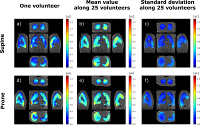 |
Nominal respiratory features in spontaneous breathing: Towards
multiparametric atlases with 3D MR spirometry in healthy
volunteers
Nathalie Barrau1,
Adrien Duwat1,
Killian Sambourg1,
Angéline Nemeth1,
Antoine Beurnier2,
Tanguy Boucneau3,
Vincent Lebon1,
and Xavier Maître1
1Université Paris-Saclay, CEA, CNRS, Inserm, BioMaps, Orsay, France, 2Hôpital Bicêtre, APHP, Le Kremlin-Bicêtre, France, 3GE Healthcare, Buc, France Keywords: Lung, Quantitative Imaging, Spirometry Three-dimensional MR spirometry fosters a double paradigm shift upon standard spirometry: from forced to free breathing and from global to local measurements. The technique makes use of voxel-wise flow-volume loops and original biomechanical markers to characterize the regional lung function. Over a diverse adult population, nominal common features showed up throughout 3D MR spirometry parametric maps in healthy volunteers spontaneously breathing in supine and prone positions. Euclidian barycenter and standard deviation maps of local tidal volumes, spontaneous expiratory peak flows, and anisotropic deformation indices are presented here as the ground for a unique atlas of the lung function. |
| 17:04 |
1406.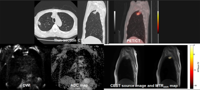 |
Comparison of Capability for Therapeutic Outcome Prediction
among CEST, DWI and PET/CT in Non-Small Cell Lung Cancer
Conservative Therapy
Yoshiharu Ohno1,2,
Masao Yui3,
Kaori Yamamoto3,
Takeshi Yoshikawa1,4,
Daisuke Takenaka1,4,
Masato Ikedo3,
Akiyoshi Iwase5,
Yuka Oshima6,
Nayu Hamabuchi6,
Satomu Hanamatsu6,
Hiroyuki Nagata2,
Takahiro Ueda1,
Hirotaka Ikeda1,
Yoshiyuki Ozawa1,
and Hiroshi Toyama1
1Radiology, Fujita Health University School of Medicine, Toyoake, Japan, 2Joint Research Laboratory of Advanced Medical Imaging, Fujita Health University School of Medicine, Toyoake, Japan, 3Canon Medical Systems Corporation, Otawara, Japan, 4Diagnostic Radiology, Hyogo Cancer Center, Akashi, Japan, 5Fujita Health University Hospital, Toyoake, Japan, 6Fujita Health University School of Medicine, Toyoake, Japan Keywords: Lung, Cancer, CEST We hypothesize that CEST imaging has a potential for therapeutic outcome prediction in NSCLC patients treated with chemoradiotherapy and may play as one of the predictors in this setting. The purpose of this study was to compare the capability for therapeutic outcome prediction among CEST imaging, DWI and FDG-PET/CT in NSCLC patients with conservative therapy. |
| 17:12 |
1407.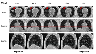 |
Respiratory Motion-Resolved 4D Pulmonary Imaging with
Stack-of-Spiral UTE at 0.55T and 3T
Pan Su1,
Waqas Majeed1,
Xin Miao1,
Josef Pfeuffer2,
Ahsan Javed3,
Rajiv Ramasawmy3,
Adrienne E. Campbell-Washburn3,
Himanshu Bhat1,
Gregor Thoermer2,
Jianing Pang1,
and Thomas Benkert2 1Siemens Medical Solutions USA, Inc., Malvern, PA, United States, 2MR Application Predevelopment, Siemens Healthcare GmbH, Erlangen, Germany, 3Cardiovascular Branch, Division of Intramural Research, National Heart, Lung, and Blood Institute, National Institutes of Health, Bethesda, MD, United States Keywords: Lung, Low-Field MRI Recently, ultrashort echo time (UTE) has gained renewed interest for lung imaging at 1.5T and 3T, which captures short T2* signal from lung parenchyma. Lower field is attractive for lung MRI due to prolonged T2* and reduced susceptibility, and it was demonstrated that high-resolution high-quality structural lung imaging can be achieved with UTE at 0.55T. In addition to anatomical information, pulmonary function such as regional ventilation is also of great clinical interest. In this study, we developed free-breathing respiratory-motion-resolved 4D pulmonary imaging using stack-of-spirals acquisition with compressed-sensing at both 0.55T and 3T, which could enable quantitative evaluation of ventilation dynamics. |
| 17:20 |
1408.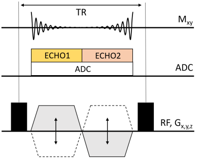 |
Submillimeter morphologic lung MRI at 0.55T using balanced
steady-state free precession with half-radial dual-echo readout
(bSTAR)
Grzegorz Bauman1,2,
Nam G Lee3,
Ye Tian4,
Oliver Bieri1,2,
and Krishna S Nayak3,4
1Deparment of Radiology, Division of Radiological Physics, University of Basel Hospital, Basel, Switzerland, 2Department of Biomedical Engineering, University of Basel, Basel, Switzerland, 3Department of Biomedical Engineering, Viterbi School of Engineering, University of Southern California, Los Angeles, CA, United States, 4Ming Hsieh Department of Electrical and Computer Engineering, Viterbi School of Engineering, University of Southern California, Los Angeles, CA, United States Keywords: Lung, Low-Field MRI, Morphology In this work we explore the potential of free-breathing balanced steady-state free precession half-radial dual-echo imaging technique (bSTAR) for morphologic lung MRI in human subjects using high-performance 0.55T MR-scanner. The technique combines an efficient minimal-TR readout sampling with interleaved wobbling Archimedean spiral pole trajectories and retrospective respiratory self-gating. Lung imaging at 0.55T helped to markedly reduce off-resonance artifacts while providing an improved signal intensity and allowed for high-quality morphologic lung MRI at a submillimeter spatial resolution.
|
| 17:28 |
1409.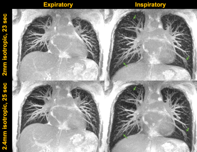 |
Rapid 3D lung imaging with bSSFP stack of spiral out-in (SoSoi)
sampling at 0.55T
Ye Tian1,
Nam G. Lee2,
Ziwei Zhao1,
and Krishna Nayak1,2
1Ming Hsieh Department of Electrical and Computer Engineering, University of Southern California, Los Angeles, CA, United States, 2Department of Biomedical Engineering, University of Southern California, Los Angeles, CA, United States Keywords: Lung, Lung MRI provides radiation-free screening of many lung diseases. Its use has been limited at field strengths >1.5T, largely due to the ultra-shot T2* and low proton density in the lung. New 0.55T systems provide improved lung MRI capability since T2* is prolonged to the order of 10ms. In this work, we propose a rapid 3D bSSFP stack-of-spiral out-in (SoSoi) pulse sequence for lung imaging. The sequence can acquire 2 mm isotropic resolution image with 23 sec breathhold or 2.4 mm isotropic resolution image with 13 sec breathhold, both show great pulmonary vessels depiction. |
| 17:36 |
1410.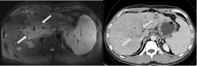 |
Direct abdominal vein thrombus imaging (DATI): a contrast free
black-blood MR technique for the diagnosis of abdominal vein
thrombosis
Liping Liao1,
Zehe Huang1,
Zeping Liu2,
Shengyuan Liang1,
Lei Qin1,
Shengzhang Pan1,
Lanbin Huang1,
Qizeng Ruan1,
Yi Sun3,
and Guoxi Xie2
1The First People’s Hospital of Qinzhou, Qinzhou, China, 2School of Biomedical Engineering, Guangzhou Medical University, Guangzhou, China, 3Siemens Healthineers, Shanghai, China Keywords: Vessels, Thrombo-Embolic Abdominal vein thrombosis (AVT) is a significant cause of morbidity. Accurate diagnosis of AVT is relevant for treatment proper decision-making. CTV or MRV requires the use of contrast medium, which may lead to patient's renal failure or allergic reaction. To address this issue, we sought to develop a direct AVT imaging (DATI) technique which is free of contrast medium. The technique is based on a respiratory navigating SPACE sequence with DANTE black-blood preparation and evaluated preliminarily on 19 AVT patients at 3.0T. Experiment results show that DATI can provide definitive thrombus detection for the diagnosis of AVT. |
| 17:44 |
1411.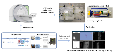 |
Phantom validation of continuous radial sampling MRI for robotic
interventional cardiovascular surgery
Sijie Zhong1,2,
Ran Tao1,
Ruoxi Wang1,2,
Hao Chen1,2,
Chen Jin1,2,
and Zhiyong Zhang1,2
1School of Biomedical Engineering, Shanghai Jiao Tong University, Shanghai, China, 2Institute of Medical Robotics, Shanghai Jiao Tong University, Shanghai, China Keywords: Vessels, Cardiovascular, Run-time MRI, Navigation, Interventional MRI Cardiovascular dilation surgery is usually performed with DSA navigation, and excessive radiation during imaging harms both the patient and the physician. Magnetic resonance imaging can avoid ionizing radiation, but its imaging efficiency severely limits the performance of real-time imaging. Therefore, we construct a complete real-time magnetic resonance navigation system through continuous radial scanning mode and corresponding reconstruction methods and formulate a group of scanning strategies. The characteristics of the special derivative catheter under magnetic resonance imaging were tested by combining the partner's magnetically compatible robotic equipment. And the results demonstrated the feasibility of this method. |
 Back to Meeting Home
 Back to the Program-at-a-Glance
The International Society for Magnetic Resonance in Medicine is accredited by the Accreditation Council for Continuing Medical Education to provide continuing medical education for physicians.
 Back to Meeting Home
 Back to the Program-at-a-Glance
 View Presentation Video