Oral Session
Renal Imaging: Structure & Function
ISMRM & ISMRT Annual Meeting & Exhibition • 03-08 June 2023 • Toronto, ON, Canada

Oral Session
Renal Imaging: Structure & Function
| 13:45 |
1283.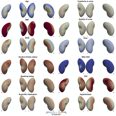 |
Three-Dimensional Kidney Shape Analysis: Associations with
Anthropometric and Disease Factors
Marjola Thanaj1,
Nicolas Basty1,
Madeleine Cule2,
Elena Sorokin2,
Jimmy Bell1,
Elizabeth Louise Thomas1,
and Brandon Witcher1
1Life Sciences, University of Westminster, London, United Kingdom, 2Calico Life Sciences LLC, South San Francisco, CA, United States Keywords: Kidney, Kidney, Mass Univariate Regression Analysis Organ MRI measurements have the potential to enhance our understanding of the precise phenotypic changes underlying many clinical conditions. Using kidney mesh-based shape analysis we were able to detect variation in specific anatomical regions of the kidney, and associate this with anthropometric traits as well as disease states including chronic kidney disease (CKD), type-2 diabetes (T2D), and hypertension. We show that CKD is associated with smaller kidneys. We also show that T2D and hypertension are associated with larger kidneys. |
| 13:53 |
1284. |
Quality Control of MRI-based Kidney Volume Estimation in ADPKD
patient in clinical practice
Chenglin Zhu1,
Arman Sharbatdaran1,
Hreedi Dev1,
Xinzi He1,
Jon D. Blumenfeld2,
James M. Chevalier2,
Daniil Shimonov2,
and Martin R. Prince1,3 1Radiology, Weill Cornell Medicine, New York, NY, United States, 2Rogosin Institute, New York, NY, United States, 3Radiology, Columbia University Vagelos College of Physicians and Surgeons, New York, NY, United States Keywords: Kidney, Data Acquisition Total kidney volume (TKV) is a critical biomarker for monitoring disease severity in autosomal dominant polycystic kidney disease (ADPKD). TKV is typically measured by manually contouring kidneys on one sequence from abdominal MRI without any quality control. Here we show that by using a deep learning model to measure kidney volume on 5 routinely acquired abdominal MRI sequences, it is possible to apply outlier analysis to find images with acquisition artifacts and to correct or exclude them from TKV estimation. This improves volume measurement consistency among the 5 sequences from 4.3% to 1.3% after quality control. |
| 14:01 |
1285.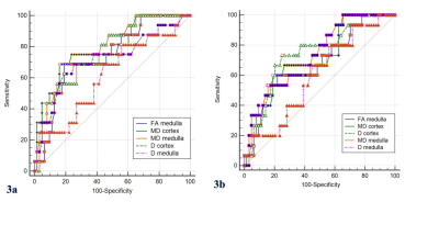 |
Diffusion weighted, intravoxel incoherent motion, diffusion
kurtosis tensor MR imaging in chronic kidney disease:
correlations with histology
Jie Zhu1,
Jia-Yin Gao1,
Pu-Yeh Wu2,
and Yan Song1
1Beijing Hospital, Beijing, China, 2GE Healthcare, Beijing, China Keywords: Data Analysis, Diffusion/other diffusion imaging techniques Noninvasive, repeatable and accurate biomarkers to identify renal histological changes for tailoring treatment and evaluating renal prognosis are demanded. In this study, we aimed to compare and probe correlations of parameters derived from standard DWI and its extending models including IVIM, DTI, and DKTI with the pathological and functional alterations in CKD. We found that the corrected diffusion-related indices, including cortical and medullary D and MD, as well as medullary FA were superior to ADC, perfusion-related and kurtosis indices for evaluating alterations of renal pathology and function in CKD patients, and these metrics were also correlated with eGFR and Scr. |
| 14:09 |
1286.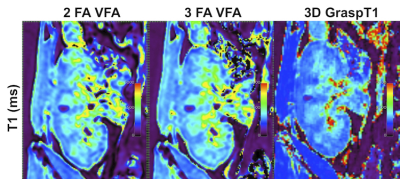 |
Magnetization-Prepared Golden-angle RAdial Sparse Parallel
(MP-GRASP) MRI for Rapid Free-Breathing 3D T1 Mapping of Renal
Allografts
Octavia Bane1,2,
Li Feng1,2,
Ding Xia1,2,
Haitham Al-Mubarak1,2,
Jordan Cuevas1,2,
Thangamani Muthukumar3,
Jonathan Dyke4,
Madhav Menon5,
Samira Farouk6,
Bachir Taouli1,2,
and Sara Lewis1,2
1Department of Diagnostic, Molecular and Interventional Radiology, Icahn School of Medicine at Mount Sinai, New York, NY, United States, 2BioMedical Engineering and Imaging Institute, Icahn School of Medicine at Mount Sinai, New York, NY, United States, 3Nephrology, Weill Cornell Medical College, New York, NY, United States, 4Citigroup Biomedical Imaging Center, Weill Cornell Medical College, New York, NY, United States, 5Nephrology, Yale School of Medicine, New Haven, CT, United States, 6Department of Medicine, Division of Nephrology, Icahn School of Medicine at Mount Sinai, New York, NY, United States Keywords: Kidney, Quantitative Imaging, T1 mapping, cortex, medulla, transplantation This work demonstrates rapid free-breathing 3D T1 mapping of the kidney using Magnetization-Prepared Golden-angle RAdial Sparse Parallel imaging (GraspT1). The accuracy and performance of GraspT1was prospectively compared with B1-corrected variable flip angle (VFA) T1 mapping in phantom and in 13 patients with renal transplant. In phantom, both GraspT1 and VFA enabled accurate T1 estimation compared to the gold standard. In patients, free-breathing 3D GraspT1 provided better spatial resolution for improved cortico-medullary differentiation compared to the VFA method, while achieving whole-kidney coverage and similar T1 quantification, at the expense of longer acquisition time. |
| 14:17 |
1287.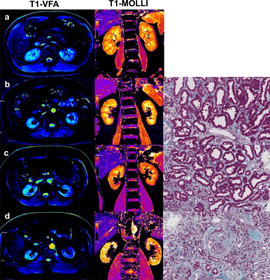 |
A preliminary study of rapid T1mapping imaging for evaluating
renal interstitial fibrosis
Chenchen Hua1,
Yi Zhuang2,
Leting Zhou1,
Lu Qiu2,
Ting Cai1,
Bin Xu1,
Shaowei Hao3,
Liang Wang1,
and Haoxiang Jiang2
1The Affiliated Wuxi People's Hospital of Nanjing Medical University, Wuxi, China, 2The Affiliated Wuxi Children's Hospital of Nanjing Medical University, Wuxi, China, 3Siemens Healthineers Digital Technology(Shanghai) CO.,Ltd., Nanjing, China Keywords: Kidney, Quantitative Imaging, T1mapping T1 mapping is a quantitative magnetic resonance imaging (MRI) technique that can reflect the extent of tissue fibrosis. Classical inversion recovery (IR) method can measure the T1 value with high accuracy, but it has long acquisition times. Therefore, the variable flip angle(VFA) method and the modified look‑locker inversion recovery method (MOLLI) are more frequently used in practical clinical applications. The objectives of this study was to compare the two rapid T1mapping imaging methods and to evaluate the diagnostic efficacy of T1 values measured by the two methods in differentiating the degree of renal interstitial fibrosis(IF). |
| 14:25 |
1288.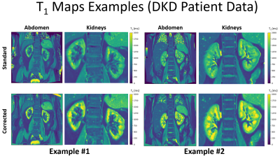 |
A shortened MOLLI for renal T1 mapping
Joao Periquito1,
Kanishka Sharma1,
Kywe Soe1,
Bashair Alhummiany2,
Jonathan Fulford3,
David Shelley4,
Kim Gooding3,
Angela Shore3,
Michael Mansfield4,
and Steven Sourbron1 1The University of Sheffield, Sheffield, United Kingdom, 2Department of Biomedical Imaging Sciences, University of Leeds, Leeds, United Kingdom, 3University of Exeter Medical School, Exeter, United Kingdom, 4Leeds Teaching Hospitals NHS Trust, Leeds, United Kingdom Keywords: Kidney, Kidney, MOLLI, T1 MAPPING A recent consensus recommends MOLLI-type methods for T1-mapping in the kidney, but these are slow due to the need for full relaxation between inversions. Acceleration can be easily achieved on routine MOLLI-sequences by repeating preparation pulses before complete relaxation, but this requires more accurate signal modelling. Here we propose a broadly applicable model-based approach which inverts a signal model built on Bloch simulations of magnetisation propagation. The method is validated on phantom data and a two-centre cohort of 50 patients with diabetic kidney disease. |
| 14:33 |
1289.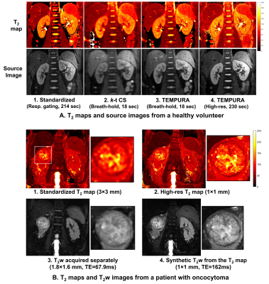 |
Highly Accelerated and High-resolution T2 Mapping in the Kidney
Based on Echo Merging Plus k-t Undersampling with Reduced
Refocusing Flip Angles
Hao Li1,2,
Andrew Nicholas Priest2,3,
Ines Horvat Menih 2,
Anne Y Warren4,
Sarah J Welsh5,
Grant D Stewart6,
Iosif A Mendichovszky3,
Susan Francis7,
and Ferdia A Gallagher2
1The Institute of Science and Technology for Brain-inspired Intelligence, Fudan University, Shanghai, China, 2Department of Radiology, University of Cambridge, Cambridge, United Kingdom, 3Department of Radiology, CUH NHS Foundation Trust, Cambridge, United Kingdom, 4Department of Histopathology, CUH NHS Foundation Trust, Cambridge, United Kingdom, 5Department of Oncology, CUH NHS Foundation Trust, Cambridge, United Kingdom, 6Department of Surgery, CUH NHS Foundation Trust, Cambridge, United Kingdom, 7Sir Peter Mansfield Imaging Centre, University of Nottingham, Nottingham, United Kingdom Keywords: Kidney, Quantitative Imaging We developed a highly accelerated multi-echo spin-echo (MESE) method based on echo merging and k-t undersampling with reduced flip angles (TEMPURA), which can be used to either reduce the acquisition time or increase spatial resolution for multi-slice kidney T2 mapping. Compared with a standardized respiratory-gated MESE sequence, fast TEMPURA reduced the acquisition time from 3–5 minutes to one breath-hold (18 s) without degrading measurement accuracy or image quality. It also outperformed using k-t undersampling alone. High-resolution TEMPURA reduced the pixel size from 3×3 mm2 to 1×1 mm2 and greatly improved the visualization of detailed structures. |
14:41 |
1290.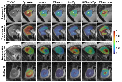 |
Initial Experience of Hyperpolarized [1-13C]pyruvate MRI in
Kidney Transplant Patients
Xiaoxi Liu1,
Ying-Chieh Lai1,2,
Shiang-Cheng Kung3,
Meyeon Park3,
Lazik Zoltan4,
Peder E.Z. Larson1,
and Zhen J. Wang1
1Department of Radiology & Biomedical Imaging, University of California, San Francisco, San Francisco, CA, United States, 2Department of Medical Imaging and Intervention, Chang Gung Memorial Hospital at Linkou, Tainwan, Taiwan, 3Department of Medicine, University of California, San Francisco, San Francisco, CA, United States, 4Department of Pathology, University of California, San Francisco, San Francisco, CA, United States Keywords: Kidney, Hyperpolarized MR (Non-Gas) We present our initial experience of applying hyperpolarized (HP) [1-13C]pyruvate MRI in three patients with renal allograft. The pyruvate metabolism to lactate and bicarbonate in two patients with well functioning allografts and normal biopsy was in the range of healthy native kidneys. The allograft pyruvate metabolism in the third patient with reduced eGFR was higher than that of healthy native kidneys. Study is ongoing to establish the range of pyruvate metabolism in normal allografts and to correlate pyruvate metabolism to allograft biopsy. This study paves the way for investigation of HP MRI in the noninvasive assessment of kidney allograft injury. |
| 14:49 |
1291. |
Time-encoded Arterial Spin Labeling for Renal Perfusion
Quantification Covering the Whole Kidneys
Zihan Ning1,
Zhensen Chen2,
Shuo Chen1,
Hualu Han1,
Long Zhao3,
Rui Wang4,
Dongyue Si1,
Huiyu Qiao1,
Rui Shen1,
and Xihai Zhao1
1Center for Biomedical Imaging Research, Department of Biomedical Engineering, Tsinghua University, Beijing, China, 2Institute of Science and Technology for Brain-Inspired Intelligence, Fudan University, Shanghai, China, 3Department of Radiology, Beijing Anzhen Hospital, Beijing, China, 4Department of Radiology, Peking University First Hospital, Beijing, China Keywords: Kidney, Perfusion, renal perfusion We performed a series of optimization on the encoding scheme, pseudo-continuous ASL (pCASL) parameters, and post-processing of time-encoded pCASL (te-pCASL), then proposed Time-encoded Arterial Spin labeling to cover the whole Kidneys (TASK) to achieve multiple time-points renal perfusion measurement efficiently. With Gave of 0.4-0.6 mT/m and Gmax/Gave around 10, Walsh-Hadamard encoding scheme, and retrospective registration, TASK was able to provide accurate and reproducible RBF and ATT measurement covering the whole kidneys with single 5-min scan. |
| 14:57 |
1292.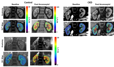 |
Quantitative BOLD MRI for Estimating Intra-renal Oxygen
Availability: Are Kidneys Hypoxemic in CKD?
Pottumarthi V Prasad1,
Lu-Ping Li1,
Bradley Hack1,
Nondas Leloudas1,
and Stuart Sprague1
1NorthShore University HealthSystem, Evanston, IL, United States Keywords: Kidney, Oxygenation Kidney BOLD MRI measurements are not specific to oxygen availability especially when comparing different cohorts because R2* also depends on fractional blood volume (fBV) and hematocrit (Hct). In this study, we have estimated fBV using ferumoxytol and Hct by blood sampling. Using these we show Quantitative BOLD MRI can characterize oxygen availability in quantitative terms. For the first time, we show that kidney cortex is normoxemic in healthy controls while moderately hypoxemic in CKD. Medulla is mildly hypoxemic in controls while moderately hypoxemic in CKD. |
| 15:05 |
1293.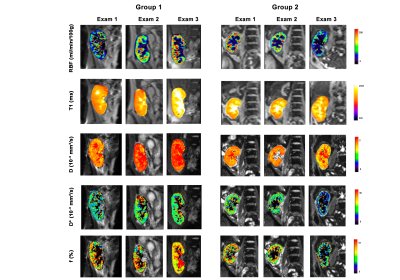 |
Potential of multiparametric MRI in the longitudinal assessment
of renal allografts after transplantation
Rebeca Echeverria-Chasco1,2,
Paloma L. Martin-Moreno2,3,
Nuria Garcia-Fernandez2,3,
Marta Vidorreta4,
Leyre Garcia-Ruiz1,
Anne Oyarzun5,
Arantxa Villanueva Larre2,5,6,
Gorka Bastarrika1,2,
and Maria A. Fernández-Seara1,2
1Radiology, Clinica Universidad de Navarra, Pamplona, Spain, 2IdiSNA, Instituto de Investigación Sanitaria de Navarra, Pamplona, Spain, 3Nephrology, Clinica Universidad de Navarra, Pamplona, Spain, 4Siemens Healthcare, Madrid, Spain, 5Electrical Electronics and Communications Engineering, Public University of Navarre, Pamplona, Spain, 6ISC, Institute of Smart Cities, Pamplona, Spain Keywords: Kidney, Transplantation A multiparametric MRI protocol (perfusion, diffusion and T1) was employed to assess longitudinally the kidney allograft at different time points after the transplatation (first week, 3rd month and one year after the surgery) in a 3T system. Patients were divided into stable and unstable function according to their evolution. Results showed that GFR and RBF increased for patients with stable function and decreased for patients with unstable function, showing significant differences between groups at Exam 3. In conclusion, multiparametric MRI can help to assess the allograft longitudinally and has the potential to predict allograft dysfunction when ASL measurements are included. |
| 15:13 |
1294.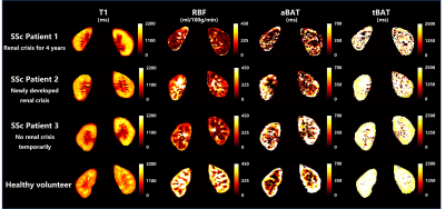 |
Renal impairment characterization of patients with systemic
sclerosis by multi-parametric magnetic resonance imaging
Xinyu Tong1,
Huilin He2,
Zihan Ning3,
Rui Shen3,
Zuoxiang He1,
Xihai Zhao3,
and Dong Xu2
1School of Clinical Medicine, Tsinghua University, Beijing, China, 2Department of Rheumatology and Clinical Immunology, Chinese Academy of Medical Sciences & Peking Union Medical College, Beijing, China, 3Center for Biomedical Imaging Research, Tsinghua University, Beijing, China Keywords: Kidney, Kidney, systemic sclerosis Renal microstructure and functional impairment in systemic sclerosis were characterized using multi-parametric quantitative MR imaging including SAMURAI, DWI and BOLD sequences. The mean values of T1, RBF, aBAT, tBAT, ADC, T2* in renal cortex and volume of renal parenchyma were quantified. Compared to healthy volunteers, patients with systemic sclerosis had significantly lower mean RBF values in both sides of renal cortex. This study revealed that decline of RBF in systemic sclerosis may indicate the pathology of microvascular impairment of renal tissues. |
| 15:21 |
1295.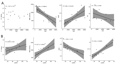 |
Functional MRI to monitor disease progression in patients with
rare kidney disease
Anna Caroli1,
Giulia Villa1,
Erica Daina2,
Paolo Brambilla3,
Sara Gamba2,
Valentina Fanny Leone4,
Camillo Carrara4,
Paola Rizzo5,
Marina Noris2,
Giuseppe Remuzzi6,
and Andrea Remuzzi7
1Bioengineering Department, Istituto di Ricerche Farmacologiche Mario Negri IRCCS, Ranica (BG), Italy, 2Department of rare diseases, Istituto di Ricerche Farmacologiche Mario Negri IRCCS, Ranica (BG), Italy, 3Unit of Radiology, ASST Papa Giovanni XXIII, Bergamo, Italy, 4Unit of Nephrology and Dialysis, ASST Papa Giovanni XXIII, Bergamo, Italy, 5Department of Molecular Medicine, Istituto di Ricerche Farmacologiche Mario Negri IRCCS, Bergamo, Italy, 6Istituto di Ricerche Farmacologiche Mario Negri IRCCS, Bergamo, Italy, 7University of Bergamo, Bergamo, Italy Keywords: Kidney, Quantitative Imaging This study investigates the correlation between MRI and histologic and clinical findings in 7 patients with C3 glomerulopathy and immune complex–associated membranoproliferative glomerulonephritis, rare diseases denoted by poor prognosis and no specific therapies. Patients underwent repeated kidney MRI, biopsy, and laboratory testing. Kidney diffusivity and perfusion were assessed by diffusion-weighted and phase-contrast MRI. Laboratory and MRI parameters changed very differently from case to case over 1 year. Perfusion biomarkers significantly correlated with histological and clinical findings. Both perfusion and diffusion biomarkers correlated with the clinical evolution of the disease. Current findings highlight MRI potential to monitor kidney disease progression. |
| 15:29 |
1296.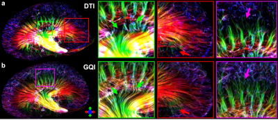 |
Improving tractography using high angular resolution diffusion
imaging in rodent kidney
Surendra Maharjan1,
Abigail Wallace1,
Megan Renate Jewett1,
Neal X Chen2,
and Nian Wang1,3
1Department of Radiology and Imaging Sciences, Indiana University School of Medicine, Indianapolis, IN, United States, 2Division of Nephrology, Department of Medicine, Indiana University School of Medicine, Indianapolis, IN, United States, 3Stark Neurosciences Research Institute, Indianapolis, IN, United States Keywords: Kidney, Diffusion Tensor Imaging, Tractography Diffusion tensor imaging (DTI) has been widely used to study renal microstructure. However, DTI-based tractography failed to track tubules throughout the kidney. Here, we aim to determine whether high angular resolution diffusion imaging (HARDI) could improve the tractography in the complicated tubular architectures of rodent kidney. |
| 15:37 |
1297.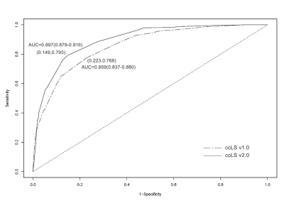 |
Diagnostic Value of Clear Cell Likelihood Score v1.0 and v2.0
for Clear Cell Renal Cell Carcinoma: A Comparative Study
Yu-wei Hao1,
Hui-yi Ye1,
and Hai-yi Wang1
1Department of Radiology, the first Medical Centre of Chinese PLA General Hospital, Beijing, China Keywords: Kidney, Tumor, Small renal masses; Clear cell likelihood score; Clear cell renal cell carcinoma ccLS provides a new tool for the diagnosis and differential diagnosis of solid renal tumors and can be used to assist radiologists in their daily diagnosis. In this study, Six radiologists were trained in the ccLS algorithm and scored independently using ccLS v1.0 and ccLS v2.0, respectively. The results show that although the interobserver agreement between ccLS v1.0 and ccLS v2.0 is comparable, the diagnostic performance of ccLS v2.0 in ccRCC is better than that of ccLS v1.0 and ccLS v2.0 reduces the percentage of ccRCC in 1-3 scores. This finding is helpful to improve the clinical universality of ccLS. |
 Back to Meeting Home
 Back to the Program-at-a-Glance
The International Society for Magnetic Resonance in Medicine is accredited by the Accreditation Council for Continuing Medical Education to provide continuing medical education for physicians.
 Back to Meeting Home
 Back to the Program-at-a-Glance
 View Presentation Video