Oral Session
Resonating with Hepatobiliary & Pancreatic Tumors: What's New?
ISMRM & ISMRT Annual Meeting & Exhibition • 03-08 June 2023 • Toronto, ON, Canada

Oral Session
Resonating with Hepatobiliary & Pancreatic Tumors: What's New?
| 08:15 |
0789.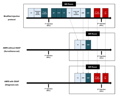 |
Abbreviated MRI with Second Shot Arterial Phase for HCC
Evaluation: Modified Version of LI-RADS and Recall Reduction
Strategy
Jeong Woo Kim1 and
Chang Hee Lee1 1Radiology, Korea University Guro Hospital, Seoul, Korea, Republic of Keywords: Liver, Liver A modified version of LI-RADS was devised for abbreviated MRI (AMRI) with second shot arterial phase (SSAP) by referring to CEUS LI-RADS. The modified LI-RADS scores using AMRI with SSAP showed a high concordance rate with the conventional LI-RADS score using full-protocol MRI. The recall rate significantly decreased when the HCC surveillance and diagnosis strategy was changed from strategy 1 (AMRI without SSAP; surveillance then recall test) to strategy 2 (AMRI with SSAP; simultaneous surveillance and diagnosis). |
08:23 |
0790.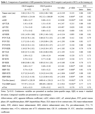 |
Preoperative Prediction of CK19 Expression and Early Recurrence
of Hepatocellular Carcinoma using T1 mapping on Gadoxetic
acid-enhanced MRI
Yue Zhao1,
Xiuhong Guan1,
Yongzhou Xu2,
Xinqing Jiang1,
and Ruimeng Yang1
1Department of Radiology, Guangzhou First People’s Hospital, Guangzhou, China, 2Philips Healthcare, Guangzhou, China Keywords: Liver, Liver, Hepatocellular carcinoma; T1 mapping. Cytokeratin 19 (CK19) is well acknowledged as a progenitor cell marker and tumor stem cell marker that plays an important role in promoting the malignant property of HCC. If the preoperative CK19 expression status in HCC can be accurately predicted noninvasively, it may provide important information for clinical decision-making. T1 mapping is useful for preoperative prediction of CK19 expression and early recurrence of HCC. The clinical-quantitative model combining alpha-fetoprotein and quantitative features showed good performance and robustness in predicting CK19 expression. T1 relaxation time on hepatobiliary phase was an independent predictor of CK19 expression and recurrence-free survival. |
| 08:31 |
0791.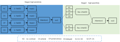 |
TPA:Two-stage Progressive Attention diagnosis framework for
hepatocellular carcinoma segmentation on Dynamic Contrast
Enhanced MRI
Lei Lei Gao1 and
Yuan-Cheng Wang1
1Zhongda Hospital Southeast University, Nanjing, China Keywords: Liver, Cancer, Deep learning We proposed a Two-stage Progressive Attention (TPA) framework by simulating the radiologists’ decision process for hepatocellular carcinoma segmentation. The study included 400 HCC patients as an internal set and 109 patients as an external test set. To obtain sensitive and specific results, our model is divided into two stages, respectively introducing attention mechanism and residual network. |
| 08:39 |
0792.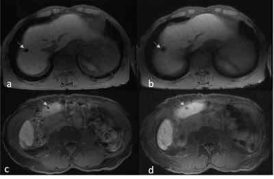 |
Application of 3D Cones Trajectory Acquisition in Hepatobiliary
MR Imaging for Detection of Hepatic Lesions
Negaur Iranpour1,
Vipul Sheth1,
Ali Syed1,
and Ryan L Brunsing1 1Radiology Body MRI, Stanford Medicine, Palo Alto, CA, United States Keywords: Liver, Liver, Free-breathing MRI, hepatobiliary imaging, 3D cones with golden-angle ordering Conventional cartesian T1 weighted 3D spoiled gradient recalled echo imaging with respiratory navigation (3D-SPGR-NAV) can help mitigate motion artifacts in post contrast liver imaging but suffer from respiratory rate dependence. 3D cones trajectories with golden angle ordering (T1g) allow continuous sampling during free-breathing liver imaging. We compared the diagnostic performance of an accelerated T1g (T1gER) versus 3D-SPGR-NAV in liver lesion detection. Two readers reviewed imaging from 35 patients containing 127 hepatic lesions. T1gER was non-inferior to 3D-SPGR-NAV for <5 mm and >=10 mm lesions. 3D-SPGR-NAV was superior for lesions 5-9 mm, possibly due to susceptibility artifact on the T1gER images. |
| 08:47 |
0793.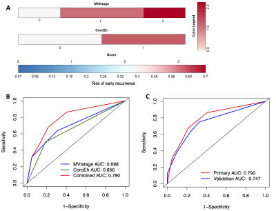 |
Corona Enhancement Combined with Microvascular Invasion for
Identification / Prognosis of Macrotrabecular-Massive
Hepatocellular Carcinoma
Lili Yang1,
Meng Wang2,
Yanyan Zhu1,
Jiahui Zhang3,
and Feng Chen1 1Radiology, The First Affiliated Hospital, Zhejiang University School of Medicine, Hangzhou, China, 2Pathology, The First Affiliated Hospital, Zhejiang University School of Medicine, Hangzhou, China, 3Radiology, Hangzhou Third Hospital, Hangzhou, China Keywords: Liver, Cancer The macrotrabecular-massive (MTM) subtype of hepatocellular carcinoma (HCC) is aggressive and associated with an unfavorable prognosis. This study aimed to characterize MTM-HCC features based on contrast‑enhanced MRI and to evaluate the prognosis of imaging characteristics combined with pathology for predicting early recurrence and overall survival after surgery. This retrospective study included 123 and 59 patients with HCC that underwent preoperative contrast‑enhanced MRI and surgery, respectively. The multivariate analysis and multiple Cox regression analysis identified that corona enhancement combined with MVI risk was significantly associated with poor outcomes after surgery. |
| 08:55 |
0794.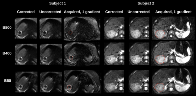 |
Motion Corrected DW-MRI With 3D Slice Level Motion Estimation In
Pediatric Liver Tumors
SERGE DIDENKO VASYLECHKO1,
LINA LU1,
CEMRE ARIYUREK1,
ONUR AFACAN1,
and SILA KURUGOL1
1RADIOLOGY, BOSTON CHILDREN'S HOSPITAL, HARVARD MEDICAL SCHOOL, BOSTON, MA, United States Keywords: Liver, Motion Correction Voxel misalignment due to unavoidable respiratory motion and bulk motion introduce large errors in DW-MRI quantitative parameter fitting. Apparent diffusion coefficient (ADC) is an effective tool for characterization of malignant tumors. In this study we evaluate the use of a motion correction method for DW-MRI imaging based on 3D slice level motion tracking using a rigid slice to volume registration and Kalman filtering. We show improvement in robustness of parameter estimation and reduction of blurring in b-value images for assessment of pediatric hepatoblastoma lesions. |
| 09:03 |
0795.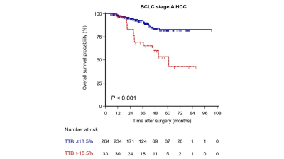 |
Subclassification of Barcelona Clinic Liver Cancer Stage A HCC
using fully automatic 3D segmentation-derived total tumor burden
at MRI
Hong Wei1,
Hanyu Jiang1,
Yidi Chen1,
Tianying Zheng1,
Ting Yang1,
Xiaolan Zhang2,
Chao Zheng2,
and Bin Song1
1West China Hospital, Sichuan University, Chengdu, China, 2Shukun (Beijing) Technology Co., Ltd, Beijing, China Keywords: Liver, Cancer, Carcinoma, hepatocellular; Barcelona Clinic Liver Cancer; Overall survival; Tumor burden In the present study of 297 patients, we evaluated the role of three-dimensional (3D) quantitative tumor burden analysis using fully automatic segmentation at magnetic resonance imaging in the subcategorization of the Barcelona Clinic Liver Cancer (BCLC) stage A hepatocellular carcinoma (HCC) after curative resection. Our results demonstrated that 3D quantitative total tumor burden (TTB) and serum a-fetoprotein were independent predictors of overall survival and could be used to subcategorize the BCLC stage A HCC. Additionally, the higher TTB (>18.5%) was correlated to more aggressive tumor behaviors (i.e., microvascular invasion and poor tumor differentiation). |
| 09:11 |
0796.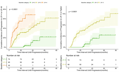 |
Long-term evolution of liver imaging reporting and Data system
Version 2018 category 2, 3, and 4 observations on MRI in
HBV-related cirrhosis
Fei Xing1,
Yi shi Wang2,
and Xian ce Zhao3
1the Third Affiliated Nantong Hospital of Nantong University, Nantong, China, 2Philips Healthcare, Beijing, China, 3Philips Healthcare, Shanghai, China Keywords: Liver, Cancer, HCC This study assessed the imaging outcomes of Liver Imaging Reporting and Data System (LI-RADS) v2018 category 2, 3, and 4 observations in prospective hepatocellular carcinoma (HCC) surveillance cohort. LI-RADS observations demonstrate increasing risk of progression to HCC with increasing category. About two-fifths of LR-4 progressed to a malignant category. LR-3 observations with APHE or threshold/subthreshold growth upgraded to LR-5 were significantly higher. Most LR-2 observations that remain stable in category for at least two years. |
| 09:19 |
0797.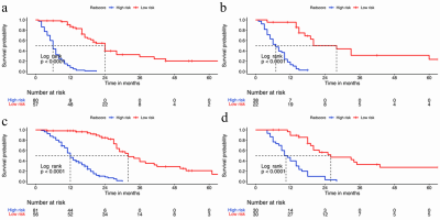 |
Incorporating diffusion weighted MRI into a radiomics model
improves diagnostic performance in survival prediction in PDAC
Piaoe Zeng1,
Jingjing Cui2,
and Huihui Yuan3 1radiology, peking university third hospital, Beijing, China, 2United Imaging Intelligence (Beijing), Beijing, China, 3Peking university third hospital, Beijing, China Keywords: Pancreas, Cancer, magnetic resonance imaging; radiomics; Radiomics features were extracted from multiparametric MRI including conventional MRI (T2WI, T1WI, arterial phase, portal venous phase images) and apparent diffusion coefficient (ADC). The radiomics score was built based on the least absolute shrinkage and selection operator regression model. Three models, including clinicopathological and radiographic characteristics (CPR) model, multiparametric MRI radiomics model and conventional MRI radiomics model, were built to predict recurrence-free survival (RFS) and overall survival (OS) in patients with resectable pancreatic ductal adenocarcinoma (PDAC). Multiparametric MRI radiomics model showed improved diagnostic performance in survival prediction than conventional MRI radiomics model and CPR model. |
| 09:27 |
0798.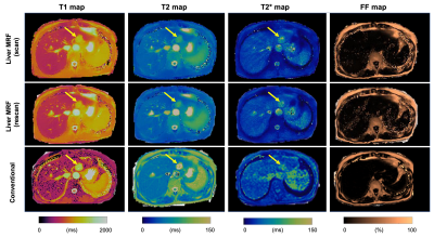 |
Magnetic resonance fingerprinting for objective and
contrast-free focal liver lesion characterization: A preliminary
study
Katsuhiro Sano1,
Shohei Fujita1,2,
Gastao Cruz3,4,
Carlos Velasco3,
Hideo Kawasaki1,
Yuki Fukumura5,
Akiyoshi Suzuki1,
Yuichi Morita1,
Koji Kamagata1,
Issei Fukunaga1,
Masami Yoneyama6,
Ryohei Kuwatsuru1,
Akio Saiura7,
Kenichi Ikejima8,
Rene Botnar3,
Claudia Prieto3,
and Shigeki Aoki1
1Department of Radiology, Juntendo University, Tokyo, Japan, 2Department of Radiology, The University of Tokyo, Tokyo, Japan, 3Department of Biomedical Engineering, School of Biomedical Engineering and Imaging Sciences, King’s College London, London, United Kingdom, 4Department of Radiology, University of Michigan, Ann Arbor, MI, United States, 5Department of Human Pathology, Juntendo University, Tokyo, Japan, 6MR Clinical Science, Philips Japan, Tokyo, Japan, 7Department of Hepatobiliary-Pancreatic Surgery, Juntendo University, Tokyo, Japan, 8Department of Gastroenterology, Juntendo University, Tokyo, Japan Keywords: Liver, MR Fingerprinting Objective and contrast-free methods for differentiating benign focal liver lesions from malignant lesions (e.g., hepatocellular carcinomas and metastases) are desired. Herein, we evaluated the diagnostic capability of liver magnetic resonance fingerprinting (MRF) in patients with focal liver lesions. Liver MRF provided repeatable T1, T2, T2*, and fat-fraction values for various focal liver lesions. The liver MRF T1 and T2 relaxation times showed high agreement with separate conventional quantitative mapping measurements. Measurements of liver MRF and a combination of MRF relaxation times provided good differentiation of focal liver lesions (AUC of 0.87 for differentiating common benign lesions from common malignant lesions). |
| 09:35 |
0799.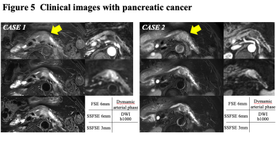 |
Utility of Thin-Slice Fat-Suppressed Single-Shot T2-Weighted MRI
with Deep Learning Image Reconstruction for Pancreatic Cancer
Ryuji Shimada1,2,
Keitaro Sofue2,
Yuichiro Somiya1,
Shintaro Horii1,
Yushi Tujita2,
Yoshiko Ueno2,
Tetsuya Wakayama3,
Akiko Kusaka1,
and Takamichi Murakami1,2
1Center of Radiology and Radiation Oncology, Kobe University Hospital, Kobe, Japan, 2Department of Radiology, Kobe University Graduate School of Medicine, Kobe, Japan, 3GE Healthcare, Hino, Japan Keywords: Pancreas, Cancer We investigated the utility of breath-hold (BH) fat-suppressed single-shot T2-weighted MRI with deep learning image reconstruction (DLIR) in 42 patients with pancreatic cancer. Three fat-suppressed T2-weighted sequences of 1) single-shot fast-spin echo (SSFSE) of 6mm thickness with 1BH, 2) SSFSE of 3mm thickness with 2BH and 3) FSE of 6mm thickness with 3BH were compared. SSFSE sequences improved signal-to-noise ratio on anatomical organs, pancreas-to-lesion contrast, and image quality in terms of motion artifacts, image sharpness, and anatomical clarity in comparison with conventional FSE. The T2-weighted SSFSE with DLIR can be a useful sequence for the evaluation of pancreatic cancer. |
| 09:43 |
0800.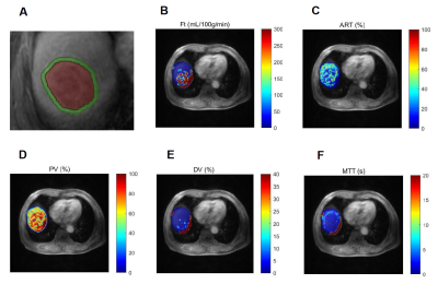 |
Prediction of microvascular invasion in solitary hepatocellular
carcinoma using bi-regional quantitative dynamic
contrast-enhanced MRI
Yongjian Zhu1,
Bing Feng1,
Wei Cai1,
Shuang Wang1,
Lizhi Xie2,
Xiaohong Ma1,
and Xinming Zhao1
1Department of Diagnostic Radiology, National Cancer Center/National Clinical Research Center for Cancer/Cancer Hospital, Chinese Academy of Medical Sciences and Peking Union Medical College, Beijing, China, 2GE healthcare, China, Beijing, China Keywords: Liver, Tumor, Pathology, Microvascular invasion The peri-tumoral region (PTR) of liver is the main-site of microvascular invasion (MVI) taken place, and contains perfusion information which could reflected the hemodynamic change during MVI. This study investigated the value of quantitative dynamic contrast-enhanced MRI (DCE-MRI) to evaluate the MVI status of hepatocellular carcinoma (HCC) in both intra-tumoral region (ITR) and PTR. The result showed quantitative DCE-MRI perfusion parameters could predict the MVI status in both ITR and PTR. Combining parameters from ITR and PTR could improve the prediction performance. Our study suggest quantitative DCE-MRI perfusion parameters could be employed as an efficient approach to predicting MVI status. |
| 09:51 |
0801.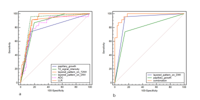 |
Differentiating Benign from Malignant Gallbladder Wall
Thickening in Non-Contrast MRI Imaging: Preliminary Study of a
Combined Indicator
Wen-Wen He1,
Hai-Ge Li1,
Jian-Guo Zhu1,
Dmytro Pylypenko2,
Fei Liu1,
Mei Wang1,
Yue-Fei Wu3,
and Jun Tian3 1Radiology, The Second Affiliated Hospital of Nanjing Medical University, Nanjing, China, 2GE Healthcare, China, Beijing, Beijing, China, 3The Second Affiliated Hospital of Nanjing Medical University, Nanjing, China Keywords: Cancer, Biliary This study evaluated the diagnostic accuracy of image features for differentiating benign from malignant gallbladder wall thickening disease with non-contrast MRI and constructed the optimal diagnostic indicator. 23 patients with wall thickening type gallbladder carcinoma and 61 patients with benign wall thickening disease were included. Six image indicators (the layered pattern on T2WI and DWI images, T2WI signal intensity, papillary growth, ADC value, and ratio of the ADC value of the lesion to that of liver parenchyma) were shown to have high diagnostic accuracy. The layered pattern on DWI combined with papillary growth was demonstrated as the optimal indicator. |
| 09:59 |
0803.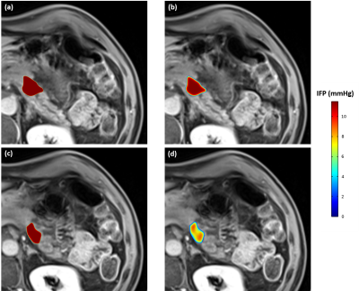 |
Patient-specific computational model of interstitial fluid flow
in pancreatic cancer based on DCE- and DW-MRI
Hooman Salavati1,2,3,
Wim Ceelen1,3,
Charlotte Debbaut2,3,
Clarisse Lecluyse4,
and Pim Pullens4,5,6
1Department of Human Structure and Repair, Ghent University, Ghent, Belgium, 2IBiTech – Biommeda, Ghent University, Ghent, Belgium, 3Cancer Research Institute Ghent (CRIG), Ghent, Belgium, 4Department of Radiology, Ghent University Hospital (UZGent), Ghent, Belgium, 5Ghent Institute of Functional and Metabolic Imaging (GIFMI), Ghent, Belgium, 6IBiTech – Medisip, Ghent University, Ghent, Belgium Keywords: In Silico, Simulations, Biomechanics
The abnormal interstitial fluid flow (IFF) in solid tumors
characterized by the elevated interstitial fluid pressure
(IFP) is one of the major barriers for treating solid
tumors. Here, we implemented DCE-MRI and IVIM DW-MRI in a
clinical study to upgrade the existing basic mathematical
framework of IFF to a patient-specific model. The results
were compared with the basic model, which showed a
noticeable difference regarding the prediction of the
heterogeneity of IFF. |
| 10:07 | 0802. | WITHDRAWN |
 Back to Meeting Home
 Back to the Program-at-a-Glance
The International Society for Magnetic Resonance in Medicine is accredited by the Accreditation Council for Continuing Medical Education to provide continuing medical education for physicians.
 Back to Meeting Home
 Back to the Program-at-a-Glance
 View Presentation Video