Oral Session
Everything Flows: Advances in Flow Imaging
ISMRM & ISMRT Annual Meeting & Exhibition • 03-08 June 2023 • Toronto, ON, Canada

Oral Session
Everything Flows: Advances in Flow Imaging
| 13:30 |
0924.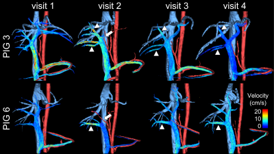 |
4D flow MRI to evaluate flow and future liver remnant growth
after portal vein embolization
Thekla Helene Oechtering1,2,
Tilman Schubert1,3,
Qianqian Zhao4,
Roxana A Alexandridis4,
Nikolaos Panagiotopoulos1,2,
Oliver Wieben1,5,
Kevin M Johnson1,6,
Alejandro Roldán-Alzate7,8,9,
and Scott B Reeder1,5,8,9,10
1Department of Radiology, University of Wisconsin-Madison, Madison, USA, WI, United States, 2Department of Radiology, Universität zu Lübeck, Lübeck, Germany, 3Department of Neuroradiology, Zurich University Hospital, Zurich, Switzerland, 4Department of Biostatistics and Medical Informatics, University of Wisconsin-Madison, Madison, WI, United States, 5Department of Medical Physics, University of Wisconsin-Madison, Madison, WI, United States, 6Department of Electrical and Computer Engineering, University of Wisconsin-Madison, Madison, WI, United States, 7Department of Radiology, University of Wisconsin-Madison, Madison, WI, United States, 8Department of Mechanical Engineering, University of Wisconsin-Madison, Madison, WI, United States, 9Department of Biomedical Engineering, University of Wisconsin-Madison, Madison, WI, United States, 10Department of Emergency Medicine, University of Wisconsin-Madison, Madison, WI, United States Keywords: Liver, Velocity & Flow Early prediction of remnant liver growth after portal vein embolization (PVE) would enable earlier surgery in patients with liver malignancies and thus decrease the risk of tumor progression. Portal blood flow after PVE holds the potential to be an important predictor for hypertrophy of the non-embolized segments. We demonstrated the feasibility of 4D flow MRI quantification of portal blood flow before and after PVE in a porcine model. Flow changes immediately after PVE were predictive of the change in liver volume 2 weeks post PVE in both the embolized and non-embolized liver lobes. |
| 13:38 |
0925.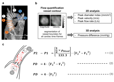 |
Estimation of Aortic Pressure Difference in Fontan Patients by
4D Flow MRI
Hsueh-Ping Hung1,
Ming-Ting Wu2,
Ken-Pen Weng3,4,
and Hsu-Hsia Peng1
1Department of Biomedical Engineering and Environmental Sciences, National Tsing Hua University, Hsinchu, Taiwan, 2Department of Radiology, Kaohsiung Veterans General Hospital, Kaohsiung, Taiwan, 3Department of Pediatrics, Kaohsiung Veterans General Hospital, Kaohsiung, Taiwan, 4Department of Pediatrics, National Yang Ming Chiao Tung University, Taipei, Taiwan Keywords: Heart, Cardiovascular We aimed to evaluate the aortic blood pressure characteristics and cardiac function in patients after the Fontan operation. The Fontan group exhibited significantly increased peak diameter index and decreased peak velocity and peak flow rate in some of planes in the aorta. The Fontan group also presented decreased pressure difference (PD) in aortic arch and descending aorta. The aortic PD might be more sensitive than cardiac index in detection of cardiovascular alteration in Fontan patients. The aortic PD measured by 4D flow MRI can be a noninvasive alternative to characterize aortic remodeling. |
13:46 |
0926.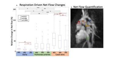 |
Respiration-resolved 5D flow MRI: Impact of the number of
respiratory states of blood flow quantification in congenital
heart disease patients
Elizabeth Weiss1,
Justin Baraboo1,
Liliana Ma1,
Mariana B. L. Falcão2,
Christopher W. Roy2,
Joshua D. Robinson3,
Matthias Stuber2,
Cynthia K Rigsby4,
and Michael Markl1
1Radiology, Northwestern University, Chicago, IL, United States, 2University of Lausanne (CHUV), Lausanne, Switzerland, 3Cardiology, Ann & Robert Lurie Children's Hospital, Chicago, IL, United States, 4Medical Imaging, Ann & Robert Lurie Children's Hospital, Chicago, IL, United States Keywords: Flow, Velocity & Flow, congenital heart disease We adapted our novel respiratory gating method to evaluate the impact of respiratory state (RS) resolution on respiratory driven flow measured by 5D flow MRI. We found that the impact of respiratory state resolution was both anatomy and vessel dependent. Caval veins and measurements in single ventricle disease patients were most impacted by the reduction of respiratory states. Shunt patients and pulmonary artery measurements were more robust to reduced RS and may be able to take advantage of the decreased acceleration associated with fewer RS. 5D flow MRI is well suited for this variable need as respiratory gating is retrospective. |
| 13:54 |
0927.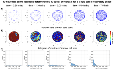 |
Autonomous 5D-flow with Radial k-Space Sampling
Ana E Rodríguez-Soto1,
Eleanor L Schuchardt2,
Sanjeet Hedge2,
Walter R Witschey3,
and Francisco Contijoch1,4 1Radiology, University of California San Diego, La Jolla, CA, United States, 2Rady Children's Hospital San Diego, San Diego, CA, United States, 3University of Pennsylvania, Philadelphia, PA, United States, 4Bioengineering, University of California San Diego, La Jolla, CA, United States Keywords: Flow, Simulations Autonomous acquisition of radial k-space data improves sample uniformity in 2D cardiac MRI. We aim to extend this approach to 5D-flow imaging (4D flow with respiratory gating). We have simulated data acquisition using an autonomous approach (ARKS) and compared it to golden-angle (GA) based spiral phyllotaxis acquisition. Simulations were based on physiologic data recorded from pediatric patients undergoing conventional 4D flow imaging. We found that a 4.5 min ARKS scan achieves a higher degree of sampling uniformity than an 8 min GA scan. Future work will focus on implementing 5D-flow ARKS in vivo and evaluating the impact on flow accuracy. |
| 14:02 |
0928.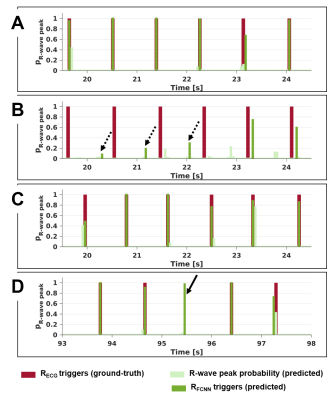 |
A deep learning framework for cardiac self-gating in
free-running radial 4D flow MRI
Mariana B.L. Falcão1,
Giulia M.C. Rossi1,
Jonas Richiardi1,
Xavier Sieber1,
Pierre Monney2,
Tobias Rutz2,
Milan Prša3,
Estelle Tenisch1,
Anna Giulia Pavon4,
Panagiotis Antiochos2,
Matthias Stuber1,5,
and Christopher W. Roy1
1Department of Radiology, Lausanne University Hospital (CHUV) and University of Lausanne (UNIL), Lausanne, Switzerland, 2Service of Cardiology, Centre de Resonance Magnétique Cardiaque (CRMC), Lausanne University Hospital (CHUV) and University of Lausanne (UNIL), Lausanne, Switzerland, 3Woman- Mother- Child Department, Lausanne University Hospital (CHUV) and University of Lausanne (UNIL), Lausanne, Switzerland, 4Division of Cardiology, Cardiocentro Ticino Institute, Ente Ospedaliero Cantonale, Lugano, Switzerland, 5Center for Biomedical Imaging (CIBM), Lausanne, Switzerland Keywords: Flow, Cardiovascular, Cardiac signal extraction Self-gating (SG) techniques improve the ease-of-use of cardiac MR by deriving cardiac signals from the data itself, obviating the need for ECG lead placement. Nonetheless, unpredictable shifts between the features of SG signals and the conventionally used R-wave peaks from ECG might hamper a direct link of reconstructed image frames with physiology. In this work, we developed a fully convolutional neural network to predict R-wave peak timepoints from SG imaging readouts in free-running radial 4D flow data, and provided a proof-of-concept of the usability of such learned R-wave peak timepoints for reconstructing cardiac-resolved 4D flow images. |
| 14:10 |
0929. |
Impact of regularization factor on quantification of flow and
turbulence in highly undersampled 4D flow MRI
Pietro Dirix1,
Stefano Buoso1,
Valery Vishnevskiy1,
and Sebastian Kozerke1
1University and ETH Zurich, Zürich, Switzerland Keywords: Flow, Image Reconstruction We investigate the impact of regularization on the quantification of flow and turbulence in highly undersampled data. To overcome the uncertainties due to the lack of ground truth, patient-specific aortic geometry and inflow conditions were extracted from in-vivo 2D cine and 2D phase-contrast MRI and used to generate synthetic personalized ground truth flow fields. Simulation results were embedded into the corresponding patient-specific 4D flow MRI effectively resulting in personalized synthetic datasets with known aortic ground truth and realistic background. The reconstruction of multiple undersampled datasets showed two distinct optimal regularization ranges for the quantification of flow velocities and turbulence. |
| 14:18 |
0930.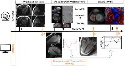 |
2.5D Flow MRI: 2D phase-contrast of the tricuspid valvular flow
with automated valve-tracking
Jerome Lamy1,
Jie Xiang1,
Felicia Seemann2,
Ricardo A Gonzales3,
Steffen Huber1,
Jeremy Steele4,
Einar Heiberg5,
and Dana C Peters1 1Radiology and Biomedical Imaging, Yale University, New Haven, CT, United States, 2National Institutes of Health, Bethesda, MD, United States, 3Oxford University, Oxford, United Kingdom, 4Internal Medicine, Yale University, New Haven, CT, United States, 5Lund University, Lund, Sweden Keywords: Flow, Data Acquisition Tricuspid regurgitant velocity is a crucial biomarker in identifying pressure overload in the right heart, associated with diastolic dysfunction and pulmonary hypertension. 2D phase-contrast cannot quantify this flow, and echocardiography is used clinically. We developed a phase-contrast method which utilizes deep-learning algorithms to track the valvular slice in a cardiac phase-dependent manner, which we call 2.5D flow. We studied its performance in nine healthy subjects and patients with tricuspid regurgitation. RV stroke volumes correlated better to forward flow volumes by 2.5D flow vs. static 2D phase-contrast (ICC=0.88 vs. 0.62). 2.5D flow characterized regurgitation in a patient. |
| 14:26 |
0931.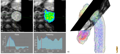 |
Deep learning based segmentation of aortic cross sections (2D+t)
in multi-vendor 4D PCMRI
Chiara Manini1,
Markus Hüllebrand1,2,
Marius Pullig3,
Titus Kühne1,4,
Sarah Nordmeyer5,
Lina Jarmatz5,
Andreas Harloff6,
Jeanette Schulz-Menger4,5,
and Anja Hennemuth1,2,4,7
1Institute of Computer-assisted Cardiovascular Medicine, Charité – Universitätsmedizin Berlin, Berlin, Germany, 2Fraunhofer MEVIS, Bremen, Germany, 3IBM Germany, Berlin, Germany, 4DZHK (German Center for Cardiovascular Research), Partner site Berlin, Germany, 5Charité – Universitätsmedizin Berlin, Berlin, Germany, 6University Hospital Freiburg, Freiburg, Germany, 7University Medical Center Hamburg-Eppendorf, Hamburg, Germany Keywords: Flow, Velocity & Flow, Aorta segmentation Standardized 4D PCMRI postprocessing protocols could enable comparable bloodflow quantification. We propose an automatic segmentation of aortic cross section over time with a residual trained data from different imaging sequences, scanner types, pathologies and position of cross section planes. Dice score, Hausdorff metric as well as flow and velocity curves for the segmented areas show good performance both in the validation and test sets. |
14:34 |
0932.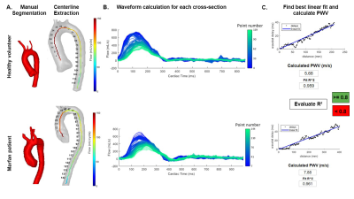 |
Repeatability of whole aorta 4D flow pulse wave velocity in
healthy volunteers and comparison with Marfan Syndrome patients
Daan Bosshardt1,2,
Mitzi M. van Andel2,
Renske Merton1,
Vivian de Waard3,
Roland R.J. van Kimmenade4,
Arthur J. Scholte5,
Moniek G.P.J. Cox6,
Aeilko H. Zwinderman7,
Aart J. Nederveen1,
Maarten Groenink2,
Pim van Ooij1,
and Eric M. Schrauben1
1Radiology and Nuclear Medicine, Amsterdam UMC, Amsterdam, Netherlands, 2Cardiology, Amsterdam UMC, Amsterdam, Netherlands, 3Medical Biochemistry, Amsterdam UMC, Amsterdam, Netherlands, 4Cardiology, Radboud University Medical Center, Nijmegen, Netherlands, 5Cardiology, Leiden University Medical Center, Leiden, Netherlands, 6Cardiology, University Medical Center Groningen, Groningen, Netherlands, 7Clinical Epidemiology, Amsterdam UMC, Amsterdam, Netherlands Keywords: Flow, Velocity & Flow, Pulse Wave Velocity Pulse wave velocity (PWV) is a well-established measure to evaluate vessel wall stiffness and increases in the aorta of patients with Marfan Syndrome (MFS). With sufficient temporal resolution, 4D-flow MRI can be used for PWV calculations. In this study, we assess the repeatability of aortic PWV calculations in healthy volunteers using an open-source software tool and apply it in a MFS cohort. Our results show high repeatability of global aortic PWV, elevated PWV in MFS patients, and an association between elevated PWV and biomarkers for advanced disease. |
| 14:42 |
0933. |
End-to-end Automation of Quantitative Processing for 4D Flow MRI
in the Aorta: Demonstration and Evaluation in 271 Subjects
Ethan M I Johnson1,
Haben Berhane1,
Elizabeth Weiss1,
Aparna Sodhi2,
Kelly Jarvis1,
Michael Scott1,
Joshua Robinson2,
Cynthia K Rigsby2,
and Michael Markl1 1Northwestern University, Chicago, IL, United States, 2Ann & Robert H. Lurie Children's Hospital, Chicago, IL, United States Keywords: Data Processing, Velocity & Flow, 4D Flow MRI, Quantitative Hemodynamics A generalized data-processing pipeline tool for performing completely automated hemodynamic assessment from raw 4D flow MR images is presented. The tool is evaluated for performance in a group of 271 subjects with mixed distribution of healthy, valve disease, and connective-tissue disorder status. A high success rate of 94% is achieved for fully-automated quantification of regional aortic peak velocities and global aortic pulse wave velocity. |
| 14:50 |
0934. |
Minimizing Background Phase in PC-MRI with the Gradient Impulse
Response Function (GIRF) and Gradient Optimized (GrOpt) Velocity
Encoding
Michael Loecher1,2 and
Daniel B Ennis1,2
1Radiological Sciences Laboratory, Stanford University, Stanford, CA, United States, 2Radiology, Veterans Administration Health Care System, Palo Alto, CA, United States Keywords: Flow, Velocity & Flow Background phase errors in PC-MRI flow measurements are caused by eddy currents and mechanical vibrations that produce unwanted magnetic fields, thereby reducing velocity measurement accuracy. In this work, we demonstrate that a GIRF measurement can be used to predict the PC-MRI background phase, then used to design gradient optimized (GrOpt) velocity encoding waveforms that minimize these errors. The method is tested in static phantoms and in vivo, where 4.8x to 6.4x reductions in background velocity are seen. Phantom results showed a reduction from 0.74±0.22 cm/s to 0.15±0.12 cm/s, and in vivo results showed reductions from 0.93±0.10 cm/s to 0.13±0.08 cm/s. |
| 14:58 |
0935. |
Phase-Contrast MRI with Hybrid One- and Two-sided Simultaneous
Three Directional Flow-Encoding and Velocity SPectrum SepAration
(HOTSPA+)
Wenjian Liu1,
Junpu Hu2,
Jiayu Zhu2,
Jian Xu3,
Zijian Zhou1,
Haikun Qi1,4,
and Peng Hu1,4
1School of Biomedical Engineering, ShanghaiTech University, Shanghai, China, 2United Imaging Healthcare, Shanghai, China, 3UIH America, Inc., Houston, TX, United States, 4Shanghai Clinical Research and Trial Center, ShanghaiTech University, Shanghai, China Keywords: Flow, Velocity & Flow Phase contrast MRI (PC MRI) has been widely used to quantify blood flow and velocity. Four-dimensional (4D) flow PC MRI needs to acquire the FC data and three-directional (3D) FE data interleaved within each cardiac k-space segment. In this work, we propose a more efficient flow encoding strategy for PC MRI using a temporal modulation technique and we showed the preliminary feasibility of quadrupling the temporal resolution or reducing the scan time by 70% compared with conventional 4D flow by redesigning and adjusting the temporal modulation strategy for under-sampled M1 space. |
| 15:06 |
0936.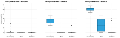 |
Phase-Unwrapping based on Graph-Cuts for Low-venc Phase-Contrast
Cine MRI
Johan Berglund1
1Dept. of Surgical Sciences, Uppsala University, Uppsala, Sweden Keywords: Flow, Velocity & Flow The use of low velocity encoding (venc) improves the velocity-to-noise ratio in phase-contrast MRI, but increases the risk of phase wrapping. A novel phase-unwrapping technique based on graph-cuts was evaluated in five cardiac patients with high-, medium-, and low-venc acquisitions. The proposed method reduced the stroke volume error compared to a LaPlacian based reference method. In retrospectively wrapped data, the proposed method demonstrated excellent unwrapping results in most cases, even for venc as low as 25 cm/s. The graph-cut based method appears promising for low-venc acquisitions combined with phase-unwrapping. |
| 15:14 |
0937.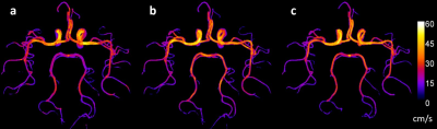 |
Intracranial Arterial Flow Velocimetry in Quantitative
Time-of-Flight MR Angiography using Deep Machine Learning
Ioannis Koktzoglou1,2 and
Rong Huang1
1Radiology, NorthShore University HealthSystem, Evanston, IL, United States, 2Pritzker School of Medicine, The University of Chicago, Chicago, IL, United States Keywords: Flow, Brain, MRA Quantitative time-of-flight (qTOF) magnetic resonance angiography (MRA) is a recently introduced technique that provides for simultaneous luminal and hemodynamic imaging of the intracranial arteries. We hypothesized that the application of a deep machine learning (DML) image analysis strategy to qTOF MRA data would improve agreement of intracranial arterial velocity measures with respect to phase contrast MRI. Compared to a more conventional image analysis procedure, we found that the application of DML image analysis to qTOF data improved agreement of component, total, and peak intracranial arterial flow velocity measures with respect to phase contrast MRI, and reduced calculation times by 35-fold. |
|
15:22 |
0938.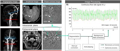 |
Effects of Free-Breathing on Cerebral Veins: study by Real-Time
Phase-Contrast MRI
Pan LIU1,2,
Heimiri Monnier1,
Kimi Piedad Owashi1,
Serge Metanbou3,
Cyrille Capel4,
and Olivier Balédent1,2
1CHIMERE UR 7516, Jules Verne University of Picardy, Amiens, France, 2Medical Image Processing Department, Amiens Picardy University Hospital, Amiens, France, 3Radiology Department, Amiens Picardy University Hospital, Amiens, France, 4Neurosurgery Department, Amiens Picardy University Hospital, Amiens, France Keywords: Head & Neck/ENT, Velocity & Flow, real time phase contrast, phase contrast, respiratory effects, cerebral veins Real-time phase-contrast sequences are increasingly used to study the effect of breathing on cerebral circulation, however, there is a lack of studies on the effect of free breathing (E-Fb) on cerebral veins. In this study, we quantified the intensity and phase shift of the E-Fb on cerebral veins in 19 healthy volunteers using a time-domain multiparameter analysis method. We analyzed the characteristics of the E-Fb on four parameters of four cerebral veins. We found that during expiration, the mean flow rate, amplitude, stroke volume, and cardiac period of internal jugulars veins and sinuses increased. |
 Back to Meeting Home
 Back to the Program-at-a-Glance
The International Society for Magnetic Resonance in Medicine is accredited by the Accreditation Council for Continuing Medical Education to provide continuing medical education for physicians.
 Back to Meeting Home
 Back to the Program-at-a-Glance
 View Presentation Video