Oral Session
Nothing Stands Still: Motion in Cardiac MRI
ISMRM & ISMRT Annual Meeting & Exhibition • 03-08 June 2023 • Toronto, ON, Canada

Oral Session
Nothing Stands Still: Motion in Cardiac MRI
13:30 |
0544.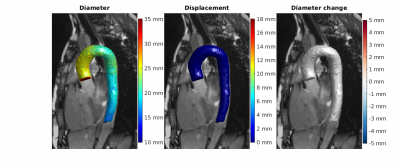 |
4D aortic motion maps from isotropic high-resolution 3D CINE
balanced steady state free precession at 3T and automated
segmentations
Renske Merton1,2,
Daan Bosshardt1,2,
Gustav J. Strijkers3,
Aart J. Nederveen1,
Eric M. Schrauben1,
and Pim van Ooij1,2
1Radiology and Nuclear Medicine, Amsterdam UMC location University of Amsterdam, Amsterdam, Netherlands, 2Atherosclerosis and Ischemic Syndromes, Amsterdam Cardiovascular Sciences, Amsterdam, Netherlands, 3Biomedical Engineering and Physics, Amsterdam UMC location University of Amsterdam, Amsterdam, Netherlands Keywords: Vessel Wall, Segmentation, 4D motion, aorta In this study we introduce 4D aortic displacement and diameter change estimations from isotropic high-resolution 3D CINE CMR as novel biomarkers for aortic stiffness. These could potentially be used as an alternative to pulse wave velocity stiffness measurements which can be unreliable for very stiff aortas as found in for example Marfan syndrome patients. Using deep learning for segmentation, visualization and quantification of aortic motion is demonstrated through 4D aortic motion maps. Time curves of displacement showed the typical cyclic behavior of cardiac motion, with a maximum displacement median of 10.3 (3.6) mm over 14 healthy volunteers. |
| 13:38 |
0545.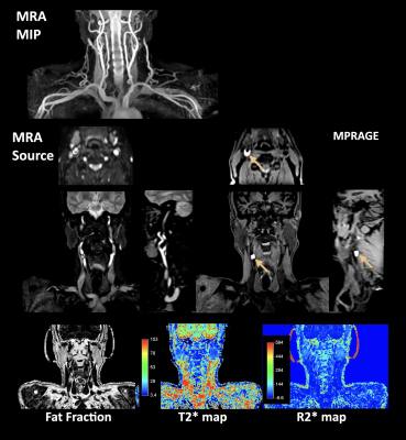 |
REACT-QUANT: simultaneous non-contrast-enhanced subclavian MRA
and vessel-wall imaging with multi-parametric quantitative
mapping
Masami Yoneyama1,
Yasuhiro Goto2,
Michinobu Nagao3,
Shuo Zhang4,
Kayoko Abe3,
Yutaka Hamatani2,
Kazuo Kodaira2,
Takumi Ogawa2,
Mana Kato2,
Isao Shiina2,
Christian Stehning4,
and Marc Van Cauteren5
1Philips Japan, Tokyo, Japan, 2Department of Radiological Services, Tokyo Women's Medical University, Tokyo, Japan, 3Department of Diagnostic Imaging and Nuclear Medicine, Tokyo Women’s Medical University, Tokyo, Japan, 4Philips Healthcare, Hamburg, Germany, 5Philips Healthcare, Best, Netherlands Keywords: Atherosclerosis, Atherosclerosis Clinical imaging procedure for vascular evaluation and tissue characterization often needs separate MR scans. Both scenarios require multiple imaging sequences with different image contrasts, which results in long exam times and image misalignment due to possible motion between the scans that needs to be corrected in post-processing. In this work, the recently proposed REACT (Relaxation-Enhanced Angiography without ContrasT) technique was further developed and combined with multi-echo data acquisitions and Dixon-based chemical-shift-based water-fat separation. Initial results in patients showed great promise in detection and assessment of vascular and vessel wall lesions in one single scan. |
| 13:46 |
0546.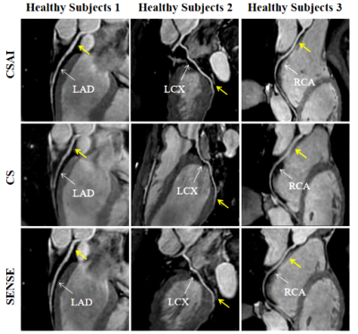 |
Feasibility of accelerated non-contrast-enhanced whole-heart
bSSFP coronary MRA by deep learning constrained compressed
sensing
Xi Wu1,2,
Jiayu Sun1,
and Xiaoyong Zhang3 1West China Hospital, Sichuan University, Chengdu, China, 2Affiliated Hospital of North Sichuan Medical College, Nanchong, China, 3Clinical Science, Philips Healthcare, Chengdu, China Keywords: Vessels, Cardiovascular, deep learning The balanced steady-state free precession (bSSFP) sequence is widely used for navigated whole-heart coronary magnetic resonance angiography (MRA) for the evaluation of coronary anatomy and abnormalities due to its inherently high blood signal intensity and blood-myocardial contrast. However, the main drawback of this approach is that the scan time is longer and prone to interference with motion artifacts. In this study, we investigated the utility of whole-heart coronary MRA using accelerated bSSFP with compressed sensing artificial intelligence (CSAI) technique at 3 Tesla. The results demonstrated the adopted CS-AI technique yielded high image quality within a clinically feasible acquisition time in healthy subjects and patients with suspected CAD. |
|
13:54 |
0547.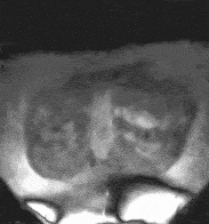 |
Whole-Heart Cardiovascular Magnetic Resonance Angiography (CMRA)
with iNAV-based Non-Rigid Motion-Corrected Reconstruction at
0.55T
Carlos Castillo-Passi1,2,3,
Michael G. Crabb1,
Camila Muñoz1,
Karl P. Kunze1,4,
Radhouene Neji 1,4,
Pablo Irarrazaval2,3,5,
Claudia Prieto1,3,5,
and Rene M. Botnar1,2,3,5
1School of Biomedical Engineering and Imaging Sciences, King's College London, London, United Kingdom, 2Institute for Biological and Medical Engineering, Pontificia Universidad Católica de Chile, Santiago, Chile, 3Millennium Institute for Intelligent Healthcare Engineering, Santiago, Chile, 4MR Research Collaborations, Siemens Healthcare Limited, Camberley, United Kingdom, 5Electrical Engineering Department, Pontificia Universidad Católica de Chile, Santiago, Chile Keywords: Heart, Low-Field MRI In this work, we demonstrate the feasibility of free-breathing whole-heart magnetic resonance angiography (CMRA) at 0.55T. We implemented an image navigator (iNAV)-based non-rigid motion-corrected reconstruction with 100% respiratory scan efficiency and patch-based low-rank (PROST) denoising. The research sequence was validated on three healthy subjects, showing that whole-heart CMRA provides good depiction of the coronary arteries at 0.55T in under 6 minutes scan time. |
| 14:02 |
0548.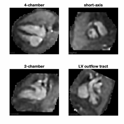 |
Fetal cardiac 3D cine MRI at low field - whole heart
slice-to-volume reconstruction from real-time spiral SSFP at
0.55T
Joshua F P van Amerom1,
Ye Tian2,
Datta S Goolaub1,
John Wood3,4,
Jon Detterich4,
Krishna S Nayak2,
and Christopher K Macgowan1,5
1Translational Medicine, Hospital for Sick Children, Toronto, ON, Canada, 2University of Southern California, Los Angeles, CA, United States, 3Department of Biomedical Engineering, University of Southern California, Los Angeles, CA, United States, 4Division of Cardiology, Children's Hospital Los Angeles, Los Angeles, CA, United States, 5Department ofMedical Biophysics, University of Toronto, Toronto, ON, Canada Keywords: Heart, Fetus, Cine, 4D, Whole Heart, Morphology Spiral SSFP of the fetal heart at low-field has the advantage of low artifact and high sampling efficiency, compared to conventional systems. This work demonstrates the feasibility of spiral SSFP on a high-performance 0.55 T scanner to produce high spatiotemporal resolution real-time 2D and motion-corrected slice-to-volume super-resolution reconstructed 3D cine images of the heart and great vessels of the human fetus in utero. Patient comfort is improved with the use of a low-field scanner, while motion-robust slice-to-volume reconstruction allows for free-breathing acquisitions that do not require precise scan plane prescriptions during acquisition. |
| 14:10 |
0549.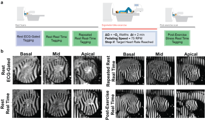 |
Highly-Accelerated Real-Time Myocardial Tagging for Exercise CMR
at 3T
Manuel A Morales1,
Siyeop Yoon1,
Ahmed Fahmy1,
Jennifer Rodriguez1,
Daniel B Herzka2,
Warren J Manning1,
and Reza Nezafat1
1BIDMC, Boston, MA, United States, 2Case Western Reserve University, Cleveland, OH, United States Keywords: Heart, Pulse Sequence Design Combined cardiac MRI and exercise (Ex-CMR) is a stress imaging test with promising applications in cardiovascular disease (CVD). Ex-CMR myocardial tagging could provide insights into myocardial deformation post-exercise. We sought to develop an SSFP-based highly-accelerate (12-fold) free-breathing ECG-free real time tagging sequence for quantification of beat-to-beat variation of myocardial deformation at rest and after exercise. The proposed real time sequence achieved a spatiotemporal resolution of 2 x 2 mm2 and 27.6 ms. Feasibility of real time imaging at 3T post-exercise was demonstrated in patients with heart failure with preserved (HFpEF) and reduced (HFrEF) ejection fraction. |
|
14:18 |
0550.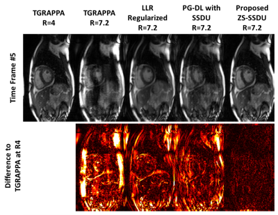 |
Highly Accelerated Cartesian Real-Time Cine CMR Using
Subject-Specific Zero-Shot Deep Learning Reconstruction
Omer Burak Demirel1,2,
Chi Zhang1,2,
Burhaneddin Yaman1,2,
Merve Gulle1,2,
Tim Leiner3,
Peter Kellman4,
and Mehmet Akçakaya1,2
1Electrical and Computer Engineering, University of Minnesota, Minneapolis, MN, United States, 2Center for Magnetic Resonance Research, University of Minnesota, Minneapolis, MN, United States, 3Department of Radiology, Mayo Clinical, Rochester, MN, United States, 4National Heart-Lung and Blood Institute, Bethesda, MD, United States Keywords: Heart, Heart Real-time cine CMR is an ECG-free free-breathing alternative for functionally assessing the heart. To achieve sufficient spatio-temporal resolutions, these require rapid imaging, e.g. compressed sensing (CS) with radial trajectories. However, at high accelerations, CS may suffer from residual aliasing and temporal blurring. Recently, deep learning (DL) reconstruction has gained immense interest for fast MRI. Yet, for free-breathing real-time cine, where subjects have different breathing and cardiac motion patterns, database learning of spatiotemporal correlations has been difficult. Here, we propose a physics-guided DL reconstruction trained in a subject-specific manner. Proposed method improves image quality compared to database-trained DL and conventional methods. |
| 14:26 |
0551.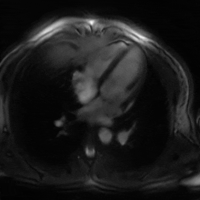 |
Interactive Cardiac Magnetic Resonance Imaging using jointly
optimized spiral acquisition and deep artifact suppression
network.
Olivier Jaubert1,
Javier Montalt-Tordera1,
Dan Knight2,
Simon Arridge3,
Jennifer Steeden1,
and Vivek Muthurangu1
1Institute of Cardiovascular Sciences, University College London, London, United Kingdom, 2Department of Cardiology, Royal Free London NHS Foundation Trust, London, United Kingdom, 3Department of Computer Science, University College London, London, United Kingdom Keywords: Heart, Machine Learning/Artificial Intelligence, Interventional, Interactive Interactive MR sequences have relatively low spatial and temporal resolution due to the limited acquisition and reconstruction time available. We propose to jointly optimize a variable density spiral acquisition and deep artifact suppression network (via a bandit-based approach) to maximize acquisition and reconstruction efficiency and provide interactive high spatio-temporal resolution images. The proposed approach was characterized in simulations and demonstrated prospectively in-vivo, offering promising performance with improved image quality and good handling of abrupt scan-plane changes. |
| 14:34 |
0552.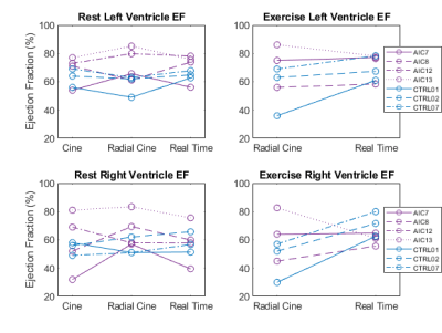 |
Real Time Cardiovascular MR During Exercise in Heart Failure
with Preserved Ejection Fraction (HFpEF)
Daniel P Seiter1,
Philip A Corrado1,
Farhan Raza2,
and Oliver Wieben1,3
1Medical Physics, University of Wisconsin-Madison, Madison, WI, United States, 2Department of Medicine, Cardiovascular Division, University of Wisconsin-Madison, Madison, WI, United States, 3Radiology, University of Wisconsin-Madison, Madison, WI, United States Keywords: Heart, Cardiovascular, Exercise, real time Right heart catheterization during exercise is the current gold standard for diagnosis of heart failure with preserved ejection fraction (HFpEF) but carries the risk of an invasive procedure. In this work, we present preliminary data using real time cardiac magnetic resonance imaging during exercise in a HFpEF cohort with comparison to healthy controls. |
| 14:42 |
0553.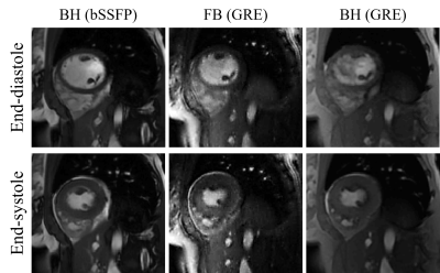 |
Deep kernel method for free-breathing and ungated cardiac MRI
reconstruction
Qing Zou1,
Sanja Dzelebdzic1,
and Tarique Hussain1
1University of Texas Southwestern Medical Center, Dallas, TX, United States Keywords: Heart, Machine Learning/Artificial Intelligence, Reconstruction We introduce a deep kernel model for the recovery of free-breathing and ungated cardiac MRI from highly undersampled measurements. The proposed scheme uses the cascade of two deep convolutional neural networks for the kernel representation of images. The parameters of the two CNNs in the proposed method are learned from the undersampled measurements directly in this work and hence the framework is unsupervised. The main benefits of the proposed scheme are (a) the elimination of the empirical choice of the feature map and kernel function in the kernel method, and (b) the unsupervised nature of the proposed framework. |
14:50 |
0554.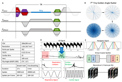 |
Self-Gated Cardiac Phase-Contrast Balanced SSFP at 0.55T
Charles McGrath1,
Oliver Bieri2,3,
Sebastian Kozerke1,
and Grzegorz Bauman2,3
1University and ETH Zurich, Zurich, Switzerland, 2Division of Radiological Physics, Department of Radiology, University Hospital Basel, Basel, Switzerland, 3Department of Biomedical Engineering, University of Basel, Basel, Switzerland Keywords: Flow, Low-Field MRI Free-running radial phase-contrast balanced steady-state free precession (PC-bSSFP) imaging including slice-select gradient inversion and respiratory and cardiac self-gating enables robust through-plane flow quantification in the aorta with excellent image signal-to-noise ratio (SNR) and contrast on a low-cost low-field 0.55T scanner. Relative to phase-contrast spoiled gradient echo (PC-GRE), PC-bSSFP offers SNR improvements by a factor of about two. Keywords: Flow, Low-Field, Balanced SSFP, Phase-Contrast, Self-gated |
| 14:58 |
0555.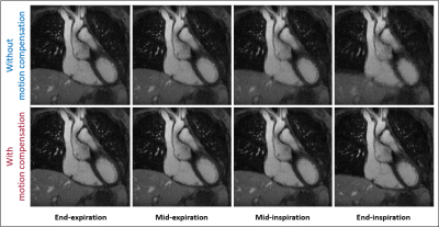 |
Intra-bin correction and inter-bin compensation of respiratory
motion in free-running 5D whole-heart MRI
Christopher W. Roy1,2,
Bastien Milani1,2,
Jérôme Yerly1,2,3,
Salim Si-Mohamed1,4,5,
Estelle Tenisch1,2,
Tobias Rutz2,6,
Milan Prsa2,7,
Ludovica Romanin1,2,8,
Aurélien Bustin9,10,
Davide Piccini1,8,
and Matthias Stuber1,2,3
1Diagnostic and Interventional Radiology, Centre Hospitalier Universitaire Vaudois (CHUV), Lausanne, Switzerland, 2University of Lausanne, Lausanne, Switzerland, 3Center for Biomedical Imaging (CIBM), Lausanne, Switzerland, 4University Lyon, Lyon, France, 5Radiology, Louis Pradel Hospital, Lyon, France, 6Service of Cardiology, Heart and Vessel Department, Centre Hospitalier Universitaire Vaudois (CHUV), Lausanne, Switzerland, 7Division of Pediatric Cardiology, Woman-Mother-Child Department, Centre Hospitalier Universitaire Vaudois (CHUV), Lausanne, Switzerland, 8Advanced Clinical Imaging Technology, Siemens Healthcare International AG, Lausanne, Switzerland, 9IHU LIRYC, Electrophysiology and Heart Modeling Institute, Université de Bordeaux, Bordeaux, France, 10Department of Cardiovascular Imaging, Hôpital Cardiologique du Haut-Lévêque, Bordeaux, France Keywords: Heart, Motion Correction In this work, we develop, validate, and apply a novel reconstruction framework for intra-bin correction and inter-bin compensation of respiratory motion in cardiac and respiratory motion-resolved free-running whole heart 5D MRI. We demonstrate respiratory resolved images with reduced artifact and without compressing the underlying physiological motion providing a means to increase the acquired resolution, accelerate the acquisition, or investigate respiratory driven changes in cardiac output and blood flow, potentially leading to new MRI driven biomarkers for cardiovascular disease |
| 15:06 |
0556.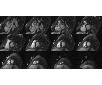 |
Free-running 5D whole-heart MRI with ferumoxytol enhancement to
evaluate cardiac function in congenital heart disease
Salim Aymeric Si-Mohamed1,2,
Ludovica Romanin3,
Mariana Falcao3,
Jerome Yerly2,
Estelle Tenisch2,
Tobias Rutz2,
Charles De Bourguignon4,
Jurg Schwitter5,
Matthias Stuber3,
Christopher Roy2,
and Milan Prsa6
1Radiology department, University of Lyon, Lyon, France, 2CIBM, CHUV, Lausanne, Switzerland, 3CIBM, CHUV, Lausanne, France, 4Research radiology department, Hospices Civiles de Lyon, Lyon, France, 5Cardiology, CHUV, Lausanne, Switzerland, 6Pediatric cardiology department, CHUV, Lausanne, France Keywords: Cardiomyopathy, Data Analysis, cardiac function Free-running 5D whole-heart imaging (5D CMR) has been proposed as a means of simplifying CMR exams by capturing the entire 3D cardiac anatomy without the need for ECG gating or breath-holds. We demonstrated that 5D CMR with ferumoxytol enhancement enables the evaluation of cardiac function in comparison to ECG gated 2D CINE images in congenital heart disease patients. In addition, it enables evaluation of the cardiac morphology with improved diagnostic quality in ~40% of the cases. This suggests that the free-running approach has the potential for replacing the conventional 2D imaging for evaluation of both cardiac function and morphology. |
| 15:14 |
0557.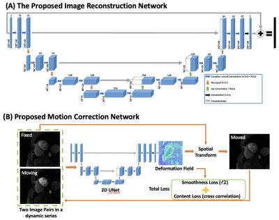 |
Deep learning-based Motion-corrected Image Reconstruction for
High-resolution Spiral First-pass Myocardial Perfusion Imaging
Marina Awad1,
Junyu Wang2,
and Michael Salerno2
1Biomedical Engineering, University of Virginia, Charlottesville, VA, United States, 2Cardiovascular Medicine, Stanford University, Stanford, CA, United States Keywords: Heart, Perfusion Cardiovascular magnetic resonance (CMR) is susceptible to motion-induced artifacts from cardiac and respiratory motion, leading to poor image quality. The inter-frame motion artifacts make quantitative analysis for cardiac function evaluation difficult. Hence motion correction is an important pre-processing step before robust quantification of myocardial perfusion. We developed a deep learning-based framework for rapid and accurate motion correction of CMR perfusion imaging using a 2D U-Net that estimates the deformation field from a moving frame to a fixed frame. |
| 15:22 |
0558.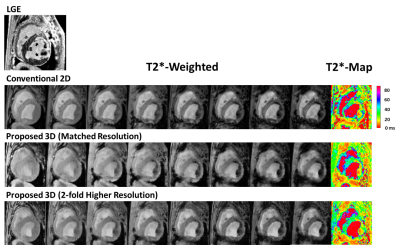 |
Fully ungated, free-breathing, 3D T2* mapping for imaging
hemorrhagic Myocardial Infarction
Xingmin Guan1,
Hsin-Jung Yang2,
Jane Sykes3,
Xinheng Zhang1,
Richard Tang1,
Anthony Christodoulou2,
Behzad Sharif1,
Debiao Li2,
Frank S Prato3,
and Rohan Dharmakumar1 1Indiana University School of Medicine, Indianapolis, IN, United States, 2Cedars-Sinai Medical Center, Los Angeles, CA, United States, 3Lawson Health Research Institute, London, ON, Canada Keywords: Image Reconstruction, Cardiovascular, T2* T2* CMR is widely used for detecting hemorrhagic myocardial infarction (MI). However, the conventional T2* CMR (2D breath-held, ECG-gated, multi-gradient-echo T2*) can suffer from limited spatial resolution and motion artifacts. We have recently developed a time-efficient, fully ungated, free breathing, 3D T2* mapping approach using a low-rank tensor (LRT) framework to address the above issues. |
 Back to Meeting Home
 Back to the Program-at-a-Glance
The International Society for Magnetic Resonance in Medicine is accredited by the Accreditation Council for Continuing Medical Education to provide continuing medical education for physicians.
 Back to Meeting Home
 Back to the Program-at-a-Glance
 View Presentation Video