Oral Session
State-of-the-Art Imaging in Human Cardiovascular Health & Disease
ISMRM & ISMRT Annual Meeting & Exhibition • 03-08 June 2023 • Toronto, ON, Canada

Oral Session
State-of-the-Art Imaging in Human Cardiovascular Health & Disease
13:45 |
1313.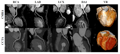 |
Deep learning-based acceleration of compressed sensing
non-contrast-enhanced coronary MRA in patients with suspected
coronary artery disease
Xi Wu1,2,
Jiayu Sun1,
and Xiaoyong Zhang3 1West China Hospital, Sichuan University, Chengdu, China, 2Affiliated Hospital of North Sichuan Medical College, Nanchong, China, 3Clinical Science, Philips Healthcare, Chengdu, China Keywords: Vessels, Cardiovascular, Deep Learning This study aims to investigate the feasibility of a compressed sensing artificial intelligence (CSAI) framework for non-contrast-enhanced coronary MRA. The image quality and the diagnostic performance of CSAI coronary MRA in patients with suspected CAD were fully evaluated using coronary computed tomography angiography (CTA) as the non-invasive clinical reference standard. The results shows that all recruited patients completed coronary MRA with high image quality and diagnostic performance within short scan time. Therefore, we conclude that the CASI coronary MRA could be a robust and safe non-invasive alternative for excluding significant disease in patients with suspected CAD. |
| 13:53 |
1314.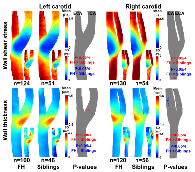 |
Carotid wall shear stress and wall thickness in patients with
familial hypercholesterolemia and their healthy siblings
Eva S. Peper1,2,
Dieuwertje Alblas3,
Renske Merton4,
Sibbeliene E. van den Bosch5,
Anne Spakman4,
Bram F. Coolen6,
Gustav J. Strijkers6,
Aart J. Nederveen4,
Albert Wiegman5,
Jelmer M. Wolterink3,
Barbara A. Hutten7,
and Pim van Ooij4
1Bern University Hospital, University of Bern, Bern, Switzerland, 2Translation Imaging Center (TIC), Swiss Institute for Translational and Entrepreneurial Medicine, Bern, Switzerland, 3Department of Applied Mathematics, Technical Medical Center, University of Twente, Enschede, Netherlands, 4Department of Radiology and Nuclear Medicine, Amsterdam Cardiovascular Sciences, Amsterdam University Medical Center, location AMC, Amsterdam, Netherlands, 5Department of Pediatrics, Amsterdam University Medical Center, location AMC, Amsterdam, Netherlands, 6Department of Biomedical Engineering & Physics, Amsterdam Cardiovascular Sciences, Amsterdam University Medical Center, location AMC, Amsterdam, Netherlands, 7Department of Epidemiology and Data Science, Amsterdam Cardiovascular Sciences, Amsterdam University Medical Center, location AMC, Amsterdam, Netherlands Keywords: Flow, Velocity & Flow, vessel wall, carotids, familial hypercholesterolemia Familial hypercholesterolemia (FH) leads to premature atherosclerosis. In this study we use 4D flow and 3D black blood (BB) MRI to investigate carotid artery wall shear stress (WSS) and wall thickness (WT) 3D maps and their relation, in FH patients on lifelong statin prescriptions and their unaffected siblings (n=234). We applied machine learning segmentation technology and 3D statistical analysis methods and found that ensemble-averaged carotid WSS and WT maps were highly similar between the groups. However, the 3D carotid correlation coefficient maps showed lower agreement between WT and WSS in patients, suggesting abnormal wall remodeling processes compared to the siblings. |
| 14:01 |
1315.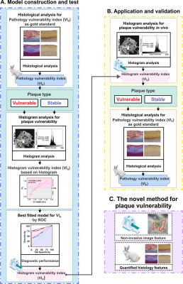 |
Predicting carotid plaque vulnerability by macrophage-targeted
nanocluster-enhanced high-resolution vessel wall imaging
Yan Gong1,
Menglin Wu2,
Dingwei Fu3,
Yu Guo4,
Xiudi Lu5,
Ying Zou6,
Xiang Zhang1,
Jinxia Zhu7,
Xianchang Zhang7,
Xue Li2,
and Shuang Xia5
1Department of Radiology, Tianjin Nankai Hospital, Tianjin, China, 2Department of Radiology, Second Hospital of Tianjin Medical University, Tianjin, China, 3Department of Radiology, First Central Clinical College, Tianjin Medical University, Tianjin, China, 4Department of Radiology, Medical Imaging Institute of Tianjin, Tianjin First Central Hospital, School of Medicine, Nankai University, Tianjin, China, 5Department of Radiology, First Teaching Hospital of Tianjin University of Traditional Chinese Medicine, Tianjin,China National Clinical Research Center for Chinese Medicine Acupuncture and Moxibustion, Tianjin, China, 6First Teaching Hospital of Tianjin University of Traditional Chinese Medicine, Tianjin,China National Clinical Research Center for Chinese Medicine Acupuncture and Moxibustion, Tianjin, China, 7MR Collaboration, Siemens Healthcare Ltd., Beijing, China Keywords: Vessel Wall, Atherosclerosis, macrophage Ischemic stroke can be attributed to the sudden rupture of high-risk atherosclerotic plaques. Therefore, identifying vulnerable plaque is essential for preventing acute vascular events. Increasing evidence has established that macrophages play a vital role in the pathogenesis of atherosclerosis. Using the black-blood technique, an imaging approach that suppresses blood signals to highlight the arterial wall, and a macrophage-targeted contrast agent, plaque vulnerability could be quantitatively assessed for the macrophage burden. This in vivo animal study non-invasively evaluated the plaque vulnerability, which may provide information on the clinical risk stratification. |
| 14:09 |
1316.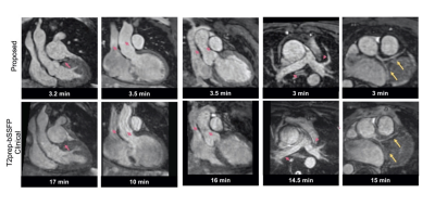 |
Highly-Efficient 3D free-breathing whole-heart MRA in 3 min:
Clinical validation in patients with adult congenital heart
disease
Anastasia Fotaki1,
Camila Munoz1,
Christopher Rush2,
Carlos Velasco1,
Karl Kunze1,3,
Radhouene Neji1,3,
Kuberan Pushparajah1,
Rene M Botnar1,4,5,6,
and Claudia Prieto1,4,5,6 1King's College London, London, United Kingdom, 2St Thomas’ Hospital, Guy’s and St Thomas’ NHS Foundation Trust, London, United Kingdom, 3MR Research Collaborations, Siemens Healthcare Limited, Camberley, United Kingdom, 4School of Engineering, Pontificia Universidad Católica de Chile, Santiago, Chile, 5Institute for Biological and Medical Engineering, Pontificia Universidad Católica de Chile, Santiago, Chile, 6Millennium Institute for Intelligent Healthcare Engineering, Santiago, Chile Keywords: Vessels, Cardiovascular, angiography Cardiovascular MRA is established for serial anatomical evaluation of patients with congenital heart disease (CHD). However, this approach is limited by diaphragmatic respiratory navigation, that leads to long acquisition times and degraded image quality due to residual motion artefacts. Here we evaluate a novel accelerated whole-heart framework in patients with adult CHD. This approach incorporates image-based navigation for translational and non-rigid motion-correction along with 3D patch-based denoising for efficient, free-breathing 3D whole-heart imaging. Comparison between the conventional and the research sequence shows superior diagnostic confidence and diagnostic accuracy for the proposed technique in significantly faster acquisition time (~3min proposed,~15min clinical). |
| 14:17 |
1317.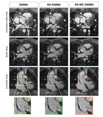 |
Similarity-driven motion-resolved reconstruction for
ferumoxytol-enhanced whole-heart MRI of congenital heart disease
patients
Ludovica Romanin1,2,
Bastien Milani1,
Christopher W. Roy1,
Aurélien Bustin1,3,4,
Salim Si-Mohamed1,5,6,
Milan Prsa7,
Tobias Rutz8,
Estelle Tenisch1,
Juerg Schwitter8,9,
Matthias Stuber1,10,
and Davide Piccini1,2
1Diagnostic and Interventional Radiology, University Hospital of Lausanne (CHUV), Lausanne, Switzerland, 2Advanced Clinical Imaging Technology, Siemens Healthineers International AG, Lausanne, Switzerland, 3IHU LIRYC, Electrophysiology and Heart Modeling Institute, Université de Bordeaux – INSERM U1045, Pessac-Bordeaux, France, 4Department of Cardiovascular Imaging, Hôpital Cardiologique du Haut-Lévêque, CHU de Bordeaux, Pessac, France, 5INSA-Lyon, CNRS, Inserm, CREATIS, Université de Lyon, Villeurbanne, France, 6Département de Radiologie, Louis Pradel Hospital, Hospices Civils de Lyon, Bron, France, 7Woman-Mother-Child Department, Lausanne University Hospital and University of Lausanne, Division of Pediatric Cardiology, Lausanne, Switzerland, 8Heart and Vessel Department, Lausanne University Hospital and University of Lausanne, Service of Cardiology, Lausanne, Switzerland, 9Cardiac MR Center, Lausanne University Hospital and University of Lausanne, Lausanne, Switzerland, 10Center for Biomedical Imaging (CIBM), Lausanne, Switzerland Keywords: Heart, Motion Correction Ferumoxytol-enhanced free-running whole-heart MRI allows for a comprehensive evaluation of the cardiovascular anatomy in 3D. A similarity-driven multi-dimensional binning algorithm (SIMBA) has been proposed as a fast and efficient reconstruction of such data, by clustering and selecting motion-consistent information. In this work, we extend the SIMBA reconstruction to make use of the inherent redundancy of motion-consistent information using a compressed-sensing reconstruction, in which sparsity is maximized by the integration of inter-cluster non-rigid 3D motion-fields. With this new framework we demonstrate improved image quality, increased coronary sharpness and vessel conspicuity. |
|
14:25 |
1318.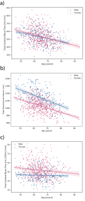 |
Sex differences between total cerebral blood flow and
age-related changes in the brain using 4D flow MRI
Alma Spahic1,
Grant S. Roberts1,
Anthony Peret2,
Rebecca L. Koscik3,
Erin Jonaitis3,4,
Carson A. Hoffman2,
Leonardo A. Rivera-Rivera1,4,
Karly A. Cody4,
Howard A. Rowley2,
Sterling C. Johnson3,4,5,
Oliver Wieben1,2,
Kevin M. Johnson1,2,
and Laura Eisenmenger2
1Medical Physics, University of Wisconsin- Madison, Madison, WI, United States, 2Radiology, University of Wisconsin- Madison, Madison, WI, United States, 3Wisconsin Alzheimer's Institute, University of Wisconsin- Madison, Madison, WI, United States, 4Wisconsin Alzheimer's Disease Research Center, University of Wisconsin- Madison, Madison, WI, United States, 5Geriatric Research Education and Clinical Center, William S. Middleton Memorial Veterans Hospital, Madison, WI, United States Keywords: Flow, Aging Vascular disease is strongly associated with Alzheimer’s disease. Therefore, it is important to establish normative cerebrovascular changes in aging populations. In this study, we assess sex-specific, age-related changes in total cerebral blood flow (tCBF) and total parenchymal volume in 754 cognitively healthy, older adults. We further investigate whether the same changes are observed in flow normalized by total parenchymal volume. We found (1) significant decrease in tCBF and total parenchymal volume with age in both sexes, (2) no correlation between age and normalized flow in male subjects, and (3) significant decrease in normalized flow with age in female subjects. |
| 14:33 |
1319.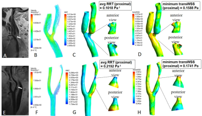 |
Tortuosity and Proximal-specific Wall Shear Stress Associated
with Plaque Location in Carotid Bulb: A High-resolution MRI and
CFD Study
Lei Ren1 and
Shuang Xia2
1Department of Radiology, First Central Clinical School, Tianjin Medical University, Tianjin, China, 2Tianjin First Central Hospital, Tianjin, China Keywords: Atherosclerosis, Blood vessels, computational fluid dynamics This study was performed from a vessel wall imaging database, and investigated plaque characteristics, as well as geometric and hemodynamic parameters among different carotid bulb plaque locations caused by atherosclerosis. The results showed that wall shear stress (WSS) magnitudes around plaque side were lower than non-plaque side. Tortuosity of stenosed region, magnitudes of relative residence time and transverse WSS in the proximal part of the lesion were the key factors independently associated with plaque location. This suggested that plaque formation was associated with local flow pattern; and tortuosity, proximal-specific hemodynamics were significantly associated with plaque location in the carotid bulb. |
|
14:41 |
1320.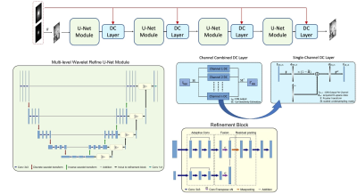 |
Multi-channel CAscaded Multi-scale WAvelet with iterative
REfinement (CAMWARE) network for accelerated whole-brain vessel
wall imaging
Junzhou Chen1,
Zhehao Hu1,
Dan Ruan2,
Fei Han3,
xiaoming Bi3,
Alexander Lerner1,
Roy Poblete1,
and Zhaoyang Fan1 1University of Southern California, Los Angeles, CA, United States, 2University of California, Los Angeles, Los Angeles, CA, United States, 3Siemens Medical Solutions, Los Angeles, CA, United States Keywords: Vessel Wall, Atherosclerosis 3D MR vessel wall imaging (VWI) is a non-invasive imaging modality for directly assessing intracranial arterial wall diseases. A typical intracranial VWI protocol requires 6-12 minutes per scan to obtain adequate spatial coverage and resolution. Such a long scan time hinders widespread use of VWI in clinical settings. We have developed a multi-channel application-ready intracranial vessel-dedicated CAscaded Multi-level WAvelet REfine (CAMWARE) network that enables a VWI scan within 4 minutes. The proposed network achieved significant improvement in vessel wall delination over conventional compressed sensing reconstruction. |
| 14:49 |
1321.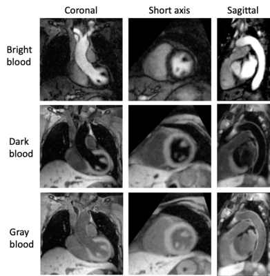 |
MR Multitasking-based Multi-dimensional Assessment of
Cardiovascular System (MT-MACS): Initial Clinical Experience
Jiayu Xiao1,
Yang Chen1,
Xin Liu2,
Zhehao Hu1,
Debiao Li3,
Anthony Christodoulou3,
Parveen Garg4,
Michael Fong4,
Alison Wilcox4,
Qi Yang2,
and Zhaoyang Fan1,5,6
1Radiology, Keck School of Medicine of USC, Los Angeles, CA, United States, 2Beijing Chaoyang Hospital, Beijing, China, 3Cedars-Sinai Medical Center, Los Angeles, CA, United States, 4Cardiology, Keck School of Medicine of USC, Los Angeles, CA, United States, 5Radiation Oncology, Keck School of Medicine of USC, Los Angeles, CA, United States, 6Viterbi School of Engineering, University of Southern California, Los Angeles, CA, United States Keywords: Myocardium, Cardiomyopathy Cardiovascular diseases are common causes of mortality and morbidity globally. An MR MultiTasking based 3D Multi-dimensional Assessment of Cardiovascular System (MT-MACS) technique has recently been developed to provide multi-contrast, cardiac phase-resolved imaging of the whole heart and thoracic aorta in a single scan without the need for ECG triggering or respiratory navigation. We aimed to assess the performance of MT-MACS in patients with suspicious cardiovascular diseases. We demonstrated the feasibility of using MT-MACS in a routine clinical setting. More importantly, it achieved good image quality in most patients with almost perfect inter-reader agreement and diagnostic accuracy. |
| 14:57 |
1322.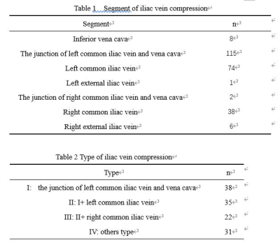 |
Relaxation-Enhanced Angiography without Contrast and Triggering
(REACT) for evaluation of iliac vein compression syndrome at 3T
Fengming Tao1,
LI Tao1,
and Xiaoyong Zhang2 1The First Affiliated Hospital of Chongqing Medical University, Chongqing, China, 2Clinical Science, Philips Healthcare, Chengdu, China Keywords: Vessels, Blood vessels Relaxation-Enhanced Angiography without Contrast and Triggering (REACT) is a novel flow-independent MR angiography technique without cardiac triggering, breath holding and contrast agent injection and has promising results to show robust blood-to-tissue contrast over multiple anatomies. This study aims to investigate the feasibility of REACT for evaluation iliac vein compression syndrome in comparison with catheter angiography. Results showed that with comparable image quality to catheter angiography and high sensitivity and specificity for the detection of stenosis, REACT was proven to be a clinically applicable method for assessing iliac vein compression syndrome. |
| 15:05 |
1323.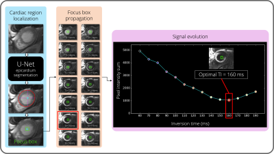 |
Automated Contrast Selection for Robust Bright- and Black-Blood
Myocardial Scar Imaging
Victor de Villedon de Naide1,
Indra Ribal1,
Pauline Gut1,2,
Valéry Ozenne1,
Géraldine Montier3,
Jean-David Maes3,
Thibault Boullé3,
Guillaume Delclaux3,
Aurélien Maillot1,
Soumaya Sridi3,
Bruno Quesson1,
Matthias Stuber1,2,4,
Hubert Cochet1,3,
and Aurélien Bustin1,2,3
1IHU LIRYC, Electrophysiology and Heart Modeling Institute, Université de Bordeaux – INSERM U1045, Avenue du Haut Lévêque, 33604, Pessac, France, Bordeaux, France, 2Department of Diagnostic and Interventional Radiology, Lausanne University Hospital and University of Lausanne, Lausanne, Switzerland, Lausanne, Switzerland, 3Department of Cardiovascular Imaging, Hôpital Cardiologique du Haut-Lévêque, CHU de Bordeaux, Avenue de Magellan, 33604 Pessac, France, Bordeaux, France, 4CIBM Center for Biomedical Imaging, Lausanne, Switzerland, Lausanne, Switzerland Keywords: Heart, Data Processing, late gadolinium enhancement, black-blood, TI scout Phase-sensitive inversion recovery is the reference imaging technique for the assessment of myocardial scars. Despite its ability to provide excellent contrast between healthy and scar tissue, small subendocardial scars can be challenging to detect due to poor scar-to-blood contrast. Joint bright- and black-blood late gadolinium enhancement techniques have been developed to provide both scar and anatomy information. Black-blood contrast is obtained after manual selection of an optimal inversion time (TI). This often results in uncertainties, variability, increased workload, and operator-dependency. In this work, we propose a method exploiting artificial intelligence to fully automate TI selection for more robust cardiac imaging. |
| 15:13 |
1324.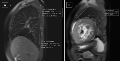 |
Lung water estimation using cardiac magnetic resonance imaging
for predicting adverse cardiovascular outcomes in patients with
heart failure
Priya Jagia1,
Mansi Verma2,
and Sanjeev Kumar3
1Dept of cardiovascular radiology and Endovascular interventions, All India institute of medical sciences, Delhi, India, 2Dept of cardiovascular radiology and Endovascular interventions, All India institute of medical sciences, Delhi, Delhi, India, 3Dept of Cardiovascular radiology and Endovascular interventions, All India institute of medical sciences, Delhi, Delhi, India Keywords: Heart, Lung, Heart failure The present study sought to determine whether CMR-derived lung water density (LWD) measurement in pulmonary congestion has prognostic relevance in predicting adverse cardiovascular outcomes in patients with heart failure. The primary endpoint was all-cause mortality or HF-related hospitalization within 6 months from CMR. It concluded - 1. The mean lung water density was significantly higher in heart failure patients compared to healthy controls. 2. Patients with “wet lungs” i.e., lung water density >18.1%, had higher incidence of adverse cardiovascular outcomes compared to patients with “dry lungs”. 3. Lung water density was an independent predictor of adverse cardiovascular outcomes |
| 15:21 |
1325.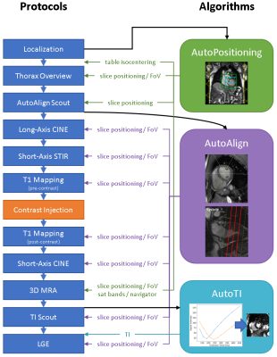 |
AI-based Single-Click Cardiac MRI Exam: Initial Clinical
Experience and Evaluation in 44 Patients
Jens Wetzl1,
Seung Su Yoon1,
Michaela Schmidt1,
Alexander Haenel2,
Alexandra-Bianca Weißgerber2,
Jörg Barkhausen2,
and Alex Frydrychowicz2
1Siemens Healthcare GmbH, Erlangen, Germany, 2Klinik für Radiologie und Nuklearmedizin, Universitätsklinikum Schleswig-Holstein Campus Lübeck, Lübeck, Germany Keywords: Machine Learning/Artificial Intelligence, Cardiovascular, Workflow We propose an Artificial Intelligence-based single-click cardiac MR exam to support technicians and increase standardization and repeatability of cardiac exams, which we evaluate in a clinical setting with 44 patients. Automations include setting of the isocenter position, slice planning in standard and cardiac orientations, adjustment volume and inversion time. Clinical sequences that can be acquired without manual planning include CINE, STIR, T1 mapping, 3D MRA and LGE. 91% (n=40) of the acquisitions could be completed without manual operator intervention, with the majority (n=3) of failed cases caused by abnormal heart geometries leading to inadequate slice planning. |
| 15:29 |
1326. |
Stability of Elevated Wall Shear Stress in Patients with
Bicuspid Aortic Valve: Insights from a Multi-Year 4D Flow MRI
Follow-Up Study
Anthony Maroun1,
Michael Scott1,
Haben Berhane1,
Justin J. Baraboo1,
Kelly Jarvis1,
Bradley D. Allen1,
Alex J. Barker2,
and Michael Markl1 1Department of Radiology, Northwestern University, Chicago, IL, United States, 2Department of Radiology, University of Colorado, Aurora, CO, United States Keywords: Flow, Valves 4D Flow MRI can identify aortic regions exposed to high wall shear stress (WSS) compared to age and sex-matched controls. This concept, known as WSS ‘heatmaps’, has recently shown potential to improve risk stratification in patients with bicuspid aortic valve (BAV). We examined the reproducibility of heatmaps in a cohort of 20 stable BAV patients with five consecutive 4D flow MRI scans and found no significant change over time. In addition, we found high reproducibility of WSS patterns and regions of elevated WSS across scans. Our findings, therefore, suggest that heatmaps can serve as a robust BAV risk measure. |
| 15:37 |
1327.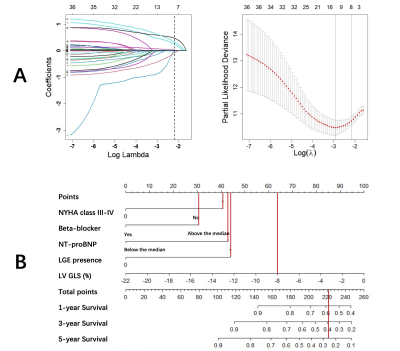 |
Factors Affecting Outcomes in Nonischemic Dilated
Cardiomyopathy: Development and Validation of a CMR-Based
Nomogram
Xiaorui Xiang1,
Xiaoqiang Lin2,
and Shihua Zhao1
1Fuwai Hospital and National Center for Cardiovascular Diseases, Beijing, China, 2Union Hospital of Fujian Medical University, Fujian, China Keywords: Cardiomyopathy, Cardiomyopathy Risk evaluation for patients with NIDCM remains an important public health challenging, and new techniques and strategies are expected to be used to prevent cardiac death. This study was designed to develop and validate a novel nomogram score to predict outcomes in patients with NIDCM over a long follow-up time period. |
 Back to Meeting Home
 Back to the Program-at-a-Glance
The International Society for Magnetic Resonance in Medicine is accredited by the Accreditation Council for Continuing Medical Education to provide continuing medical education for physicians.
 Back to Meeting Home
 Back to the Program-at-a-Glance
 View Presentation Video