Oral Session
Novel Methods in fMRI
ISMRM & ISMRT Annual Meeting & Exhibition • 03-08 June 2023 • Toronto, ON, Canada

| 13:45 |
1268.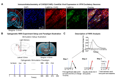 |
Short single pulse optogenetic fMRI mapping of downstream
targets in thalamo-cortical pathways
Linshan Xie1,2,
Xunda Wang1,2,
Teng Ma1,2,3,
Hang Zeng1,2,
Junjian Wen1,2,
Peng Cao3,
Ed X. Wu1,2,4,
and Alex T.L. Leong1,2
1Laboratory of Biomedical Imaging and Signal Processing, The University of Hong Kong, Hong Kong SAR, China, 2Department of Electrical and Electronic Engineering, The University of Hong Kong, Hong Kong SAR, China, 3Department of Diagnostic Radiology, Li Ka Shing Faculty of Medicine, The University of Hong Kong, Hong Kong SAR, China, 4School of Biomedical Sciences, Li Ka Shing Faculty of Medicine, The University of Hong Kong, Hong Kong SAR, China Keywords: fMRI (task based), fMRI (task based) Short single pulse stimulation is advantageous to map the downstream neural targets compared to pulse train stimulation because it can minimize the excessive neural synchronization and avoid numerous series of complex neural events. It is desirable for fMRI studies to investigate the properties of neural circuits via delivering single pulse stimulation. However, the subtle BOLD responses evoked by short stimuli are hard to detect due to the sensitivity issue. Here, we employed fMRI to examine the long-range downstream targets of the somatosensory thalamus with 10ms single pulse stimulation. A model-free fMRI analysis was utilized to visualize the spatiotemporal activity propagation. |
13:53 |
1269.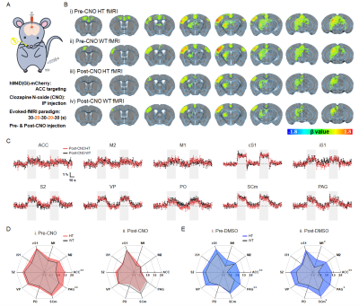 |
Somatosensory-evoked fMRI with chemogenetic modulation reflects
behavioral normalization in hypersensitized mice
Won Beom Jung1,2,
Soowon Lee3,
Geun Ho Im1,
Taeyi You1,
Eunjoon Kim4,5,
and Seong-Gi Kim1,6
1Center for Neuroscience Imaging Research (CNIR), Institute for Basic Science (IBS), Suwon, Korea, Republic of, 2Medical Imaging AI Research Center, Canon Medical Systems Korea, Seoul, Korea, Republic of, 3Graduate School of Medical Science and Engineering,, Korea Advanced Institute of Science and Technology (KAIST), Daejeon, Korea, Republic of, 4Department of Biological Sciences, Korea Advanced Institute for Science and Technology (KAIST), Daejeon, Korea, Republic of, 5Center for Synaptic Brain Dysfunctions, Institute for Basic Science (IBS), Daejeon, Korea, Republic of, 6Department of Biomedical Engineering, Sungkyunkwan University, Suwon, Korea, Republic of Keywords: fMRI (task based), Animals The fMRI mapping with selective modulation of local neural population at the manipulated region is a powerful approach that can causally link circuit-specific interactions to behavioral performance. However, most fMRI studies combined with chemogenetics were conducted in the resting state, which is difficult to elucidate whether and how differences in functional activity by neuromodulation induce behavioral changes. Here, we demonstrated the effects of fMRI-guided focal chemogenetic modulation on both somatosensory-evoked network and its relevant behaviors in mice. |
| 14:01 |
1270.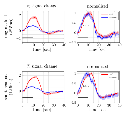 |
Impact of EPI readout duration on the diffusion-fMRI response
onset time
Shota Hodono1,
Jonathan R Polimeni2,3,
and Martijn A Cloos1
1Centre for Advanced Imaging, The University of Queensland, Brisbane, Australia, 2Athinoula A. Martinos Center for Biomedical Imaging, Massachusetts General Hospital, Charlestown, MA, United States, 3Harvard-MIT Division of Health Sciences and Technology, Massachusetts Institute of Technology, Cambridge, MA, United States Keywords: fMRI (task based), Diffusion/other diffusion imaging techniques We investigated the impact of the EPI readout duration on the onset time of activation observed with diffusion functional MRI (DfMRI). Both long (28.5-ms) and short (12.5-ms) readouts showed a clear functional response, even in individual subjects. However, using a long readout the onset time was shifted towards SE-BOLD (1.6-s [short readout] vs 1.2-s [long readout]), suggesting that longer readouts make the DfMRI signal more similar to SE-BOLD, presumably due to increased BOLD contamination. |
| 14:09 |
1271.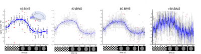 |
High Temporal Resolution Blood Oxygen Level Dependent functional
MRI.
Martyna Dziadosz1,2,3,4,
Tom Hilbert2,5,6,
Jérôme Yerly2,6,
Matthias Stuber2,6,
Matthias Nau7,
Micah M. Murray1,2,3,6,8,
Eleonora Fornari2,6,
and Bendetta Franceschiello2,3,4 1Laboratory for Investigative Neurophysiology (The LINE), Lausanne University Hospital and University of Lausanne (CHUV-UNIL), Lausanne, Switzerland, 2Department of Diagnostic and Interventional Radiology, Lausanne University Hospital and University of Lausanne, Lausanne, Switzerland, 3The Sense Innovation and Research Centre, Lausanne and Sion, Switzerland, 4Institute of Systems Engineering, School of Engineering, HES-SO Valais-Wallis, Sion, Switzerland, 5Advanced Clinical Imaging Technology, Siemens Healthineers International AG, Lausanne, Switzerland, 6CIBM Center for Biomedical Imaging, Lausanne, Switzerland, 7Laboratory of Brain and Cognition, National Institute of Mental Health, National Institutes of Health, Bethesda, MD, United States, 8Department of Hearing and Speech Sciences, Vanderbilt University, Nashville, TN, United States Keywords: Brain Connectivity, fMRI (task based), hemodynamic response, high temporal resolution, visual cortex We demonstrate the feasibility and robustness of a new method to measure blood-oxygen-level-dependent functional MRI(BOLD-fMRI) signals at high temporal resolutions(up to 250ms). Whole-brain data at 1x1x1 mm3 were acquired uninterruptedly during a blocked-design ON/OFF visual paradigm(checkerboard vs. grey image). Images were reconstructed with a 4D x-y-z-t dimensions at 2.5s, 1.0s, 500ms and 250ms temporal resolution, allowing to retrieve the expected %signal change(2%) in visual cortices. We also evaluated the effect of compressed-sensing(CS) application in BOLD-fMRI reconstruction schemes, finding that CS improves the obtained signal in the calcarine sulcus(around 23k(a.u) in pixel intensity of BOLD map TCFE corrected vs 10k(a.u)). |
| 14:17 |
1272.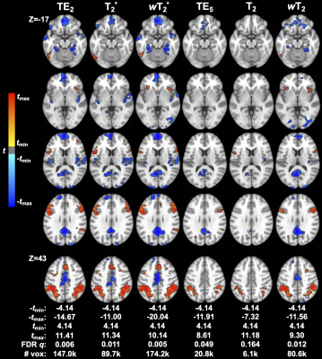 |
Multi-echo, multi-contrast functional activation: validation of
combined spin- and gradient-echo EPI in fMRI
Elizabeth G. Keeling1,2,
Maurizio Bergamino1,
Sudarshan Ragunathan1,3,
C. Chad Quarles1,4,
and Ashley M. Stokes1
1Barrow Neurological Institute, Phoenix, AZ, United States, 2Arizona State University, Tempe, AZ, United States, 3Hyperfine, Inc., Guilford, CT, United States, 4MD Anderson Cancer Center, Houston, TX, United States Keywords: Data Acquisition, fMRI (task based), Neuro, multi-echo, multi-contrast, EPI Standard functional MRI (fMRI) suffers from susceptibility-induced dropout near air-tissue interfaces and is sensitive to larger vessels. Conversely, a combined spin- and gradient-echo (SAGE) acquisition can provide sensitivity to functional activation across macro- and microvascular scales with reduced signal dropout. Multi-echo analysis of SAGE-fMRI data was performed by using quantitative and relaxation-weighted T2* and T2. In a task-based experiment, SAGE relaxation-weighted analyses showed increased contrast- and temporal signal-to-noise ratios (CNR and tSNR, respectively), especially for microvascular analysis. SAGE-fMRI provides improvements over standard fMRI in image quality and robustness of functional activation, as well as inclusion of microvascular sensitivity. |
| 14:25 |
1273.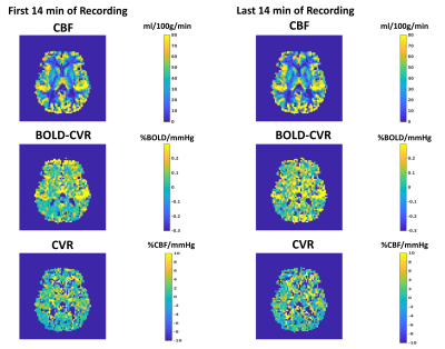 |
Mapping Grey Matter Cerebrovascular Reactivity and Oxygen
Consumption Using Resting-State BOLD-ASL Functional MRI
Antonio Maria Chiarelli1,
Eleonora Patitucci2,
Michael Germuska3,
Alessandra Stella Caporale1,
Emma Biondetti4,
Hannah Chandler3,
Kevin Murphy5,
Valentina Tomassini4,5,
and Richard Goeffrey Wise4,5
1Neuroscience, Imaging and Clinical Sciences, University G. D'Annunzio of Chieti Pescara, Chieti Scalo, Italy, 2Department of Psychology, Cardiff University Brain Research Imaging Centre (CUBRIC), Cardiff, United Kingdom, 3School of Psychology, Cardiff University Brain Research Imaging Centre (CUBRIC), Cardiff, United Kingdom, 4University G. D'Annunzio of Chieti Pescara, Chieti Scalo, Italy, 5Cardiff University Brain Research Imaging Centre (CUBRIC), Cardiff, United Kingdom Keywords: fMRI (resting state), Quantitative Imaging, Cerebrovascular Reactivity BOLD and ASL CBF fMRI can measure cerebrovascular reactivity (CVR) following a vasodilatory stimulus such as hypercapnia. BOLD and ASL CVR measurements allow us to extract the maximum BOLD signal modulation from which, through biophysical modelling, OEF and CMRO2 can be inferred. We measured these physiological variables in grey matter by assessing the resting-state coupling between fMRI and end-tidal CO2 (reflecting arterial CO2) recordings. In-vivo evaluation of two, sequentially acquired, 14-min recordings at rest demonstrated the method’s good repeatability. This simplified calibrated fMRI approach does not require an exogenous hypercapnic stimulus and thus holds promise for future applications. |
| 14:33 |
1274.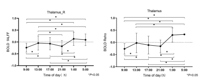 |
Resting-state fMRI study of vigilance level under endogenous
modulation based on ReHo and fALFF in humans under normal
entrained conditions
Hanqi Xing1,
Zhiwei Wu1,
Mengya Ma1,
Ziyang Song1,
Yang Song2,
Yunzhu Wu2,
Yue Chang1,
and Hui Dai1
1The First Affiliated Hospital of Soochow University, Suzhou, China, 2MR Scientific Marketing, Siemens Healthcare, Shanghai China., Shanghai, China Keywords: fMRI (resting state), Brain, sleep Time-dependent neuromodulation mechanisms in human cognitive-behavioral tasks are not yet fully established. The present study aimed to analyze the changes in resting-state functional magnetic resonance imaging (rs-fMRI) blood oxygen level dependent (BOLD) signals over the 24-h day and their correlation with vigilance level. We recruited 20 healthy volunteers to be scanned at six-time points 24 hours a day. Compared to 9:00, 13:00, 17:00, and 21:00h, thalamic BOLD signals increased and vigilance level decreased at 1:00 and 5:00h. We speculate that the BOLD signals increased in the thalamus may represent a compensatory mechanism for maintaining relative vigilance level. |
| 14:41 |
1275.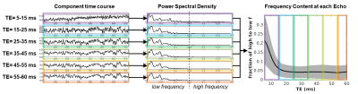 |
Data-driven analysis of echo planar time-resolved MRI suggests
frequency-specific mechanisms of brain fluctuations with unique
TE signatures
Lisa C. Krishnamurthy1,
Venkatagiri Krishnamurthy2,
Fuyixue Wang3,
Lawrence L Wald3,
and Vince D Calhoun4
1Georgia State University, Stone Mountain, GA, United States, 2Emory University, Atlanta, GA, United States, 3Harvard University, Combridge, MA, United States, 4Georgia State University, Atlanta, GA, United States Keywords: fMRI (resting state), Brain A novel analysis of EPTI rsfMRI to assess frequency contributions in unique echoes. |
| 14:49 |
1276.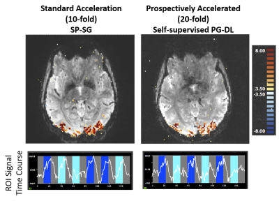 |
Unlocking 20-fold acceleration towards 0.5-second whole-brain
HCP-style fMRI
Omer Burak Demirel1,2,
Luca Vizioli2,3,
Burhaneddin Yaman1,2,
Steen Moeller2,
Logan Dowdle2,3,
Essa Yacoub2,
Kamil Ugurbil2,
and Mehmet Akçakaya1,2
1Electrical and Computer Engineering, University of Minnesota, Minneapolis, MN, United States, 2Center for Magnetic Resonance Research, University of Minnesota, Minneapolis, MN, United States, 3Department of Neurosurgery, University of Minnesota, Minneapolis, MN, United States Keywords: fMRI, fMRI Functional MRI (fMRI) is acquired with simultaneous multi-slice (SMS) imaging and in-plane acceleration to provide sufficient coverage and spatio-temporal resolutions. However, further accelerations are desirable to achieve BRAIN initiative targets. In this work, we investigate self-supervised deep learning reconstruction at 20-fold (SMS×in-plane=5×4) retrospective and prospective accelerations. Results show DL at 20-fold retrospective acceleration is similar to split slice-GRAPPA at 10-fold acceleration. Furthermore, we show that DL method trained on retrospective 20-fold acceleration generalizes well and successfully reconstructs prospectively 20-fold accelerated fMRI data. |
| 14:57 |
1277.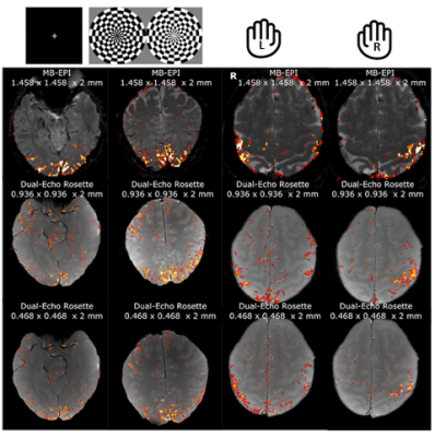 |
Submillimeter fMRI Acquisition using a dual-echo Rosette-k-space
trajectory at 3T
Gianna Nossa1,
Humberto Monsivais1,
Seokkyoon Hong2,
Taewoong Park2,
Fethi Sila Erdil1,
Xin Shen3,
Ali Caglar Özen4,
Serhat Ilbey4,
Mark Chiew5,
Cecilia Steinwurzel6,
Zoe Kourtzi6,
Yen-Yu Ian Shih7,
and Uzay Emir1,2
1School of Health Science, Purdue University, West Lafayette, IN, United States, 2Weldon School of Biomedical Engineering, Purdue University, West Lafayette, IN, United States, 3Radiology and Biomedical Imaging, University of California, San Francisco, San Francisco, CA, United States, 4Department of Radiology, Medical Center-University of Freiburg, Freiburg, Germany, 5Department of Medical Biophysics, University of Toronto, Toronto, ON, Canada, 6Department of Psychology, University of Cambridge, Cambridge, United Kingdom, 7Biomedical Research Imaging Center, University of North Carolina Chapel Hill, Chapel Hill, NC, United States Keywords: fMRI (task based), Brain In this study, we overcome the technological barrier against acquiring submillimeter resolution (~ 0.5 mm) fMRI data at 3T via a novel dual-echo Rosette k-space design. This design results in fine representation of activation maps in two different functional tasks and might be a springboard in neuroimaging by providing a very high-resolution spatiotemporal dynamics of neural networks. The method will be further evolved with the feedback from the MRI community via the github platform as such for the further acceleration, inflow saturation and 3D coverage via 3D sampling and/or multiband approaches. |
15:05 |
1278.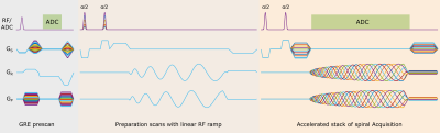 |
Rapid and high-resolution bSSFP fMRI using 3D stack of spirals
at 9.4 T
Praveen Iyyappan Valsala1,
Philipp Ehses2,
Marten Veldmann2,
and Klaus Scheffler1,3
1Magnetic Resonance Center, Max Planck Institute for Biological Cybernetics, Tübingen, Germany, 2German Center for Neurodegenerative Diseases (DZNE), Bonn, Germany, 3Department of Biomedical Magnetic Resonance, Eberhard Karls University, Tübingen, Germany Keywords: Data Acquisition, fMRI (task based), bSSFP, Spiral, non-cartesian, sub-millimeter, whole-brain We explored the spatio-temporal resolution and coverage of bSSFP BOLD contrast using a highly segmented 3D spiral readout. The functional activation maps acquired with the novel functional contrast for full visual field checkerboard stimulus is presented at submillimeter resolutions (0.6 mm3 & 0.8 mm3) and at 1.2 mm3 for whole brain coverage. We also demonstrated rapid slice-selective water excitation using binomial pulses. |
| 15:13 |
1279.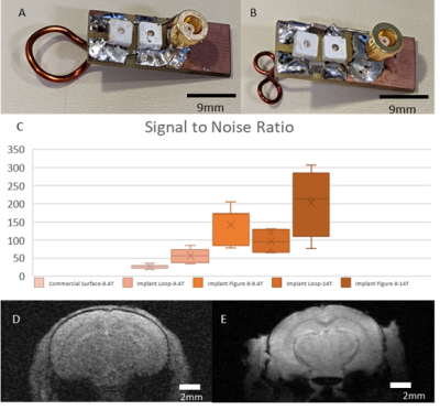 |
Implantable Coils Enable High-Resolution Functional MRI in Awake
Mice
David Hike1,
Xiaochen Liu1,
Zeping Xie1,2,
Bei Zhang1,
Wenchao Yang1,
Alyssa Murstein1,3,
Andy Liu1,3,
Daniel Glen4,
Richard Reynolds4,
and Xin Yu1
1Athinoula A. Martinos Center for Biomedical Imaging, Department of Radiology, Massachusetts General Hospital, Boston, MA, United States, 2School of Traditional Medicine, Southern China University, Guangzhou, China, 3Neuroscience, Boston University, Boston, MA, United States, 4NIMH, National Institutes of Health, Bethesda, MD, United States Keywords: fMRI (task based), Preclinical, Implantable Coil This study utilizes implantable RF coils affixed to mouse heads which are used as head fixation points to minimize motion and remove B1-related artifacts due to motion-induced loading change during scanning. This method increases SNR significantly and enables high-resolution EPI-based functional imaging in awake mice at 14T, highlighting both cortical and subcortical activation following visual and whisker stimulation. Furthermore, this high-resolution awake mouse fMRI setup enables high sensitivity to map brain activation from subcortical nuclei and extends the detection of associated brain regions related to the stimulation. |
| 15:21 |
1280.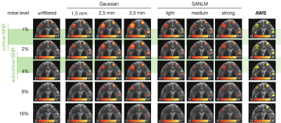 |
En Route to Fine-Grained Neurosignatures in the Individual
Brain: Evaluating Methodology to Boost Spatial Accuracy &
Sensitivity of BOLD fMRI
Igor Fabian Tellez Ceja1,
Thomas Gladytz1,
Ludger Benedikt Starke1,
Karsten Tabelow2,
Thoralf Niendorf1,3,
and Henning Matthias Reimann1
1Berlin Ultrahigh Field Facility (B.U.F.F.), Max Delbrück Center for Molecular Medicine, Berlin, Germany, 2Weierstrass Institute for Applied Analysis and Stochastics, Berlin, Germany, 3Experimental and Clinical Research Center, a joint cooperation between the Charité Medical Faculty and the Max Delbrück Center for Molecular Medicine in the Helmholtz Association, Berlin, Germany Keywords: High-Field MRI, fMRI In recent years, fMRI at ultrahigh magnetic field strengths (≥7T) has shifted from group analyses to probing neural processing in the individual brain. Identifying neurosignatures requires detection of BOLD effects with high sensitivity and spatial accuracy. Yet, it remains a challenge to enhance the sensitivity of fMRI for the BOLD effect without blurring the spatial details. Here, we assess the quality of the Gaussian, spatial adaptive non-local means (SANLM) and the adaptive weights smoothing (AWS) filters by employing a synthetic fMRI dataset as ground truth. AWS provides superior localization of the BOLD activations with high sensitivity at reasonable noise levels. |
|
15:29 |
1281.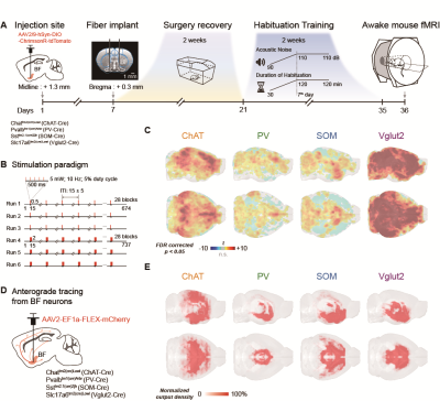 |
Cell-type specific basal forebrain modulation shapes global
functional network organizations supporting behavioral
variability
Chuanjun Tong1,2,
Yijuan Zou2,
Yanqiu Feng1,
and Zhifeng Liang2
1Southern Medical University, Guangzhou, China, 2Institute of Neuroscience, Chinese Academy of Sciences, Shanghai, China Keywords: fMRI, fMRI, cell-type specific optogentics; basal forebrain Activations of the basal forebrain (BF) were associated with arousal fluctuations1, and the regulation of the default model network2,3. However, it remains ambiguous how the cell-type specific BF neurons shape the behavioral performances. We developed an awake mouse fMRI setup with simultaneous cell-type specific optogenetic stimulations in BF. Combined with the anterograde tracing data4, we revealed weak structural-functional correspondence of BF neurons, and demonstrated the cell-type specific BF modulations shaped global functional network organizations supporting behavioral variability. Our results made great sense on the understanding the cerebral regulations and behaviors from BF neurons in a macroscopic whole-brain view. |
|
15:37 |
1282.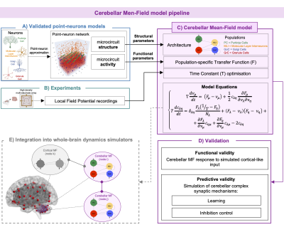 |
A complete cerebellar mean-field model ready to be integrated
into whole-brain dynamic simulators
Roberta Maria Lorenzi1,
Alice Geminiani1,
Yann Zerlaut2,
Alain Destexhe3,
Claudia A.M. Gandini Wheeler Kingshott1,4,5,
Fulvia Palesi1,
Claudia Casellato1,
and Egidio D'Angelo1,5
1Department of Brain and Behavioral Sciences, Università di Pavia, Pavia, Italy, 2Institut du Cerveau - Paris Brain Institute, ICM, Inserm, CNRS, APHP, Hôpital de la Pitié Salpêtrière, Paris, France, 3CNRS, Paris-Saclay University, Saclay, France, 4Department of Neuroinflammation, UCL Queen Square Institute of Neurology, NMR Research Unit, Queen Square Multiple Sclerosis Centre, London, United Kingdom, 5Brain Connectivity Center, IRCCS Mondino Foundation, Pavia, Italy Keywords: In Silico, New Devices, Mean field Whole-brain dynamics can be reproduced in silico by simulating Blood Oxygen Level Dependent (BOLD) signals, typically recorded with fMRI, using cortical and subcortical mean-field models, which provide a population-level description of the underlying neuronal dynamics. Notably, a mean-field model specific for the cerebellum is missing given its structural and functional specific properties. We present the first biologically-grounded cerebellar mean-field model optimized on experimental data. Our model reproduces cerebellar activity and synaptic mechanisms characterizing physiological and pathological conditions. The cerebellar mean-field model is a new device ready to be integrated in whole-brain dynamic simulator, improving understanding of brain function and dysfunction. |
 Back to Meeting Home
 Back to the Program-at-a-Glance
The International Society for Magnetic Resonance in Medicine is accredited by the Accreditation Council for Continuing Medical Education to provide continuing medical education for physicians.
 Back to Meeting Home
 Back to the Program-at-a-Glance
 View Presentation Video