Oral Session
Advances in ASL & BBB Mapping
ISMRM & ISMRT Annual Meeting & Exhibition • 03-08 June 2023 • Toronto, ON, Canada

08:15 |
0367.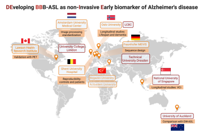 |
DEveloping Blood-Brain barrier arterial spin labeling as a
non-Invasive Early biomarker (DEBBIE)
Beatriz Padrela1,
Mervin Tee2,
Markus Sneve3,
Amnah Mahroo4,
Oliver Geier5,
David Thomas6,
Catherine Morgan7,
Paulien Moyaert8,9,
Esin Ozturk10,
Wibeke Nordhøy11,
Lene Pålhaugen12,
Jennifer Linn13,
Per Selnes12,
Klaus Eickel4,14,
Simon Konstandin4,14,
Joost Kuijer1,
Daniel Hoinkiss4,
Nora Breutigam4,
Mareike Buck4,
Rik Achten9,
Frederik Barkhof1,15,
Saima Hilal2,
Tormod Fladby12,16,
Udunna Anazodo8,
Jan Petr17,
Henk J.M.M. Mutsaerts1,
and Matthias Günther4
1Department of Radiology and Nuclear Medicine, Amsterdam UMC, Amsterdam, Netherlands, 2National University of Singapore and National University Health System, Saw Swee Hock School of Public Health, Singapore, Singapore, 3Department of Psychology, University of Oslo, Center for Lifespan Changes in Brain and Cognition, Oslo, Norway, 4Fraunhofer-Insitute for Digital Medicine MEVIS,, Bremen, Germany, 5Clinics of Radiology and Nuclear Medicine, University of Oslo, Department of Physics and Computational Radiology, Oslo, Norway, 6University College London, Dementia Research Center, London, United Kingdom, 7University of Auckland, School of Psychology and Centre for Brain Research, Auckland, New Zealand, 8Lawson Health Research Institute, London, ON, Canada, 9Department of Medical Imaging, Ghent University Hospital, Gent, Belgium, 10Bogazici University, Institute of Biomedical Engineering, Istanbul, Turkey, 11Clinics of Radiology and Nuclear Medicine, Oslo University Hospital, Department of Physics and Computational Radiology, Oslo, Norway, 12Department of Neurology, Akershus University Hospital, Lørenskog, Norway, 13Technische Universität Dresden, Department of Neuroradiology, University Hospital Carl Gustav Carus, Dresden, Germany, 14mediri GmbH, Heidelberg, Germany, 15University College London, Queen Square Institute of Neurology and Centre for Medical Image Computing (CMIC), London, United Kingdom, 16University of Oslo, Institute of Clinical Medicine, Campus Ahus, Lørenskog, Norway, 17Institute of Radiopharmaceutical Cancer Research, Helmholtz-Zentrum Dresden-Rossendorf, Dresden, Germany Keywords: Alzheimer's Disease, Aging One of the earliest signs of Alzheimer’s disease (AD) is the loss of blood-brain barrier (BBB) integrity. Arterial spin labeling (ASL) MRI is a non-invasive way to measure perfusion and several other hemodynamic and physiological parameters, including vascular permeability. The DEveloping BBB-ASL as non-Invasive Early biomarker (DEBBIE) consortium aims to develop and integrate innovative techniques to allow robust BBB permeability assessments by ASL to develop a sensitive, non-invasive, and early biomarker for AD and related dementias. This work summarizes our planned efforts to develop and establish an MRI-based BBB permeability biomarker. |
| 08:23 |
0368.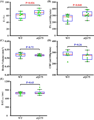 |
Non-contrast assessment of blood-brain barrier permeability to
water in mice: An arterial spin labeling study
Zhiliang Wei1,2,
Hongshuai Liu3,
Zixuan Lin1,
Minmin Yao3,
Ruoxuan Li3,
Chang Liu3,
Yuguo Li1,2,
Jiadi Xu1,2,
Wenzhen Duan3,4,
and Hanzhang Lu1,2,5
1Russell H. Morgan Department of Radiology and Radiological Science, Johns Hopkins University School of Medicine, Baltimore, MD, United States, 2F. M. Kirby Research Center for Functional Brain Imaging, Kennedy Krieger Research Institute, Baltimore, MD, United States, 3Department of Psychiatry and Behavioral Sciences, Johns Hopkins University School of Medicine, Baltimore, MD, United States, 4The Solomon H. Snyder Department of Neuroscience, Johns Hopkins University School of Medicine, Baltimore, MD, United States, 5Department of Biomedical Engineering, Johns Hopkins University School of Medicine, Baltimore, MD, United States Keywords: Arterial spin labelling, Animals Blood-brain barrier (BBB) plays a critical role in brain health and diseases. However, there is a scarcity of tools to assess BBB integrity, particularly in mouse models. Here, we aimed to develop a non-contrast arterial-spin-labeling-based MRI technique to estimate BBB permeability in mice by measuring relative fractions of labeled water in cerebral veins. Systematic optimizations were performed to enhance signal sensitivity with a further study investigating reproducibility. The proposed method revealed significant BBB dysfunctions in a mouse model of Huntington’s disease, which were further validated with histology. Our method may open new avenues for preclinical mechanistic research or therapeutic trails. |
| 08:31 |
0369.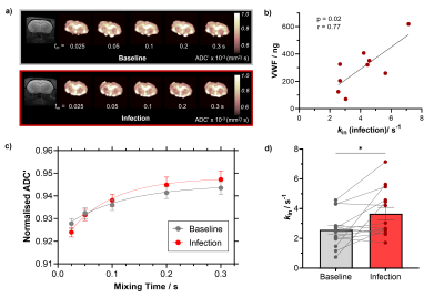 |
FEXI MRI detects increased blood-brain barrier water
permeability in response to mild lung infection
Yolanda Ohene1,2,
Will Harris 3,
Elizabeth Powell4,
Nina W. Wycech3,
Samo Lasič5,6,
Kieron South3,
Graham Coutts3,
Andrew Sharp7,
Catherine B. Lawrence 3,
Hervé Boutin 3,
Geoff J. M. Parker4,8,
Laura M. Parkes1,2,
and Ben R. Dickie2,9
1Division of Psychology, Communication and Human Neuroscience, University of Manchester, Manchester, United Kingdom, 2Geoffrey Jefferson Brain Research Centre, University of Manchester, Manchester, United Kingdom, 3Division of Neuroscience, University of Manchester, Manchester, United Kingdom, 4UCL, London, United Kingdom, 5Danish Research Centre for Magnetic Resonance, Copenhagen, Denmark, 6Random Walk Imaging, Åkarp, Sweden, 7Evotec (UK) Ltd., Cheshire, United Kingdom, 8Bioxydyn Limited, Manchester, United Kingdom, 9Division of Informatics, University of Manchester, Manchester, United Kingdom Keywords: Diffusion/other diffusion imaging techniques, Permeability Non-disruption alterations to the blood-brain barrier (BBB) can be difficult to detect and therefore require highly sensitive tools for reliable measurement. Here, we apply a BBB filter exchange imaging (BBB-FEXI) technique to assess the rat brain in response to mild Streptococcus pneumoniae lung infection. We observe a significant 78 ± 39 % increase in BBB water permeability during infection. Higher water exchange measures were associated with higher levels of vascular inflammation, while BBB tight junction proteins remained unchanged. The expression of aquaporin-4 water channel was 38% higher in infected animals, which may drive the increase in water exchange during infection. |
| 08:39 |
0370.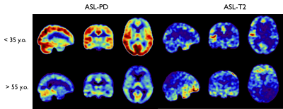 |
Simultaneous imaging of cerebral perfusion and blood-CSF
exchange using dual-TE split echo train Fast-Spin-Echo Arterial
Spin Labeling
Manuel Taso1 and
David C Alsop1 1Division of MRI Research, department of Radiology, Beth Israel Deaconess Medical Center, Harvard Medical School, Boston, MA, United States Keywords: Arterial spin labelling, Neurofluids The recent years have seen a growing interest in the study of brain waste clearance mechanisms, also referred to as glymphatics. Recently, ASL has been used to image choroid plexus perfusion but also the exchange between blood and CSF by taking advantage of the long CSF T2 compared to blood and brain tissue. We evaluate the possibility to measure this using reduced flip-angles FSE with variable-density k-space sampling, using a split echo-train strategy to collect in the same echo-train a short-TE PD-weighted as well as long-TE T2-weighted ASL datasets. We evaluate in a pilot group potential age-related differences. |
| 08:47 |
0371.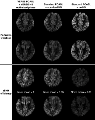 |
Minimizing SAR for SNR-Efficient Pseudo-Continuous Arterial Spin
Labeling at 7T
Joseph G. Woods1,
Mark Chiew1,2,3,
and Thomas W. Okell1
1Wellcome Centre for Integrated Neuroimaging, FMRIB, Nuffield Department of Clinical Neuroscience, University of Oxford, Oxford, United Kingdom, 2Department of Medical Biophysics, University of Toronto, Toronto, ON, Canada, 3Physical Sciences, Sunnybrook Research Institute, Toronto, ON, Canada Keywords: Arterial spin labelling, High-Field MRI
Interest in 7T PCASL
is increasing due to the large potential increases in SNR.
However, PCASL is a relatively high SAR technique, requiring
TRs to be extended with deadtime at 7T, which reduces
SNR-efficiency and some of the benefit of moving to 7T.
Here, we demonstrate that using VERSE to reduce the SAR of
both the PCASL and background suppression pulses can reduce
TRs by almost 25%, leading to an improvement in
tSNR-efficiency compared to an equivalent non-VERSE scan. We
reduced the off-resonance sensitivity that VERSE introduces
to the background suppression pulses by carefully optimizing
the phase waveform. |
| 08:55 |
0372.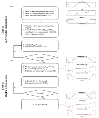 |
Hybrid Adiabatic Pulse with asYmmetry (HAPY): an asymmetric
adiabatic pulse with an application in pulsed ASL at 7T
Didi Chi1,2,
Yasmin Blunck1,2,
Rebecca Glarin2,
Catherine E. Davey1,2,
Daniel Stäb3,
Josef Pfeuffer4,
Leigh A. Johnston1,2,
and Jin Jin5
1Department of Biomedical Engineering, University of Melbourne, Parkville, Australia, 2Melbourne Brain Centre Imaging Unit, University of Melbourne, Parkville, Australia, 3MR Research Collaborations, Siemens Healthcare Pty Ltd, Melbourne, Australia, 4MR Research Collaborations, Siemens Healthcare Pty Ltd, Sydney, Australia, 5MR Research Collaborations, Siemens Healthcare Pty Ltd, Brisbane, Australia Keywords: Arterial spin labelling, Arterial spin labelling, Pulsed ASL The increased power deposition and inhomogeneity of $$$\Delta{B_0}$$$ and $$$B_1^+$$$ fields limit the application of ASL at 7T. A new type of asymmetric adiabatic pulses, Hybrid Adiabatic Pulses with asYmmetry (HAPY) along with its optimisation scheme, is proposed in this study. In simulation and phantom experiments, the proposed HAPY pulse demonstrates RF labelling pulse energy reduction and high labelling efficiency under challenging $$$\Delta{B_0}$$$ and $$$B_1^+$$$ conditions. In vivo experiments show the successful application of PICORE-PASL with HAPY labelling pulses in 7T ASL with increased temporal resolution. |
| 09:03 |
0373.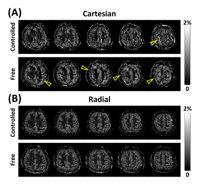 |
Balanced Steady-State Free Precession and Radial Sampling for
Arterial Spin Labeled Perfusion Imaging
Paul Han1,2,
Thibault Marin1,2,
Yue Zhuo1,2,
Jinsong Ouyang1,2,
Georges El Fakhri1,2,
and Chao Ma1,2
1Gordon Center for Medical Imaging, Department of Radiology, Massachusetts General Hospital, Boston, MA, United States, 2Department of Radiology, Harvard Medical School, Boston, MA, United States Keywords: Arterial spin labelling, Perfusion, Brain Arterial spin labeling (ASL) is a non-invasive MRI technique that allows to quantitatively measure cerebral blood flow. However, the major limitation of ASL is in the intrinsically low signal-to-noise ratio (SNR). Balanced steady-state free precession (bSSFP) sequence has been proposed to mitigate this limitation, however, bSSFP is sensitive to off-resonance effects. ASL perfusion imaging with bSSFP can be sensitive to effects from motion and flow when Cartesian sampling scheme is used. This work proposes and investigates radial sampling scheme for ASL with bSSFP to allow perfusion imaging with relatively high SNR and robustness to motion and off-resonance effects. |
| 09:11 |
0374.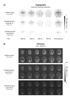 |
Simultaneous 4D ASL angiography and perfusion MRI using
time-encoded pCASL preparation with Stack of Spirals readout
Merlijn C.E. van der Plas1,2,
Kirsten Koolstra3,
Martijn Nagtegaal2,
Emiel Hartsema2,
Lena Vaclavu2,
Sophie Schmid2,
Leoni Petitclerc2,
Peter Bornert4,
and Matthias van Osch2 1University Medical Center Utrecht, Utrecht, Netherlands, 2C.J. Gorter Center for high field MRI, Leiden University Medical Center, Leiden, Netherlands, 3Phillips, Best, Netherlands, 4Phillips, Hamburg, Germany Keywords: Arterial spin labelling, Perfusion In this study, a framework was set up for the simultaneous acquisition of 4D MRA and perfusion ASL data while maintaining whole brain coverage. A Hadamard-8 preparation was combined with a 3D Stack of Spirals readout which resulted in 7 timepoints. By combining these two techniques we were able to obtain both angiography and perfusion from a single dataset. By improving the efficiency of the sampling scheme i.e. using variable density spirals, the total scan time could be reduced further while improving the SNR during the perfusion phase, albeit at the expense of the quality of the 4D MRA. |
| 09:19 |
0375.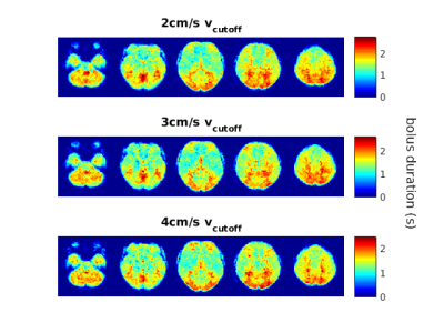 |
Velocity-selective arterial spin labelling bolus duration
measurements: Implications for consensus recommendations
Ian D Driver1,
Hannah L Chandler1,
Eleonora Patitucci2,
Richard G Wise1,3,4,
and Michael Germuska2
1Cardiff University Brain Research Imaging Centre (CUBRIC), School of Psychology, Cardiff University, Cardiff, United Kingdom, 2Cardiff University Brain Research Imaging Centre (CUBRIC), School of Physics and Astronomy, Cardiff University, Cardiff, United Kingdom, 3Department of Neurosciences, Imaging and Clinical Sciences, ‘G. d’Annunzio University’ of Chieti-Pescara, Chieti, Italy, 4Institute for Advanced Biomedical Technologies (ITAB), ‘G. d’Annunzio University’ of Chieti-Pescara, Chieti, Italy Keywords: Arterial spin labelling, Brain, cerebrovascular reactivity Velocity-selective ASL (VSASL) measurement of CBF is relatively insensitive to arterial arrival times, unlike other ASL techniques, making it suitable for measuring CBF in patient groups with long arrival times. However, VSASL can underestimate CBF when the trailing edge of the labelled bolus arrives before imaging. Our study finds substantial spatial heterogeneity in bolus duration in Fourier Transform velocity-selective inversion (FT-VSI), with short bolus durations in anterior regions and longer bolus durations in posterior regions. These results build on a recent VSASL consensus paper contributing to recommendations for cerebrovascular reactivity mapping experiments. |
| 09:27 |
0376.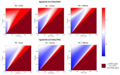 |
Analytical model for determination of exchange times in multi-TE
velocity-selective Arterial Spin Labeling
Mareike Alicja Buck1,2,
Klaus Eickel1,3,
and Matthias Günther1,2,3
1Fraunhofer MEVIS, Bremen, Germany, 2University Bremen, Bremen, Germany, 3mediri GmbH, Heidelberg, Germany Keywords: Arterial spin labelling, Arterial spin labelling, velocity-selective Arterial Spin Labeling, multi-TE In this work, for the first time, an analytical model for determining the exchange times for obtaining vascular permeability information in velocity-selective Arterial Spin Labeling (VSASL) with multiple echo times (TE) is presented. Signal intensity comparisons were performed between different multi-TE VSASL techniques with each of the standard multi-TE pCASL and multi-TE PASL techniques for different sampling times. This demonstrates that, particularly for longer arterial transit times, the VSASL signal is twice as high compared with standard multi-TE measurements and thus shows promise in the field of non-invasive permeability measurements. |
| 09:35 |
0377.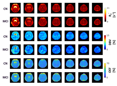 |
Oxygen Extraction Fraction mapping between normal cognition and
mild cognitive impairment in an elderly cohort using
quantitative BOLD
Linh N. N. Le1,
Gregory J. Wheeler1,
Evan Fletcher2,
Nicholas P. Blockley3,
and Audrey P. Fan1,2
1Department of Biomedical Engineering, University of California, Davis, Davis, CA, United States, 2Department of Neurology, University of California, Davis, Davis, CA, United States, 3School of Medicine & Health Sciences, University of Nottingham, Nottingham, United Kingdom Keywords: Oxygenation, Aging Oxygen Extraction Fraction (OEF) is a valuable indicator of brain physiology influenced by cognitive impairment and vascular pathology in older adults. Our objective is to explore age-related OEF change for cognitively normal (CN) and Mild-Cognitive-Impairment (MCI) subjects. In this study, 56 elderly participants were recruited. Individuals with MCI resulted in higher OEF compared to CN participants, especially in the occipital lobe (p=0.007). In parietal lobe, we observed an inverse correlation between OEF and age (p=0.026) in CN and positive association between OEF and executive function (p=0.023) in MCI. The results showed an effect of aging and cognitive impairment on OEF |
| 09:43 |
0378.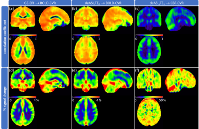 |
Investigating the applicability of dual echo ASL for
simultaneous BOLD- and CBF-based mapping of cerebrovascular
reactivity
Gabriel Hoffmann1,2,
Franziska Richter1,
Jan Kufer1,
Lena Schmitzer1,
Carina Gleißner1,
Jens Göttler1,
Stephan Kaczmarz1,2,3,
and Christine Preibisch1,2
1Department of Diagnostic and Interventional Neuroradiology, Klinikum rechts der Isar, Technical University of Munich, Munich, Germany, 2TUM-Neuroimaging Center, Klinikum rechts der Isar, Technical University of Munich, Munich, Germany, 3Philips GmbH Market DACH, Hamburg, Germany Keywords: Multi-Contrast, Validation, Cerebrovascular reactivity Combined measurement of BOLD- and CBF-based CVR by means of deASL would be valuable for assessment of cerebrovascular diseases. To investigate the applicability of deASL for CVR mapping, we acquired GE-EPI, deASL and pCASL data from 21 healthy volunteers during a hypercapnia challenge. BOLD-CVR from deASL was slightly but significantly less sensitive to hypercapnia-induced BOLD signal changes than GE-EPI, while differences between deASL- and pCASL-based CBF-CVR were rather insignificant. However, deASL failed in 5 of 21 subjects, while pCASL and GE-EPI merely failed in two and one subjects, respectively. Thus, we expect an elevated deASL failure rate in patient populations. |
| 09:51 |
0379.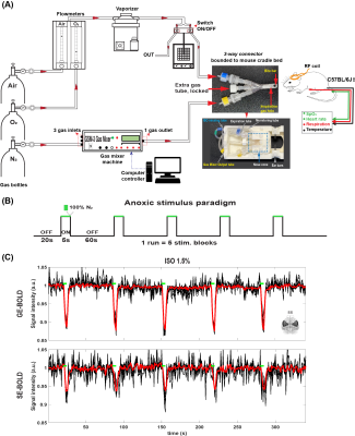 |
Quantitative perfusion mapping by BOLD-DSC MRI with transient
hypoxia in mouse under volatile anesthetics
THI THUY LE1,2,3,
GEUN HO IM1,
CHAN HEE LEE1,
and SEONG-GI KIM1,2,3
1Center for Neuroscience Imaging Research (CNIR), Institute for Basic Science (IBS), Suwon, 16419, Republic of Korea, Suwon, Korea, Republic of, 2Department of Biomedical Engineering, Sungkyunkwan University, Suwon, 16419, Republic of Korea, Suwon, Korea, Republic of, 3Department of Intelligent Precision Healthcare Convergence, Sungkyunkwan University, Suwon, 16419, Republic of Korea, Suwon, Korea, Republic of Keywords: Hematologic, DSC & DCE Perfusion Transient hypoxia-induced BOLD-DSC perfusion imaging approach can noninvasively map cerebral blood flow (CBF) and cerebral blood volume (CBV) in animals with injectable anesthetics. Since animal models are frequently investigated in translational research under volatile anesthetics, we developed a simple gas delivery system for inducing transient hypoxia, and performed perfusion studies of mouse under isoflurane and dexmedetomidine. Highly sensitive and reproducible perfusion metrics were found, and regional impact of anesthesia to basal perfusion metrics was assessed. |
| 09:58 |
0380.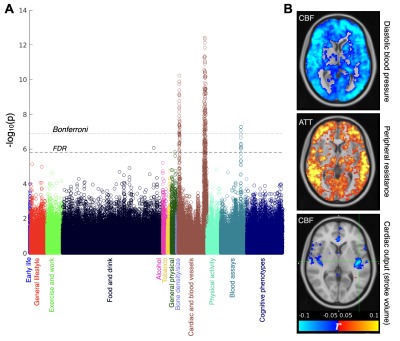 |
Population Brain Perfusion Imaging: Arterial Spin Labeling in UK
Biobank
Thomas W Okell1,
Karla L Miller1,
Martin Craig2,
Fidel Alfaro-Almagro1,
David L Thomas3,4,
Enrico De Vita5,
Matthias Günther6,
Paul M Matthews7,
Stephen M Smith1,
and Michael A Chapell2
1Wellcome Centre for Integrative Neuroimaging, FMRIB, Nuffield Department of Clinical Neurosciences, University of Oxford, Oxford, United Kingdom, 2Sir Peter Mansfield Imaging Centre, School of Medicine, University of Nottingham, Nottingham, United Kingdom, 3Dementia Research Centre, UCL Queen Square Institute of Neurology, University College London, London, United Kingdom, 4Dept of Brain Repair and Rehabilitation, UCL Queen Square Institute of Neurology, University College London, London, United Kingdom, 5MR Physics group, Radiology Department, Great Ormond Street Hospital, London, United Kingdom, 6Fraunhofer Institute for Digital Medicine MEVIS, Bremen, Germany, 7Department of Brain Sciences, Imperial College London, London, United Kingdom Keywords: Arterial spin labelling, Arterial spin labelling UK Biobank is an ongoing study which will acquire brain imaging in 100,000 participants, along with detailed lifestyle, biochemical and genetic information, and long-term health records. Here we describe the recent addition of arterial spin labeling (ASL) perfusion imaging to UK Biobank, with preliminary analyses of the first ~3,500 subjects. These revealed a range of highly significant associations between ASL metrics and non-imaging measures (e.g., blood pressure), as well as other imaging measures (e.g., white matter hyperintensity volume). This should become a useful resource for studying perfusion differences across a population and prior to disease onset. |
 Back to Meeting Home
 Back to the Program-at-a-Glance
The International Society for Magnetic Resonance in Medicine is accredited by the Accreditation Council for Continuing Medical Education to provide continuing medical education for physicians.
 Back to Meeting Home
 Back to the Program-at-a-Glance
 View Presentation Video