Oral Session
1H Spectroscopy
ISMRM & ISMRT Annual Meeting & Exhibition • 03-08 June 2023 • Toronto, ON, Canada

13:30 |
0489.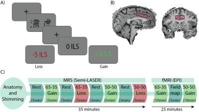 |
Combined fMRS and fMRI During Reinforcement Learning in a Large
Cohort at 7T: When Does Cognitive Processing Occur?
Tal Finkelman1,
Edna Furman-Haran2,
Kristoffer Carl Mikael Aberg3,
Rony Paz3,
and Assaf Tal1 1Chemical and Biological Physics, Weizmann Institute of Science, Rehovot, Israel, 2life sciences core facilities, Weizmann Institute of Science, Rehovot, Israel, 3Brain Sciences, Weizmann Institute of Science, Rehovot, Israel Keywords: Spectroscopy, fMRI (task based), functional MRS We present multimodal functional MRS-fMRI-Behavioral data, which demonstrates how the E/I balance changes in the dACC during a reinforcement learning paradigm. The E/I balance decreases during rest periods between tasks, supporting a consolidation phase that is invisible to BOLD-fMRI. Additionally, we find a significant negative correlation between both GABA and glutamate, and the mean z-score of the BOLD signal from the spectroscopic voxel, during the decision-making game. We suggests that the elevation in Glu is related to cellular activity rather than neuronal activity, indicating a GABAergic activation during the task. |
|
13:38 |
0490.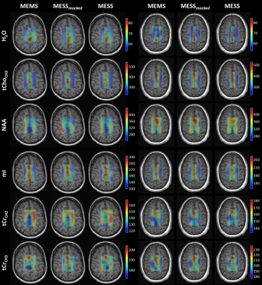 |
Simultaneous concentration and T2 mapping of brain metabolites
by multi-echo spectroscopic imaging
Rudy Rizzo1,2,
Angeliki Stamatelatou3,
Arend Heerschap3,
Tom Scheenen3,
and Roland Kreis1,2
1Magnetic Resonance Methodology, Institute of Diagnostic and Interventional Neuroradiology, University of Bern, Bern, Switzerland, 2Translational Imaging Center (TIC), Swiss Institute for Translational and Entrepreneurial Medicine, Bern, Switzerland, 3Department of Medical Imaging, Radboud University Medical Center, Nijmegen, Netherlands Keywords: Spectroscopy, Data Acquisition, T2-mapping A multi-parametric MR Spectroscopic Imaging (MRSI) experiment (Multi-Echo Single-Shot MRSI, MESS-MRSI) deploys partially sampled multi-echo trains from single readouts combined with simultaneous multi-parametric model fitting to produce metabolite-specific T2 and concentration maps in 7min. It was tested in-vivo on a cohort of 5 subjects. Cramer-Rao Lower-Bounds (CRLBs) are used as measure of performance. The novel scheme was compared with the (i) traditional Multi-Echo Multi-Shot (MEMS) method and (ii) a truncated version of MEMS, which mimics the MESS acquisition (MESS-mocked). Results extended former findings for single voxel measurements with improvements in CRLB ranging from 17-45% for concentrations and 10-23% for T2s. |
| 13:46 |
0491.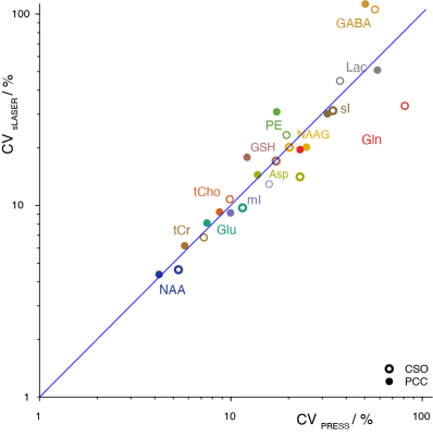 |
sLASER Performed Similarly to PRESS at Revealing Metabolite-Age
Correlations
Steve C.N. Hui1,2,
Tao Gong3,4,
Helge J. Zöllner1,2,
Kathleen Hupfeld1,2,
Aaron Gudmundson1,2,
Yulu Song1,2,
Saipavitra Murali-Manohar1,2,
Christopher Davies-Jenkins1,2,
Georg Oeltzschner1,2,
Guangbin Wang3,4,
and Richard A. E. Edden1,2
1Radiology and Radiological Sciences, Johns Hopkins University School of Medicine, Baltimore, MD, United States, 2F.M. Kirby Research Center for Functional Brain Imaging, Kennedy Krieger Institute, Baltimore, MD, United States, 3Department of Radiology, Shandong Provincial Hospital Affiliated to Shandong First Medical University, Jinan, China, 4Department of Radiology, Shandong University, Jinan, China Keywords: Spectroscopy, Data Analysis, magnetic resonance spectroscopy, sLASER, PRESS, localization, aging Recent MRS community consensus recommended sLASER over PRESS for reduced chemical-shift displacement. There is very little evidence supporting this consensus in terms of ability to reveal in vivo biochemistry. sLASER- and PRESS-localized spectra were collected in gray- and white-matter regions in 102 adult subjects (aged 20-69). sLASER showed slightly higher SNR than PRESS (by 4% on average), but improved SNR and localization did not convert into reduced variance or improved detection of metabolite-age correlations. Between-subject CVs of 13 modeled metabolites were remarkably consistent, and the pattern of metabolite-age correlations was also similar. |
| 13:54 |
0492.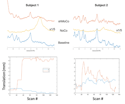 |
Prospective motion-corrected spectroscopy in the human cervical
spinal cord
Isaac M Adanyeguh1,
Pierre-Gilles Henry1,
and Dinesh K Deelchand1
1Center for Magnetic Resonance Imaging, University of Minnesota, Minneapolis, MN, United States Keywords: Spectroscopy, Spinal Cord Proton magnetic resonance spectroscopy in the spinal cord is particularly challenging and motion remains one of biggest obstacles. Although several prospective motion correction techniques currently exist in the human brain, none have yet been reported in the spinal cord. Here we report prospective motion correction in the spinal cord using reduced-field-of-view 2DRF excitation. Results show than spectral quality were similar with and without motion. This study demonstrates the feasibility of prospective motion correction for spinal cord MRS. |
| 14:02 | 0493. |
Use of fast B1 shimming to enable localized MR spectroscopy in
different regions of the human brain on a clinical platform at 7
Tesla
Özlem Ipek1,2,
Jeremie Clement1,3,
Marzena Arridge4,
Philippa Bridgen2,
Tom Wilkinson2,
Laura Mancini4,
Raphael Tomi-Tricot2,5,
Enrico de Vita2,6,
and Ralf Mekle7 1Biomedical Engineering, King's College London, London, United Kingdom, 2London Collaborative Ultra high field System (LoCUS), King's College London, London, United Kingdom, 3System Technologies, Siemens Healthcare GmbH, Erlangen, Germany, 4Lysholm Department of Neuroradilogy, National Hospital for Neuroradiology and Neurosurgery, London, United Kingdom, 5MR Research Collaborations, Siemens Healthiness, Frimley, United Kingdom, 6Radiology, Great Ormond Street Hospital, London, United Kingdom, 7Center for Stroke Research Berlin, Charité Universitätsmedizin Berlin, Berlin, Germany Keywords: Spectroscopy, High-Field MRI, Paralel Transmit and B1 shimming We demonstrated that applying a fast implementation of an RF phase shimming method enabled the acquisition of localized MR spectra in different regions of the human brain including B1 challenging areas, such as the hippocampus and prefrontal cortex, using a commercially available pTx coil on a clinical platform at 7T. RF efficiency was increased by ~100% cerebellum and PFC while ~50-75% for pons and hippocampus. The most favorable flip angle distributions were obtained for the cerebellum and the prefrontal cortex. Fast B1 shimming with advanced MRS methodology enables high-quality single voxel spectra at 7T using a commercially available setup. |
| 14:10 |
0494.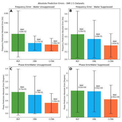 |
Frequency and Phase Correction of GABA-edited MR Spectroscopy
using Complex-valued Convolutional Neural Network
Hanna Bugler1,2,3,4,
Rodrigo Berto1,2,3,4,
Roberto Souza1,3,5,
and Ashley D. Harris2,3,4
1Biomedical Engineering Department, University of Calgary, Calgary, AB, Canada, 2Department of Radiology, University of Calgary, Calgary, AB, Canada, 3Hotchkiss Brain Institute, University of Calgary, Calgary, AB, Canada, 4Alberta Children's Hospital Research Institute, University of Calgary, Calgary, AB, Canada, 5Electrical and Software Engineering Department, University of Calgary, Calgary, AB, Canada Keywords: Spectroscopy, Machine Learning/Artificial Intelligence, Brain Edited Magnetic Resonance Spectroscopy (Edited-MRS) is important for the quantification of ɣ-amino butyric acid (GABA) in vivo. However, during acquisition, data may suffer phase and frequency shifts, which affects the quality of the output spectrum. Frequency and phase correction (FPC) is necessary to account for these shifts, and deep learning models have obtained recent success in this task. Still, current methods do not take into consideration that MRS data is complex-valued. We propose a complex-valued convolutional neural network model for FPC. Our results showed that our model compares favorably against two recently proposed deep learning methods. |
14:18 |
0495.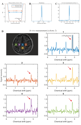 |
Single-shot SLOW-editing for Downfield α-Glucose MRSI at 7T
Utilizing SNR-Enhancing Glucose Mutarotation Effect
Guodong Weng1,2,
Piotr Radojewski1,2,
and Johannes Slotboom1,2
1Support Center for Advanced Neuroimaging (SCAN), University of Bern, Bern, Switzerland, 2Translational Imaging Center, sitem-insel, Bern, Switzerland Keywords: Pulse Sequence Design, Spectroscopy, Spectral Editing Changes in brain glucose occur in many neurological disorders as well as during aging. Most studies on the uptake of glucose in the brain use positron emission tomography, which requires injection of a radioactive tracer. Our study shows that ultra-high-field 1H-MRS single-shot SLOW-editing can be used to measure α-D-glucose at 5.22 ppm in vivo, and thus that α-D-glucose might have the potential to be used as a an economic radiation free tracer in the human brain. |
| 14:26 |
0496.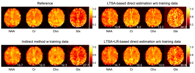 |
Direct estimation of metabolite maps from undersampled k-space
data using linear tangent space alignment
Chao Ma1,2,
Thibault Marin1,2,
Paul K. Han1,2,
and Georges El Fakhri1,2
1Radiology, Massachusetts General Hospital, Boston, MA, United States, 2Radiology, Harvard Medical School, Boston, MA, United States Keywords: Spectroscopy, Image Reconstruction, MRSI quantification Conventional MRSI methods perform MRSI image reconstruction and spectral quantification in two separate steps. This work presents a novel direct estimation method for MRSI that reconstructs high-resolution metabolite concentration maps from undersampled k-space data, leveraging a linear tangent space alignment (LTSA) model for spectral quantification and a low-rank (LR) model for denoising. Furthermore, the proposed framework allows estimating the temporal basis functions of the LR model from the undersampled, noise-corrupted k-space data, thus eliminating the need for experiment-dependent or pre-acquired spectral training data. The performance of the proposed method was validated using numerical simulation phantom and in vivo MRSI data. |
| 14:34 |
0497. |
Reconstruction of High-Resolution Metabolite Maps from Noisy
MRSI Data by Incorporating Spatiospectral Constraints through
Learned Kernels
Yudu Li1,2,
Chao Ma3,4,
Shirui Luo2,
Wen Jin1,5,
Ruihao Liu1,
Georges El Fakhri3,4,
Yao Li6,
Maria Jaromin2,
Volodymyr Kindratenko2,5,
Brad Sutton1,2,7,8,
and Zhi-Pei Liang1,2,5
1Beckman Institute for Advanced Science and Technology, University of Illinois at Urbana-Champaign, Urbana, IL, United States, 2National Center for Supercomputing Applications, University of Illinois at Urbana-Champaign, Urbana, IL, United States, 3Radiology, Harvard Medical School, Boston, MA, United States, 4Radiology, Massachusetts General Hospital, Boston, MA, United States, 5Department of Electrical and Computer Engineering, University of Illinois at Urbana-Champaign, Urbana, IL, United States, 6School of Biomedical Engineering, Shanghai Jiao Tong University, Shanghai, China, 7Department of Bioengineering, University of Illinois at Urbana-Champaign, Urbana, IL, United States, 8Carle Illinois College of Medicine, University of Illinois at Urbana-Champaign, Urbana, IL, United States Keywords: Spectroscopy, Image Reconstruction High-resolution MR spectroscopic imaging (MRSI) suffers from very low signal-to-noise ratio, which is often addressed using a priori information/constraints. Existing constrained reconstruction methods utilize spectral constraints in the form of spectral subspaces/manifolds, while impose spatial constraints though spatial regularization. This paper presents a novel kernel-based partial separability model for reconstruction of high-resolution of metabolite maps from noisy MRSI data. The proposed model uses spectral basis functions to absorb spectral prior and a learned kernel function to absorb spatial prior. Experimental results demonstrated very encouraging reconstruction performance. |
| 14:42 |
0498.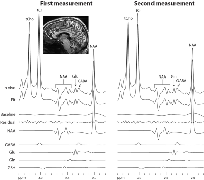 |
Single-shot GABA Editing at 7 Tesla
Li An1,
Maria Ferraris Araneta1,
Inna Loutaev1,
Tara Turon1,
Christopher S Johnson1,
Sungtak Hong1,
and Jun Shen1 1National Institute of Mental Health, National Institutes of Health, Bethesda, MD, United States Keywords: Data Acquisition, Spectroscopy, MRS, GABA, 7 Tesla Two-step editing techniques have been widely used to detect the GABA H4 signal at 3.01 ppm with the dominant creatine methyl proton signal cancelled by subtraction. However, subtraction is inherently sensitive to patient movements and system instability. In this work, a single-shot spectral editing technique is developed to detect the GABA H2 resonances at 2.28 ppm in the human brain at 7 Tesla. Although GABA H2 is partially overlapped by glutamate H4, we demonstrate that GABA-glutamate correlation originating from spectral overlap can be reduced to practically zero, therefore suppressing the overlapping effect of glutamate H4 on GABA H2 without subtraction. |
| 14:50 |
0499.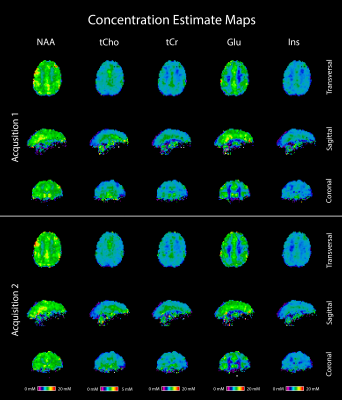 |
Intra-Subject Stability and Reproducibility of 7 T FID-CRT-MRSI
Philipp Lazen1,
Benjamin Spurny-Dworak2,
Wolfgang Bogner3,4,
Lukas Hingerl3,
Bernhard Strasser3,
Rupert Lanzenberger2,
Karl Rössler1,
Siegfried Trattnig3,4,5,
and Gilbert Hangel1,3
1Department of Neurosurgery, Medical University of Vienna, Vienna, Austria, 2Division of General Psychiatry, Department of Psychiatry and Psychotherapy, Medical University of Vienna, Vienna, Austria, 3High-field MR Center, Department of Biomedical Imaging and Image-guided Therapy, Medical University of Vienna, Vienna, Austria, 4Christian Doppler Laboratory for MR Imaging Biomarkers, Vienna, Austria, 5Institute for Clinical Molecular MRI, Karl Landsteiner Society, St. Pölten, Austria Keywords: Spectroscopy, Brain We evaluated the intra-subject stability of CRT-FID-MRSI at 7 T in a cohort of 15 volunteers. Two magnetic resonance spectroscopic imaging acquisitions were performed consecutively and metabolite concentration estimates (CEs) were calculated using internal water referencing. The resulting metabolic maps were segmented in 55 brain regions. For analysis, the CEs of both acquisitions were compared and coefficients of variations (CVs) were calculated. We saw high stability in almost all volunteers, with CVs being well below 5% in the brain regions generally associated with good data quality. |
| 14:58 |
0500.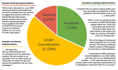 |
Toward the Use of MRS Methodological Consensus by the Clinical
Research Community - An Early Assessment of Dissemination
Results
Jodi J. Weinstein1,2,
Abigail Dalton1,
Jack Kaufman1,
In-Young Choi3,4,
Roland Kreis5,6,
and Christoph Juchem7,8
1Department of Psychiatry and Behavioral Health, Renaissance School of Medicine at Stony Brook University, Stony Brook, NY, United States, 2New York State Psychiatric Institute and Department of Psychiatry, Columbia University Irving Medical Center, New York City, NY, United States, 3Department of Neurology, University of Kansas Medical Center, Kansas City, KS, United States, 4Hoglund Biomedical Imaging Center, University of Kansas Medical Center, Kansas City, KS, United States, 5Magnetic Resonance Methodology, Institute of Diagnostic and Interventional Neuroradiology, University of Bern, Bern, Switzerland, 6Translational Imaging Center, sitem-insel, Bern, Switzerland, 7Department of Biomedical Engineering, Columbia University School of Engineering and Applied Science, New York, NY, United States, 8Department of Radiology, Columbia University Irving Medical Center, New York, NY, United States Keywords: Spectroscopy, Translational Studies The MRS community recently established a set of expert Consensus Recommendations. To date, however, knowledge on their implementation beyond anecdotal reports is largely lacking. Here we report a preliminary assessment of the achieved dissemination and, more importantly, the level of acceptance of the established guidelines and standards by the clinical MRS research community. We compiled feedback from 22 editors-in-chief of major clinical journals, together accounting for more than 100 (>12%) of the in vivo clinical MRS publications from 2021. |
| 15:06 |
0501.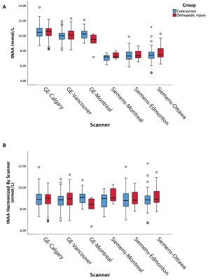 |
Differential MRS results in a clinical multisite study depending
on site harmonization and analysis approach
Parker L La1,
Tiffany K Bell1,
William Craig2,
Quynh Doan3,
Miriam H Beauchamp4,
Roger Zemek5,
Keith O Yeates6,
and Ashley D Harris1
1Radiology, University of Calgary, Calgary, AB, Canada, 2University of Alberta and Stollery Children's Hospital, Edmonton, AB, Canada, 3University of British Columbia, Vancouver, BC, Canada, 4University of Montreal and St Justine Hospital, Montreal, QC, Canada, 5University of Ottawa and Children's Hospital of Eastern Ontario, Ottawa, ON, Canada, 6Psychology, University of Calgary, Calgary, AB, Canada Keywords: Spectroscopy, Brain, Traumatic Brain Injury The use of multiple sites and scanners is commonly used to increase sample size; however, this introduces unwanted variance into spectroscopy data. In this work we compare statistical co-variate techniques in combination with ComBat harmonization using data from a concussion study. We show traditional statistical methods of accounting for multi-site variance (using covariates to control for site and vendor) could result in erroneous interpretation of clinical data. Our data supports the use of ComBat harmonization and suggests how to best control for multisite data statistically. |
| 15:14 |
0502.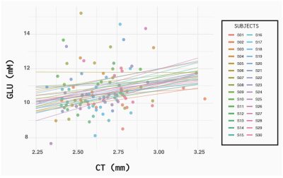 |
Glutamate and its relation to cortical thickness and perfusion
across the cingulate cortex
Jessica Archibald1,
Erin L. MacMillan2,3,
Niklaus Zölch4,
and John L.K. Kramer5 1Experimental Medicine, University of British Columbia, Vancouver, BC, Canada, 2ImageTech, Simon Fraser University, Surey, BC, Canada, 3Philips, Markham, ON, Canada, 4Psychiatry and Forensic Medicine, Universität Zürich, Zürich, Switzerland, 5Anesthesiology, Pharmacology and Therapeutics, University of British Columbia, Vancouver, BC, Canada Keywords: Spectroscopy, Neuro, neuroscience Investigating the relationships between morphological, functional, and metabolic changes in the brain can help reveal subtle changes caused by disease. To establish an understanding of the relationship between brain glutamate levels with cortical thickness (CT) and cerebral blood flow (CBF) in the healthy brain, magnetic resonance spectroscopy (MRS), arterial spin labeled MRI and anatomical MRI were obtained from 4 sub-divisions of the cingulate cortex. A linear mixed effects model revealed that glutamate was significantly correlated with CT (p=0.05, R2=0.30) but not with CSF (p=0.21, R2=0.17) within healthy participants. These results will help inform studies of disease or therapeutic interventions. |
| 15:22 |
0503.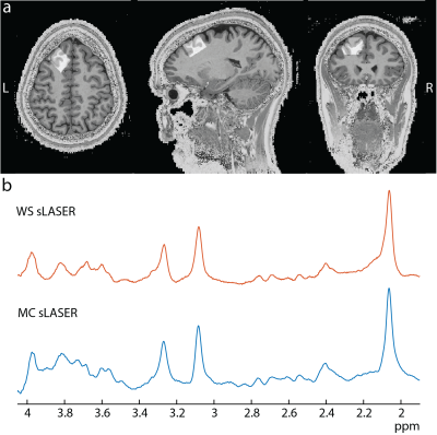 |
Prospective frequency correction with metabolite cycling for
improved spectral quality in the prefrontal cortex at 7T
Kimberly L Chan1,
Ivan E Dimitrov1,2,
Madhukar H Trivedi3,
and Anke Henning1,4 1Advanced Imaging Research Center, The University of Texas Southwestern, Dallas, TX, United States, 2Philips Healthcare, Gainesville, FL, United States, 3Center for Depression Research and Clinical Care, Department of Psychiatry, The University of Texas Southwestern, Dallas, TX, United States, 4Max Planck Institute for Biological Cybernetics, Tübingen, Germany Keywords: Spectroscopy, Brain The prefrontal cortex plays a central role in controlling executive functions such as decision-making and emotional-monitoring and has thus been the subject of several 1H-MRS studies on psychiatric illnesses. This region, however, is sensitive to motion-induced increases in linewidths due to the voxel moving away from the originally shimmed location. Here, we combined metabolite cycling (MC) with continuous prospective frequency correction to better account for frequency instabilities when performing single-voxel brain MRS at 7T. We find that MC sLASER with continuous prospective frequency correction reduces frequency instability, water linewidths, and N-acetylaspartate linewidths relative to semi-LASER acquisitions without prospective frequency correction. |
 Back to Meeting Home
 Back to the Program-at-a-Glance
The International Society for Magnetic Resonance in Medicine is accredited by the Accreditation Council for Continuing Medical Education to provide continuing medical education for physicians.
 Back to Meeting Home
 Back to the Program-at-a-Glance
 View Presentation Video