Oral Session
Brain Microstructure: Restriction & Exchange
ISMRM & ISMRT Annual Meeting & Exhibition • 03-08 June 2023 • Toronto, ON, Canada

Oral Session
Brain Microstructure: Restriction & Exchange
Videos Unavailable
| 15:45 |
0679.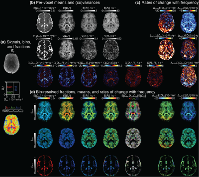 |
Frequency-dependent diffusion-relaxation multidimensional MRI of
the human brain
Jessica TE Johnson1,
Thomas J Ross2,
Yihong Yang2,
Daniel Topgaard3,
and Dan Benjamini1 1National Institute on Aging, Baltimore, MD, United States, 2National Institute on Drug Abuse, Baltimore, MD, United States, 3Lund University, Lund, Sweden Keywords: Microstructure, Diffusion/other diffusion imaging techniques Using an integrative acquisition and processing pipeline that joins concepts from oscillating gradients, tensor-valued encoding, and diffusion-relaxation correlation, we comprehensively explored microstructure and local chemical composition in the human brain. Using both frequency-dependent and tensorial aspects of the encoding spectrum b(ω), we designed an in vivo, whole brain, 40-min 7D D(ω)-R1-R2 distribution acquisition protocol at 2mm isotropic resolution. Scanning eleven healthy participants, we demonstrated frequency/time-dependent changes of diffusion-relaxation correlations measures in the human brain. Finally, intra-scan test–retest repeatability of a range of reconstructed parametric maps was investigated. |
| 15:53 |
0680.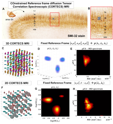 |
COnstrained Reference frame diffusion TEnsor Correlation
Spectroscopic (CORTECS) MRI
Alexandru V Avram1,2,3,
Kadharbatcha Saleem1,
and Peter J Basser1 1Eunice Kennedy Shriver National Institute of Child Health and Human Development, National Institutes of Health, Bethesda, MD, United States, 2Center for Neuroscience and Regenerative Medicine, Bethesda, MD, United States, 3Henry M. Jackson Foundation for the Advancement of Military Medicine Inc., Bethesda, MD, United States Keywords: Diffusion/other diffusion imaging techniques, Microstructure, diffusion tensor distribution, DTD, gray matter, cortical layers, DTI We propose a practical new framework for mapping non-parametric diffusion tensor distributions (DTDs). For diffusion MRI data with sufficiently high spatial resolution, we can constrain all microscopic diffusion tensors of the DTD to be diagonalized using a single orthonormal reference frame estimated from the entire mesoscopic voxel. The constrained DTD is determined by the correlation spectrum of the corresponding microscopic principal diffusivities and can be measured very efficiently using Inverse Laplace Transform methods and single diffusion encoded measurements. cDTD spectral components measured in cortical tissue show good sensitivity to cytoarchitectonic domains and reveal lamination patterns observed in corresponding histological images. |
| 16:01 |
0681.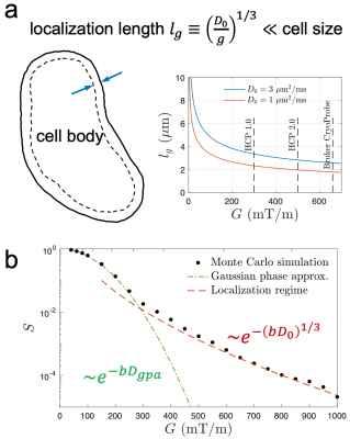 |
Localization Regime of Diffusion (LoRD) in mouse and pig
cortical gray matter and its sensitivity to soma size
Hong-Hsi Lee1,2,
Nian Wang3,4,
Els Fieremans5,
Susie Y Huang1,2,
and Dmitry S Novikov5 1Radiology, Athinoula A. Martinos Center for Biomedical Imaging, Massachusetts General Hospital, Charlestown, MA, United States, 2Radiology, Harvard Medical School, Boston, MA, United States, 3Radiology and Imaging Sciences, Indiana University School of Medicine, Indianapolis, IN, United States, 4Stark Neurosciences Research Institute, Indianapolis, IN, United States, 5Center for Advanced Imaging Innovation and Research, New York University School of Medicine, New York, NY, United States Keywords: Microstructure, Gray Matter It is non-trivial to decompose the diffusion MRI signal, as conventionally measured using relatively weak gradients, into contributions from multiple compartments and estimate the cell sizes. Instead, using strong diffusion gradients, a universal compartment-independent localization regime emerges, as they saturate all magnetization except that near cell membranes. The signal sensitivity to the membrane and its surface-to-volume ratio enables the soma size estimation without modeling multiple compartments. Here, we observed the unique functional form of localization regime and estimate the soma size in mouse and pig brains cortical gray matter on a Bruker CryoProbe system. |
| 16:09 |
0682.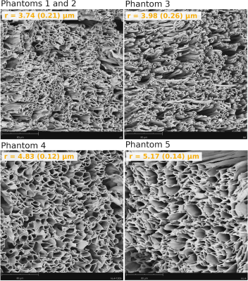 |
Using diffusion-relaxation MRI to estimate the inner radius of
co-electrospun axon-mimicking fibres
Erick Jorge Canales-Rodríguez1,2,
Marco Pizzolato2,3,
Feng-Lei Zhou4,
Muhamed Barakovic5,6,7,
Jean- Philippe Thiran1,8,9,
Derek K. K. Jones10,
Geoffrey J.M. Parker4,11,12,
and Tim B. Dyrby2,3
1Signal Processing Laboratory (LTS5), École Polytechnique Fédérale de Lausanne, Lausanne, Switzerland, 2Danish Research Centre for Magnetic Resonance, Centre for Functional and Diagnostic Imaging and Research, Copenhagen University Hospital, Amager & Hvidovre, Copenhagen, Denmark, 3Department of Applied Mathematics and Computer Science, Technical University of Denmark, Kongens, Lyngby, Denmark, 4Centre for Medical Image Computing, Department of Medical Physics and Biomedical Engineering , University College London, London, United Kingdom, 5Translational Imaging in Neurology (ThINk) Basel, Department of Biomedical Engineering, University Hospital Basel and University of Basel, Basel, Switzerland, 6MS Center and Research Center for Clinical Neuroimmunology and Neuroscience Basel, University Hospital Basel and University of Basel, Basel, Switzerland, 7Roche Pharma Research & Early Development, Neuroscience and Rare Diseases, Roche Innovation Center, Basel, Switzerland, 8Radiology Department, Centre Hospitalier Universitaire Vaudois and University of Lausanne, Lausanne, Switzerland, 9Centre d’Imagerie Biomédicale (CIBM), EPFL, Lausanne, Switzerland, 10Cardiff University Brain Research Imaging Centre, Cardiff University, Cardiff, Wales, United Kingdom, 11Department of Neuroinflammation, Queen Square Institute of Neurology, University College London, London, United Kingdom, 12Bioxydyn Limited, Manchester, United Kingdom Keywords: Diffusion/other diffusion imaging techniques, Validation A new approach for estimating inner axon radii from intra-axonal T2 relaxation times was recently proposed. However, further validations are required before this technique can be used widely. The main aim of this study is to validate this T2-based pore size estimation technique in phantoms comprising co-electrospun hollow axon-mimicking fibres designed to have non-circular cross-sections and different radii distributions. For this purpose, a diffusion-relaxation MRI dataset was acquired with a 7T preclinical scanner, from which the intra-fibre T2 times and pore sizes were estimated. The resulting pore sizes were compared to those measured from Scanning Electron Microscope images. |
16:17 |
0683.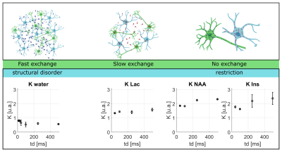 |
Probing lactate exchange in Gray Matter via time-dependent
DW-MRS
Eloïse Mougel1,
Sophie Malaquin1,
Marco Palombo2,3,
and Julien Valette1 1Université Paris-Saclay, CEA, CNRS, MIRCen, Laboratoire des Maladies Neurodégénératives, Fontenay-aux-roses, France, 2Cardiff University Brain Research Imaging Centre (CUBRIC), School of Psychology, Cardiff University, Cardiff, United Kingdom, 3School of Computer Science and Informatics, Cardiff University, Cardiff, United Kingdom Keywords: Microstructure, Diffusion/other diffusion imaging techniques Time-dependent DW-MRS can probe the underlying tissue microstructure. However, it has been previously shown that time-dependent apparent diffusivity and apparent kurtosis exhibit different behaviors for water and intracellular metabolite. These differences may be largely explained by exchange between intra- and extracellular spaces occurring for water. The aim of this work is to measure time-dependent diffusion of lactate which, like water, is present in and exchanges between both compartments, but for which the exchange rate is unknown. Comparison with water and intracellular metabolites indicates that lactate exchange is slow (relative to the probed diffusion times up to 500 ms). |
| 16:25 |
0684.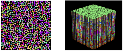 |
Exchange between structurally-disordered compartments
Dmitry S Novikov1,
Ricardo Coronado-Leija1,
and Els Fieremans1
1Radiology, NYU School of Medicine, New York, NY, United States Keywords: Microstructure, Modelling, exchange, diffusion, structural disorder Which signatures of the diffusion signal are responsible for non-Gaussian diffusion inside cells or extra-cellular space, and which ones are the hallmark of the exchange between compartments? Answering this unresolved question is vital for mapping tissue microstructure in brain (especially in gray matter) and body. Here we employ the effective medium formalism to extend the multi-site exchange approach onto the case when diffusion in each tissue compartment is arbitrarily complex. We find the time-dependent diffusivity and kurtosis of a general system of exchanging non-Gaussian compartments for all times, validate with Monte Carlo simulations, and discuss practical implications. |
|
16:33 |
0685.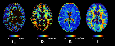 |
Quantifying human gray matter microstructure using NEXI and 300
mT/m gradients
Quentin Uhl1,
Tommaso Pavan1,
Malwina Molendowska2,
Derek K Jones2,
Marco Palombo2,
and Ileana Jelescu1 1Dept. of Radiology, Lausanne University Hospital (CHUV), Lausanne, Switzerland, 2CUBRIC, Cardiff University, Cardiff, United Kingdom Keywords: Microstructure, Modelling For the first time, we report Neurite Exchange Imaging (NEXI) microstructure model parameters estimates in human cortex in vivo. We also investigate the performance of two extensions of this model, the addition of a dot compartment and a correction for wide pulses. Parameter estimates are consistent with previous findings in the rat cortex in vivo. Importantly, NEXI estimates displayed good scan-rescan reproducibility while retaining sensitivity to inter-subject differences. Future work will focus on improving the precision, in particular of the exchange time estimates, possibly leveraging multi-dimensional diffusion MRI acquisitions. |
| 16:41 |
0686.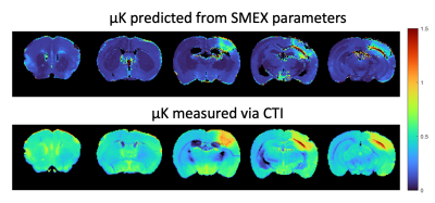 |
Exchange-driven Microscopic Kurtosis in Correlation Tensor MRI
Sune Nørhøj Jespersen1,2,
Rita Alves3,
Jonas Olesen1,2,
Rafael Neto Henriques3,
and Noam Shemesh3
1Department of Physics and Astronomy, Aarhus University, Aarhus, Denmark, 2Center of Functionally Integrative Neuroscience (CFIN) and MINDLab, Department of Clinical Medicine, Aarhus University, Aarhus, Denmark, 3Champalimaud Research, Champalimaud Foundation, Lisboa, Portugal Keywords: Microstructure, Microstructure Correlation Tensor MRI (CTI) provided strong evidence for non-vanishing microscopic kurtosis (µK) in neural tissues, both in animals and in humans. However, µK sources remain to be elucidated. Standard Model with Exchange (SMEX) contrasts have been recently proposed by Olesen et al. for mapping exchange properties, especially in gray matter. Here, we derive an expression for µK originating from the SMEX biophysical model due to exchange, and in a model of stroke compare µK derived from SMEX to µK measured with CTI. Our findings suggest that µK(CTI)>µK(SMEX), suggesting a degree of microstructural origin for the CTI contrast. |
| 16:49 |
0687.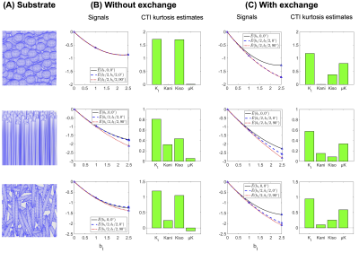 |
Diffusional exchange versus microscopic kurtosis from CTI: Two
conflicting interpretations of the same data
Arthur Chakwizira1,
Filip Szczepankiewicz1,
and Markus Nilsson2
1Medical Radiation Physics, Lund, Lund University, Lund, Sweden, 2Clinical Sciences Lund, Lund University, Lund, Sweden Keywords: Diffusion/other diffusion imaging techniques, Microstructure Correlation tensor magnetic resonance imaging (CTI) is a recently proposed technique that uses diffusion MRI with double diffusion encoding to disentangle microscopic from isotropic and anisotropic kurtosis. One assumption of the method is that intercompartmental exchange is negligible. Another approach that assumes multi-Gaussian exchange (MGE) can also be applied to data acquired with a CTI protocol, but it interprets the contrast as exchange rather than microscopic kurtosis. Using Monte Carlo simulations in substrates of permeable spheres, cylinders and ellipsoids, we observe that both the CTI parameter μK and the estimated exchange rate from MGE increase with the underlying exchange rate. |
| 16:57 |
0688. |
3D diffusion MRI with twin-navigator-based GRASE for cortical
gray matter time-dependency measurements in the human brain
Haotian Li1,
Qinfeng Zhu1,
Yi-Cheng Hsu2,
Yi Sun2,
and Dan Wu1 1Key Laboratory for Biomedical Engineering of Ministry of Education, Department of Biomedical Engineering, College of Biomedical Engineering & Instrument Science, Zhejiang University, Hangzhou, China, 2Siemens Healthineers Ltd, Hangzhou, China Keywords: Diffusion/other diffusion imaging techniques, Brain 3D oscillating gradient sequences enable diffusion measurement at short diffusion-time (td), but it suffered from low resolution and low SNR in clinical systems, and thus, the td-dependency in complex structures, such as cortical gray matter, was not characterized. Here we proposed a twin-navigator-based 3D oscillating gradient diffusion-weighted gradient spin-echo sequence for high-resolution whole-brain td–dMRI acquisition. We demonstrated that different cortical regions exhibited distinct td-dependency patterns with different diffusion dispersion exponent (θ) based on the power-law. We found θ was greater than 0.5 with the highest dispersion in the pre- and post-central cortex and lowest value in the frontal region. |
| 17:05 |
0689.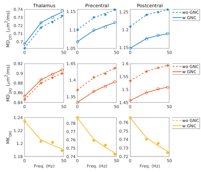 |
Measuring time-dependent diffusion on a high-performance
head-only gradient system at 3T: influence and correction of
gradient nonlinearity
Erpeng Dai1,
Ante Zhu2,
Grant Yang1,3,
Kristin Quah1,3,
Ek T Tan4,
Eric Fiveland2,
Thomas K Foo2,
and Jennifer A McNab1 1Department of Radiology, Stanford University, Stanford, CA, United States, 2GE Research, Niskayuna, NY, United States, 3Department of Electrical Engineering, Stanford University, Stanford, CA, United States, 4Department of Radiology and Imaging, Hospital For Special Surgery, New York, NY, United States Keywords: Microstructure, Diffusion/other diffusion imaging techniques Oscillating gradient spin echo (OGSE) sequence is an effective approach to measure time-dependent diffusion processes at short diffusion times. High-performance, head-only gradient systems open new opportunities for using OGSE to study time-dependent diffusion with both high b-values and oscillating frequencies, such as diffusion kurtosis imaging (DKI). One source of error that can be more problematic for the head-only gradients is gradient nonlinearity (GNL). Here, we measure the time dependence of diffusion tensor imaging (DTI)/DKI in human brains with OGSE on a head-only MAGNUS gradient and investigate the influence of GNL on the time dependence measures of diffusivity/kurtosis. |
| 17:13 |
0690.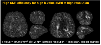 |
High-SNR whole-brain microstructure diffusion MRI using
Romer-EPTI
Fuyixue Wang1,2,
Zijing Dong1,2,
Hong-Hsi Lee1,2,
Susie Y. Huang1,2,3,
Jonathan R. Polimeni1,2,3,
and Lawrence L. Wald1,2,3 1Athinoula A. Martinos Center for Biomedical Imaging, MGH, Charlestown, MA, United States, 2Department of Radiology, Harvard Medical School, Charlestown, MA, United States, 3Harvard-MIT Health Sciences and Technology, MIT, Cambridge, MA, United States Keywords: Diffusion/other diffusion imaging techniques, Microstructure Diffusion MRI is a widely-used non-invasive imaging method for studying tissue microstructure but often suffers from low signal-to-noise ratios when using high b values. Here, we present a diffusion MRI acquisition technique that can achieve significantly higher SNR efficiency while providing distortion-free high-quality images. It achieves high robustness to phase variations and motion and provides an efficient acquisition technique for microstructural diffusion imaging. Ultra-high b-value and time-dependent experiments were performed to evaluate the improved SNR efficiency of Romer-EPTI. |
| 17:21 |
0691.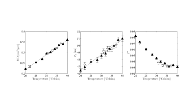 |
Detecting temperature-driven microstructural modulations in
tissue using diffusion MRI
Jelle Veraart1,
Daniel Nunes2,
and Noam Shemesh2 1Center for Biomedical Imaging, Dept. Radiology, NYU Grossman School of Medicine, New York, NY, United States, 2Champalimaud Research, Champalimaud Centre for the Unknown, Lisbon, Portugal Keywords: Diffusion/other diffusion imaging techniques, Microstructure In this study, we uncover that the signal fraction of a fully restricted diffusion compartment in fixed white matter tissue, the so-called dot fraction, is dependent on the tissue temperature. In particular, we report a sudden increase in its nominal value of this fraction when the tissue temperature drops below approximately 32°C. The underlying mechanism remains unexplained, but its impact on modeling of ex vivo diffusion MRI cannot be underestimated. The observation corroborates our hypotheses of a temperature-dependent disruption of the multilamellar structure of the myelin sheath, leading to water trapped in vacuoles that are formed in the myelin sheath. |
| 17:29 |
0692.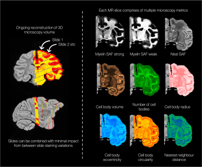 |
Towards whole-brain, quantitative characterisation of
microscopy-derived microstructure in the BigMac dataset
Amy FD Howard1,
Istvan N Huszar1,
Silei Zhu1,
Daniel ZL Kor1,
Lea Roumazeilles2,
Saad Jbabdi1,
and Karla L Miller1 1Wellcome Centre for Integrative Neuroimaging (FMRIB Centre), Nuffield Department of Clinical Neurosciences, University of Oxford, Oxford, United Kingdom, 2Wellcome Centre for Integrative Neuroimaging, Experimental Psychology, Medical Sciences Division, University of Oxford, Oxford, United Kingdom Keywords: Validation, Diffusion/other diffusion imaging techniques, Microstructure characterisation, microscopy The BigMac dataset is an open access resource combining in vivo MRI, postmortem MRI and multi-contrast microscopy data in a single, whole macaque brain. Here we perform data-driven segmentation of the BigMac histology slides to extract quantitative microscopy metrics for myelin, cell density, and cellular morphology (e.g. soma size and packing). Utilising high-quality MRI-microscopy registrations, we work towards building 3D volumes of quantitative microscopy derived metrics obtained at high resolution. This “ground truth” atlas of how cellular distributions vary across the brain is then directly related to co-registered diffusion MRI acquired in the same tissue. |
| 17:37 |
0693.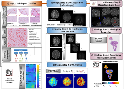 |
Establishing a Correlation between Diffusion Heterogeneity and
Tissue Structural Heterogeneity Using Stereotactic Biopsies on
Brain Tumors
Muge Karaman1,
Stefania Maraka1,
Tibor Valyi-Nagy1,
Syed Khalid1,
Konstantin Slavin1,
Gursant Atwal1,
Ahmad Daher1,
Guangyu Dan1,
Alessandro Scotti1,
Dan Schonfeld1,
and X. Joe Zhou1 1University of Illinois College of Medicine at Chicago, Chicago, IL, United States Keywords: Diffusion/other diffusion imaging techniques, Brain, Tissue heterogeneity Non-Gaussian diffusion MRI with a continuous-time random-walk (CTRW) model offers a unique avenue to probing tissue microstructural heterogeneity. The CTRW parameters, α and β, corresponding to temporal and spatial intravoxel diffusion heterogeneities, have empirically been linked to tumor tissue heterogeneity in clinical studies. This study aims at directly establishing the correlation between the CTRW parameters from patients suspected of glioma and tissue microstructural heterogeneity revealed by histology on stereotactic brain biopsies. We developed a practical protocol that integrates quantitative imaging techniques and surgical procedures to perform MR-histology correlation, and demonstrated that lower CTRW parameters correspond to increased tissue microstructural heterogeneity. |
 Back to Meeting Home
 Back to the Program-at-a-Glance
The International Society for Magnetic Resonance in Medicine is accredited by the Accreditation Council for Continuing Medical Education to provide continuing medical education for physicians.
 Back to Meeting Home
 Back to the Program-at-a-Glance
 View Presentation Video