Oral Session
Tissue Properties: Electrical, Magnetic & Microstructural Effects
ISMRM & ISMRT Annual Meeting & Exhibition • 03-08 June 2023 • Toronto, ON, Canada

Oral Session
Tissue Properties: Electrical, Magnetic & Microstructural Effects
| 15:45 |
1039.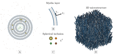 |
Towards microstructure-informed QSM: A digital phantom study
Anders Dyhr Sandgaard1,
Valerij G. Kiselev2,
Noam Shemesh3,
and Sune Nørhøj Jespersen1,4
1Center for functionally integrative neuroscience, department of clinical medicine, Aarhus University, Aarhus, Denmark, 2Division of Medical Physics, Department of Radiology, University Medical Center Freiburg, Freiburg, Germany, 3Champalimaud Research, Champalimaud Centre for the Unknown, Lisbon, Portugal, 4Department of Phsysics and Astronomy, Aarhus University, Aarhus, Denmark Keywords: Susceptibility, Quantitative Susceptibility mapping Magnetic susceptibility can provide valuable information about chemical composition and microstructural organization in tissues. However, its estimation from the MRI signal phase is particularly difficult, as it depends on both magnetic tissue properties on all length scales. Here we investigate the feasibility of inverting our recently presented model of WM magnetic microstucture to estimate susceptibility. This is done on a digital brain phantom based on actual dMRI measurements of an ex-vivo mouse brain at ultra-high field. |
| 15:53 |
1040.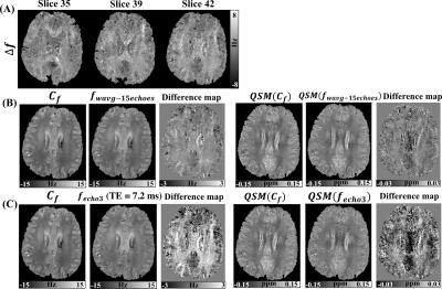 |
The impact of white matter microstructure with multi-fiber
populations on gradient-echo frequency maps and QSM
Lin Chen1,2,
Deng Mao3,
Guillaume Gilbert4,
Maarten Versluis5,
Peter van Zijl1,2,
and Xu Li1,2
1Department of Radiology and Radiological Sciences, Johns Hopkins University School of Medicine, Baltimore, MD, United States, 2F.M. Kirby Research Center for Functional Brain Imaging, Kennedy Krieger Institute, Baltimore, MD, United States, 3MR Clinical Science, Philips Healthcare, Baltimore, MD, United States, 4MR Clinical Science, Philips Healthcare, Mississauga, ON, Canada, 5MRI Clinical Science, Philips Healthcare, Best, Netherlands Keywords: Microstructure, White Matter A hollow cylinder fiber model (HCFM) with two orientation-dispersed fiber populations was used to characterize white matter microstructure-based susceptibility effects. The resulting TE-dependent frequency shifts were fitted with a curve function parameterized by a microstructure induced frequency difference (Δf) and bulk susceptibility induced frequency shift (Cf). Local frequency and QSM reconstruction using the fitted Cf versus using weighted echo averaging were compared by a head phantom and in vivo human brain data. Δf provided a useful contrast to illustrate the underlying white matter microstructure and Cf helped to improve the quantification accuracy of tissue magnetic susceptibility using QSM. |
| 16:01 |
1041.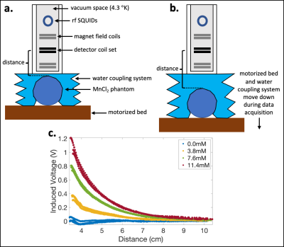 |
Superconducting Quantum Interference Device (SQUID-) Based
Susceptometry as a Reference Measurement for Quantitative
Susceptibility Mapping
Gregory Simchick1,2,
Ronald T Wakai2,
Roland Fischer3,
Oswaldo Baffa2,4,
Kevin Pratt5,
Doug Paulson5,
Scott B Reeder1,2,6,7,8,
and Diego Hernando1,2
1Radiology, University of Wisconsin-Madison, Madison, WI, United States, 2Medical Physics, University of Wisconsin-Madison, Madison, WI, United States, 3University Medical Center Hamburg-Eppendorf, Hamburg, Germany, 4Physics, University of Sao Paulo, Ribeirao Preto, Brazil, 5Tristan Technologies, Inc, San Diego, CA, United States, 6Medicine, University of Wisconsin-Madison, Madison, WI, United States, 7Biomedical Engineering, University of Wisconsin-Madison, Madison, WI, United States, 8Emergency Medicine, University of Wisconsin-Madison, Madison, WI, United States Keywords: Validation, Susceptibility Many quantitative susceptibility mapping (QSM) studies lack independent reference measurements of susceptibility (χ) for validation. In this work, superconducting quantum interference device (SQUID-) based susceptometry and QSM data were acquired for phantoms with varying manganese chloride (MnCl2) concentrations. χSQUID demonstrated low uncertainty and excellent agreement with the nominal susceptibility values. χQSM and R2* agreed relatively well with the nominal values and previously published results. However, χQSM demonstrated higher uncertainty in comparison to χSQUID. SQUID susceptometry has the potential to identify sources of bias and variability in QSM methods by serving as an independent reference measurement of susceptibility. |
| 16:09 |
1042.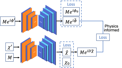 |
Constrained Dipole Inversion Using Jointly Learned Local Field
and Susceptibility Priors
Xi Peng1 1Mayo Clinic, Rochester, MN, United States Keywords: Susceptibility, Quantitative Susceptibility mapping The inversion of tissue magnetic susceptibility from a single-orientation phase measurement is an ill-posed problem. In this work, we propose a novel learning-based constrained reconstruction to integrate the jointly learned tissue field and susceptibility priors with the physics-based dipole inversion formalism such that the non-Gaussian model biased caused by the substantial errors in the estimated tissue field and the streaking artifacts in the tissue susceptibility can be effectively reduced. An efficient solver was also developed to solve the optimization problem. We demonstrated the superior performance of the proposed method over the state-of-the-art dipole inversion method using in vivo datasets. |
| 16:17 |
1043.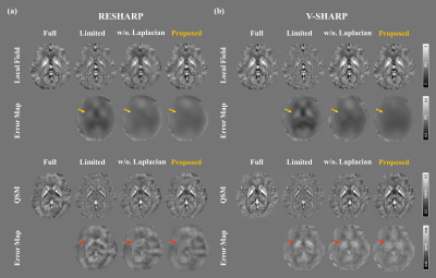 |
Harmonic Field Extension for QSM with Reduced Spatial Coverage
using Physics-informed Generative Adversarial Network
Siyun Jung1,
Soohyun Jeon1,
and Dong-Hyun Kim1
1Department of Electrical and Electronic Engineering, Yonsei University, Seoul, Korea, Republic of Keywords: Susceptibility, Quantitative Susceptibility mapping Acquisition of whole brain Quantitative Susceptibility Mapping (QSM) is time-consuming that requires the patient’s effort. On the other hand, limiting the QSM field-of-view to only the region of interest (e.g., deep grey matter) can shorten scan time and lessen the patient’s burden. However, reducing spatial coverage degrades conventional background field removal performance, resulting in an underestimation of susceptibility values. To overcome these limitations, we propose a harmonic field extension method using a physics-informed generative adversarial network. According to the experimental results, the proposed method significantly improves the accuracy of both the local field and QSM at reduced spatial coverage. |
| 16:25 |
1044.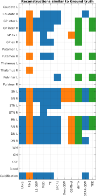 |
Region-of-Interest Based Statistical Analysis of the 2019 QSM
Challenge
Patrick Fuchs1,
Carlos Milovic2,
and Karin Shmueli1
1Medical Physics and Biomedical Engineering, University College London, London, United Kingdom, 2School of Electrical Engineering, Pontificia Universidad Catolica de Valparaiso, Valparaiso, Chile Keywords: Susceptibility, Quantitative Susceptibility mapping Since its inception, the most recent QSM challenge and corresponding dataset have helped to improve dipole-inversion algorithms. To date, only global metrics have been used to evaluate performance of these algorithms. Typically, in clinical applications of QSM, the susceptibility value in a specific brain region or structure is of interest. Therefore, we compared the accuracy of QSM algorithms in 28 regions in several ways including regional metrics and median differences, which is important to facilitate clinical translation of QSM. We found that, according to regional metrics, some direct QSM methods are surprisingly accurate considering their low global scores. |
| 16:33 |
1045.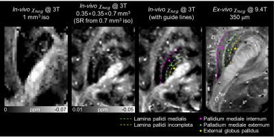 |
In-vivo delineation of fine structures in the human brain using
high resolution deep learning-powered chi-separation
Sooyeon Ji1,
Juhyung Park1,
Hyeong-Geol Shin2,3,
Minjun Kim1,
and Jongho Lee1
1Department of Electrical Computer Engineering, Seoul National University, Seoul, Korea, Republic of, 2Department of Radiology, Johns Hopkins University School of Medicine, Baltimore, MD, United States, 3F.M. Kirby Research Center for Functional Brain Imaging, Kennedy Krieger Institute, Baltimore, MD, United States Keywords: Susceptibility, Data Processing Fine structures in the human brain are delineated in the positive and negative susceptibility maps reconstructed in less than 25 min of scan time. The reconstruction pipeline consists of four deep learning networks, each of which performs multi-echo denoising, QSM reconstruction, χ-separation, and super-resolution. The reconstructed maps delineate two laminar structures within globus pallidus, the fibers of internal capsule, and nigrosome structures in the substantia nigra. |
| 16:41 |
1046.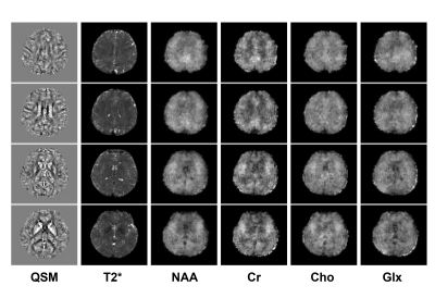 |
Fast Submillimeter QSM for Simultaneous QSM/MRSI of the Brain at
7T
Rong Guo1,2,
Yudu Li2,3,
Yibo Zhao2,4,
Aaron Anderson2,
Pallab Bhattacharyya5,
Mark Lowe5,
Yao Li6,
Brad Sutton2,3,4,7,8,
and Zhi-Pei Liang2,3,4
1Siemens Medical Solutions USA, Inc., Urbana, IL, United States, 2Beckman Institute for Advanced Science and Technology, University of Illinois at Urbana-Champaign, Urbana, IL, United States, 3National Center for Supercomputing Applications, University of Illinois at Urbana-Champaign, Urbana, IL, United States, 4Department of Electrical and Computer Engineering, University of Illinois at Urbana-Champaign, Urbana, IL, United States, 5Imaging Institute, Cleveland Clinic, Cleveland, OH, United States, 6School of Biomedical Engineering, Shanghai Jiao Tong University, Shanghai, China, 7Department of Bioengineering, University of Illinois at Urbana-Champaign, Urbana, IL, United States, 8Carle Illinois College of Medicine, University of Illinois at Urbana-Champaign, Urbana, IL, United States Keywords: Susceptibility, Quantitative Susceptibility mapping The feasibility of simultaneous QSM and MRSI has been recently demonstrated using SPICE. But the resolution of QSM obtained by SPICE at 7T is limited to only 3 mm due to bandwidth requirements. To overcome the resolution limitation, this work proposes a fast acquisition sequence for high-resolution encoding, and a model-based method to reconstruct from sparse sampling. As a result, submillimeter QSM can be achieved within only 1 minute, which enables simultaneous QSM (at 0.8 mm) and MRSI (at 3.0 mm) in an 8-minute scan at 7T. |
16:49 |
1047.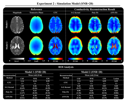 |
A Deep learning informed Polynomial Fitting Approach for
Electrical Properties Tomography
Kyu-Jin Jung1,
Thierry G.Meerbothe2,3,
Chuanjiang Cui1,
Mina Park4,
Jaeuk Yi1,
Cornelis A.T. van den Berg2,3,
Dong-Hyun Kim1,
and Stefano Mandija2,3
1Department of Electrical and Electronic Engineering, Yonsei University, Seoul, Korea, Republic of, 2Department of Radiotherapy, UMC Utrecht, Utrecht, Netherlands, 3Computational Imaging Group for MR Therapy and Diagnostics, UMC Utrecht, Utrecht, Netherlands, 4Department of Radiology, Gangnam Severance Hospital, Yonsei University College of Medicine, Seoul, Korea, Republic of Keywords: Electromagnetic Tissue Properties, Electromagnetic Tissue Properties This work presents a neural network informed fitting approach for conductivity reconstructions in MR-Electrical Properties Tomography. First, an artificial neural network is used to predict weights from T2-weighted images. These weights are used in a weighted fitting approach to calculate polynomial coefficients that parametrize the phase map. The conductivity is finally reconstructed from these coefficients. The reconstruction approach is tested on simulated data and in-vivo data and shows more accurate results than conventional fitting methods. |
| 16:57 |
1048.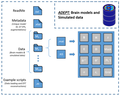 |
In silico brain data in ADEPT, A Database for MR-Electrical
Properties Tomography applications
Thierry G. Meerbothe1,2,
Cornelis A.T. van den Berg1,2,
and Stefano Mandija1,2
1Department of Radiotherapy, UMC Utrecht, Utrecht, Netherlands, 2Computational Imaging Group for MR Therapy and Diagnostics, UMC Utrecht, Utrecht, Netherlands Keywords: Electromagnetic Tissue Properties, Electromagnetic Tissue Properties This work presents ADEPT, A Database for MR-Electrical Properties Tomography applications. ADEPT is an open database that contains simulated MRI field data from electromagnetic simulations on brain models with and without tumor inclusion for EPT reconstructions. These data can serve as common ground to benchmark EPT reconstruction methods developed in different centers and will alleviate the computational burden for the creation of simulated data for training data-driven EPT methods. In the future, the database will be openly accessible and will be extended with phantom and in-vivo data. Other centers are also encouraged to share their data. |
| 17:05 |
1049.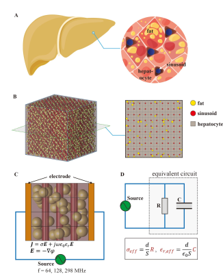 |
Probing underlying biophysical mechanisms of electrical
properties change by pathogenesis at the microscopic cellular
level
Henghui Liu1,
Guofang Xu1,
Yinhao Ren1,
Feng Liu2,
Xiang Nan3,
and Jijun Han1
1School of Biomedical Engineering, Anhui Medical University, Hefei, China, 2School of Information Technology and Electrical Engineering, University of Queensland, Brisbane, Australia, 3Department of Anatomy, Anhui Medical University, Hefei, China Keywords: Electromagnetic Tissue Properties, Liver Magnetic resonance electrical properties tomography provides a non-invasive approach to extracting pixel-wise electrical properties by B1 field mapping. However, there is a void in our understanding of the underlying biophysical mechanism of electrical properties changes accompanied by pathogenesis at the microscopic cellular level. In this work, we build a microscopic tissue model to study fatty liver disease, which focuses on exploring the relationship between fat fraction and electrical properties. Our findings could bridge microscopic lesions and pixel-wise EPT images and may offer a novel strategy for retrieving electrical properties via fat quantification techniques such as Dixon directly. |
|
17:13 |
1050.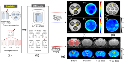 |
Imaging of Irradiation Effects in Brain Tissues by Electrical
Conductivity Using MRI
Nitish Katoch1,
Bup Kyung Choi1,
Ji Ae Park2,
Tae Hoon Kim3,
Young Hoe Hur4,
Jin Woong Kim5,
and Hyung Joong Kim1
1Department of Biomedical Engineering, Kyung Hee University, Seoul, Korea, Republic of, 2Division of Applied RI, Korea Institute of Radiological and Medical Science, Seoul, Korea, Republic of, 3Medical Convergence Research Center, Wonkwang University Hospital, Iksan, Korea, Republic of, 4Department of Hepato-Biliary-Pancreas Surgery, Chonnam National University Medical School, Gwangju, Korea, Republic of, 5Department of Radiology, Chosun University Hospital and Chosun University College of Medicine, Gwangju, Korea, Republic of Keywords: Electromagnetic Tissue Properties, Electromagnetic Tissue Properties, Electrical Conductivity, MREPT, Radiation Therapy, High-field MRI Radiation-induced injury can damage normal tissues caused by unintentional exposure to ionizing radiation. Image-based evaluation of tissue damage by irradiation has an advantage for the early assessment of therapeutic effects. This study aims to non-invasively evaluate tissue response following radiation therapy in phantoms and in vivo mouse brains at 3T and 9.4T MRI scanner. Due to changes in ionic strength in tissue after radiation, magnetic resonance (MR)-based electrical properties tomography could be a suitable imaging tool to assess the radiation therapy response on biological tissues. |
| 17:21 |
1051.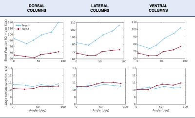 |
Tissue orientation effects on T2 relaxation in fresh and fixed
spinal cord white matter
Michelle Medina1,2,3,
Lara Bartels1,2,3,
Jonathan Doucette1,2,3,
Andrew Yung4,
Kirsten Bale4,
Piotr Kozlowski4,5,
Christoph Birkl4,6,
and Alexander Rauscher1,2,3,7
1Physics and Astronomy, University of British Columbia, Vancouver, BC, Canada, 2UBC MRI Research Center, Vancouver, BC, Canada, 3Pediatrics, University of British Columbia, Vancouver, BC, Canada, 4UBC MRI Research Centre, Vancouver, BC, Canada, 5Radiology and Urologic Sciences, University of British Columbia, Vancouver, BC, Canada, 6Neuroradiology, Medical University of Innsbruck, Innsbruck, Austria, 7Djavad Mowafaghian Centre for Brain Health, University of British Columbia, Vancouver, BC, Canada Keywords: Microstructure, White Matter, Relaxometry In this study, we investigated the orientation dependence of T2 in fresh and fixed spinal cord white matter (WM). Scans from three pig spinal cord tissues were acquired at 7T at 6 different orientations with respect to the main magnetic field. We found a considerable orientation dependence in the short fraction relaxation rate R2 (=1/T2) in fresh WM as opposed to a weak orientation effect in fixed WM. To our knowledge, this is the first direct comparison of orientation dependence in a WM tissue sample in the fresh and fixed state. Orientation dependence was not observed in long fraction R2. |
|
17:29 |
1052.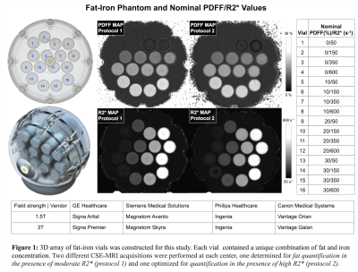 |
Multi-center, multi-vendor validation of PDFF-R2* mapping in an
Optimized Fat-Iron Phantom
Jitka Starekova1,
David Rutkowski2,
Won C Bae3,
Hung Do4,
Ananth Madhuranthakam5,
Vadim Malis3,
Sujoy Mukherjee5,
Sheng Qing Lin5,
Suraj Serai6,
Takeshi Yokoo5,
Scott B Reeder1,7,8,9,10,
Jean H Brittain2,
and Diego Hernando1,8
1Radiology, University of Wisconsin-Madison, Madison, WI, United States, 2Calimetrix, Madison, Wisconsin, Madison, WI, United States, 3Radiology, University of California, San Diego, San Diego, CA, United States, 4Canon Medical Systems, Tustin, CA, United States, 5Radiology, UT Southwestern, Medical Center, Dallas, TX, United States, 6Radiology, Children’s Hospital of Philadelphia, Philadelphia, PA, United States, 7Biomedical Engineering, University of Wisconsin-Madison, Madison, WI, United States, 8Medical Physics, University of Wisconsin-Madison, Madison, WI, United States, 9Medicine, University of Wisconsin-Madison, Madison, WI, United States, 10Emergency Medicine, University of Wisconsin-Madison, Madison, WI, United States Keywords: Validation, Phantoms Reliable, quantitative assessment of fat and iron is important in the management of chronic liver diseases. Confounder-corrected chemical-shift-encoded (CSE)-MRI estimates proton-density fat-fraction (PDFF) and R2* as quantitative biomarkers for fat and iron, which have been shown to be highly reproducible across centers, field strengths and manufacturers. However, reproducibility in the setting of concomitantly high levels of fat and iron is poorly understood. To ensure the fidelity of CSE-MRI in clinical routine, a validation study was performed under controlled conditions on a phantom that modulates fat and iron simultaneously. Excellent multi-center and multi-vendor reproducibility of CSE-MRI PDFF and R2* was found. |
17:37 |
1053.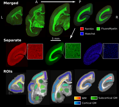 |
Histological validation of myelin-sensitive MRI metrics in the
common marmoset
Christopher D Rowley1,2,
Daryan Chitsaz2,
Ilana R. Leppert1,
Jennifer S.W. Campbell1,
Stephen Nuara3,
Timothy E. Kennedy2,
G. Bruce Pike4,
and Christine L. Tardif1,2,5 1McConnell Brain Imaging Centre, Montreal Neurological Institute and Hospital, McGill University, Montreal, QC, Canada, 2Department of Neurology and Neurosurgery, McGill University, Montreal, QC, Canada, 3Comparative Medicine and Animal Resources Center, McGill University, Montreal, QC, Canada, 4Hotchkiss Brain Institute and Departments of Radiology and Clinical Neuroscience, University of Calgary, Calgary, AB, Canada, 5Department of Biomedical Engineering, McGill University, Montreal, QC, Canada Keywords: Validation, Brain, Marmoset Several studies have compared myelin-sensitive MRI maps to myelin staining to demonstrate the degree of correlation of the MRI metric with myelin content. This study in the common marmoset compares six myelin-sensitive MRI metrics acquired in vivo to stains for myelin, cell nuclei, and ferritin. T2*-based myelin water fraction (MWF) and inhomogeneous magnetization transfer saturation (ihMTsat) presented the greatest specificity for myelin content, with ihMTsat having a higher signal in grey matter regions. T1w/T2w had the strongest correlation with the iron-storage protein ferritin, and T2* presented the greatest correlation with the cell nuclei stain. |
 Back to Meeting Home
 Back to the Program-at-a-Glance
The International Society for Magnetic Resonance in Medicine is accredited by the Accreditation Council for Continuing Medical Education to provide continuing medical education for physicians.
 Back to Meeting Home
 Back to the Program-at-a-Glance
 View Presentation Video