Oral Session
Cartilage
ISMRM & ISMRT Annual Meeting & Exhibition • 03-08 June 2023 • Toronto, ON, Canada

| 08:15 |
1109. |
Bariatric Surgery Effects on Knee Articular Cartilage and
Osteoarthritis Symptoms – a 12-month Follow-up Using T2
Relaxation Time and WOMAC Index
Sami Lehtovirta1,2,
Ahti Kemppainen1,2,
Marianne Haapea2,3,4,
Jaro Karppinen1,5,6,
Eveliina Lammentausta2,3,
Vesa Koivukangas7,
Eero Kyllönen8,
Mika Nevalainen1,2,3,
Petri Lehenkari2,7,9,
Victor Casula1,2,
and Miika T. Nieminen1,2,3
1Research Unit of Health Science and Technology, University of Oulu, Oulu, Finland, 2Medical Research Center, University of Oulu and Oulu University Hospital, Oulu, Finland, 3Department of Diagnostic Radiology, Oulu University Hospital, Oulu, Finland, 4Research Service Unit, Oulu University Hospital, Oulu, Finland, 5Center for Life Course Health Research, University of Oulu, Oulu, Finland, 6Finnish Institute of Occupational Health, Oulu, Finland, 7Department of Surgery, Oulu University Hospital, Oulu, Finland, 8Department of Physical Medicine and Rehabilitation, Oulu University Hospital, Oulu, Finland, 9Research Unit of Translational Medicine, University of Oulu, Oulu, Finland Keywords: Cartilage, Osteoarthritis Obesity is a common risk factor for osteoarthritis (OA). We set out to study knee cartilage using T2 relaxation time, and knee OA symptoms with WOMAC OA index in obese individuals. Bariatric surgery patients were split into successful and unsuccessful weight loss groups, and compared with a control group of obese individuals over a 12-month follow-up. The lesser weight loss group displayed improvement of cartilage with lower T2 values in the lateral compartment of femoral cartilage, compared to the higher weight loss. We also observed improvement of knee symptoms in both successful and unsuccessful weight loss groups after 12 months. |
| 08:23 |
1110.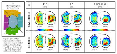 |
Utilization of an MRI compatible loading device for the
evaluation of unloaded-to-loaded changes in region specific
tibial cartilage T1ρ and T2:
Erin C Argentieri1,
Andrew C Zhu2,
Arden Wach2,
Ashley Pekmezian2,
Sonia Bansal2,
Ryan E Breighner1,
Hollis G Potter1,
Suzanne A Maher2,
and Matthew F Koff1 1Radiology and Imaging, Hospital for Special Surgery, New York, NY, United States, 2Biomechanics, Hospital for Special Surgery, New York, NY, United States Keywords: Cartilage, Quantitative Imaging Evaluation of unloaded-to-loaded changes within cartilage revealed that tibial cartilage thickness, T1ρ, and T2 metrics all decreased/shortened following application of 50% BW axial load. Shortening of loaded qMRI values is likely attributed to water loss from the cartilage matrix due to PG/matrix damage and attendant loss of cartilage FCD within this cadaveric model. As both the static and dynamic responses of cartilage to load are impacted by degeneration, quantification of unloaded-to-loaded T1ρ and T2 values may provide additional insight into cartilage health. Future work will evaluate unloaded-to-loaded cartilage T2* metrics to better elucidate movements of free and bound water pools. |
| 08:31 |
1111.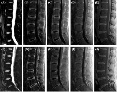 |
High Contrast Cartilaginous Endplate Imaging Using
Dual-Inversion Recovery Prepared Ultrashort Echo Time (DIR-UTE)
Sequence
Jiyo Srinivasan Athertya1,
James Lo1,2,
Alicia Ji1,
Charles Ding1,
Xiaojun Chen1,
Soo Hyun Shin1,
Bhavsimran Singh Malhi 1,
Saeed Jerban1,
Micael Carl3,
Monica Guma4,5,
Eric Y Chang1,5,
Jiang Du1,2,5,
and Yajun Ma1
1Radiology, UCSD, San Diego, CA, United States, 2Bioengineering, UCSD, San Diego, CA, United States, 3GE Healthcare, San Diego, CA, United States, 4Medicine, UCSD, San Diego, CA, United States, 5Radiology Service, VA, San Diego, CA, United States Keywords: MSK, Contrast Mechanisms, Cartilaginous endplate, Spine The cartilaginous endplate (CEP) plays a key role in maintaining the normal function of the intervertebral disc (IVD) by acting as a bridge for the transport of nutrients into the IVD cells. In this study, we developed a 3D dual inversion recovery prepared ultrashort echo time (DIR-UTE) sequence for high contrast CEP imaging and compared its performance with previously developed techniques on a clinical 3T scanner. We found that the proposed DIR-UTE sequence demonstrated the best image contrast for CEP imaging, which is highly promising for future clinical use. |
| 08:39 |
1112.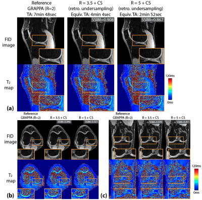 |
Rapid Isotropic 3D T2 Mapping of the Knee using Dual-Echo
Steady-State MRI with Compressed Sensing Reconstruction
Shu-Fu Shih1,2,
Zhaohuan Zhang1,2,
Ashmita Deb1,2,
Xiaodong Zhong3,
Timothy W. Ryan1,
and Holden H. Wu1,2
1Radiological Sciences, David Geffen School of Medicine, University of California Los Angeles, Los Angeles, CA, United States, 2Bioengineering, University of California Los Angeles, Los Angeles, CA, United States, 3MR R&D Collaborations, Siemens Medical Solutions USA, Inc., Los Angeles, CA, United States Keywords: Cartilage, Cartilage MRI T2 mapping of cartilage has been shown to be useful in characterization and monitoring of osteoarthritis (OA). Compared to conventional spin echo-based sequences, the 3D dual-echo steady-state (DESS) sequence can provide faster T2 mapping. Previous works proposed 3D DESS T2 mapping in the knee using anisotropic resolution (e.g., in-plane resolution of 0.3x0.3mm2 and slice thickness of 1.5-3mm), which limits the depiction of fine structures in 3D multiplanar reformatted images. This work investigated 3D DESS T2 mapping in the knee with isotropic acquired resolution of 0.66x0.66x0.66 mm3 and used compressed sensing (CS) to accelerate the acquisition time to <5 minutes. |
| 08:47 |
1113.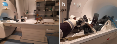 |
Patellofemoral cartilage compression and recovery in response to
loading measured with dynamic MRI using prospective motion
correction
Thomas Lange1,
Philipp Rovedo1,
Patrick Hucker1,
Elham Taghizadeh2,3,
Kaywan Izadpanah4,
Maxim Zaitsev1,
and Hans Meine2,3
1Division of Medical Physics, Department of Diagnostic and Interventional Radiology, Medical Center - University of Freiburg, Freiburg, Germany, 2Fraunhofer Institute for Digital Medicine MEVIS, Bremen, Germany, 3Medical Image Computing Group, Department of Informatics, University of Bremen, Bremen, Germany, 4Department of Orthopedic and Trauma Surgery, Medical Center – University of Freiburg, Freiburg, Germany Keywords: Cartilage, Motion Correction, dynamic MRI, loading, compression, recovery The patellofemoral cartilage compression and recovery is measured with dynamic MRI in response to a bout of in situ loading in a cohort of ten healthy subjects. To mitigate motion artifacts arising from the loading paradigm, the experiments are performed with prospective motion correction based on optical tracking. The measured cartilage compression and recovery time course in response to loading and unloading is characterized by a larger fully elastic compression component adapting instantaneously to applied load changes, and a smaller compression component, which only gradually adapts to load changes and exhibits in particular a very protracted recovery after unloading. |
| 08:55 |
1114.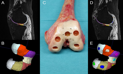 |
Characterization of the Textural Features from Quantitative MRI
for Determination of Cartilage Degeneration
Vladimir Juras1,
Stefan Toegel2,3,
Benedikt Hager4,5,6,
Markus Schreiner7,
Veronika Janacova1,
Pavol Szomolanyi4,
Didier Laurent8,
Franziska Saxer9,
Rahel Heule10,
Oliver Bieri11,
Esther Raithel12,
Christoph Fuchssteiner13,
Wolfgang Weninger13,
Reinhard Windhager2,
and Siegfried Trattnig4
1Medical University of Vienna, Vienna, Austria, 2Karl Chiari Lab for Orthopaedic Biology, Department of Orthopedics and Trauma Surgery, Medical University of Vienna, Vienna, Austria, 3Ludwig Boltzmann Institute for Arthritis and Rehabilitation, Vienna, Austria, 4High Field MR Centre, Department of Biomedical Imaging and Image-guided Therapy, Medical University of Vienna, Vienna, Austria, 5Austrian Cluster for Tissue Regeneration, Ludwig Boltzmann Institute for Experimental and Clinical Traumatology, Viennq, Austria, 6CD Laboratory for MR Imaging Biomarkers (BIOMAK), Vienna, Austria, 7Department of Orthopedics and Trauma Surgery, Medical University of Vienna, Vienna, Austria, 8Department of Translational Medicine, Novartis Institutes for Biomedical Research, Basel, Switzerland, 9Department of translational Medicine, Novartis Institutes for Biomedical Research, Basel, Switzerland, 10Center for MR Research, University Children's Hospital, Zurich, Switzerland, 11Division of Radiological Physics, Department of Radiology, University of Basel Hospital, Basel, Switzerland, 12Siemens Healthcare GmbH, Erlangen, Germany, 13Center for Anatomy and Cell Biology, Division of Anatomy, Medical University of Vienna, Vienna, Austria Keywords: Cartilage, Osteoarthritis, cartilage; texture analysis, histology Texture features derived from quantitative MRI maps of cartilage have attracted increasing attention from the osteoarthritis (OA) community in recent years. In this work, texture analysis was used on T2 maps and validated using histological analysis. Some texture features (autocorrelation, contrast and entropy) correlated with the Mankin score; autocorrelation also correlated with collagen orientation calculated from PLM images. The correlation between image-derived features with histological quality scores can be a decisive step towards monitoring of cartilage regeneration in-vivo and help to identify therapies that restore articular cartilage quantity and quality. |
| 09:03 |
1115.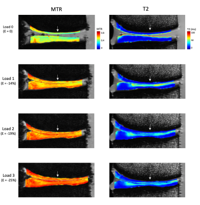 |
Assessment of knee cartilage strain using the magnetization
transfer ratio and T2: an ex vivo study at 9.4T MRI
Emily Sullivan1,2,
Andrew Yung3,
Jessica Küpper2,4,
Kirsten Bale3,
Piotr Kozlowski3,
and David Wilson2,4
1School of Biomedical Engineering, University of British Columbia, Vancouver, BC, Canada, 2Centre for Hip Health and Mobility, University of British Columbia, Vancouver, BC, Canada, 3UBC MRI Research Centre, University of British Columbia, Vancouver, BC, Canada, 4Orthopaedics, University of British Columbia, Vancouver, BC, Canada Keywords: Cartilage, Osteoarthritis The magnetization transfer ratio (MTR) was investigated as an alternative biomarker for cartilage strain compared to T2. Six ex vivo bovine knee specimens were compressed in a 9.4T scanner and scanned at several physiological step loads. Average strain, MTR, and T2 were calculated for each voxel column throughout the full depth of cartilage, and depth-dependent trends were identified. ΔMTR changed uniformly across the cartilage volume with strain, while ΔT2 showed a more localized yet inconsistent response. This suggests that MTR may be more sensitive in measuring cartilage strain in clinical applications where image resolution is lower. |
| 09:11 |
1116.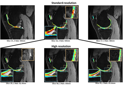 |
Reliable high-resolution in vivo human knee T1ρ imaging
quantification with robust fitting methods
Zhiyuan Zhang1,2,3,
Jeehun Kim1,3,4,
Richard Lartey1,3,
Carl Scherman Winalski1,3,5,
and Xiaojuan Li1,3,5
1Program of Advanced Musculoskeletal Imaging (PAMI), Cleveland Clinic, Cleveland, OH, United States, 2Biomedical Engineering, Case Western Reserve University, Cleveland, OH, United States, 3Department of Biomedical Engineering, Lerner Research Institute, Cleveland Clinic, Cleveland, OH, United States, 4Electrical, Computer, and Systems Engineering, Case Western Reserve University, Cleveland, OH, United States, 5Department of Diagnostic Radiology, Imaging Institute, Cleveland Clinic, Cleveland, OH, United States Keywords: Cartilage, Quantitative Susceptibility mapping Quantitative MR T1ρ and T2 imaging are promising methods to detect osteoarthritis at its early stage. Current T1ρ and T2 mapping in human subjects is limited to a relatively low resolution which has limited sensitivity to focal lesions due to partial-volume effects. One of the hurdles to achieving high resolution is the increase in fitting bias when using a conventional nonlinear least-squares fitting with low SNR images. In this study, we evaluated T1ρ quantification with in-vivo high-resolution imaging with different fitting methods. |
| 09:19 |
1117.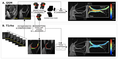 |
Layers-Based Analysis of Knee Articular Cartilage: Comparing T1ρ
Imaging Against Quantitative Susceptibility Mapping at 3 Tesla
ALLEN A CHAMPAGNE1,
TAYLOR M ZULEGER2,3,4,5,
DANIEL R SMITH2,4,5,
ALEXIS B SLUTSKY-GANESH2,4,5,6,
SHAYLA M WARREN2,4,5,
LEXIE M SENGKHAMMEE2,4,5,
SAGAR MANDAVA7,
HONGJIANG WEI8,
DAVIDE D BARDANA9,
GREG D MYER2,4,5,10,
and JED A DIEKFUSS2,4,5
1School of Medicine, Queen's University, Kingston, ON, Canada, 2Emory Sports Performance And Research Center, Flowery Branch, GA, United States, 3Neuroscience Graduate Program, University of Cincinnati, Cincinnati, OH, United States, 4Emory Sports Medicine Center, Atlanta, GA, United States, 5Department of Orthopaedics, Emory University School of Medicine, Atlanta, GA, United States, 6Department of Kinesiology, University of North Carolina at Greensboro, Greensboro, NC, United States, 7GE Healthcare, Atlanta, GA, United States, 8School of Biomedical Engineering, Shanghai Jiao Tong University, Shanghai, China, 9Department of Orthopaedics, Queen's University, Kingston, ON, Canada, 10The Micheli Center for Sports Injury Prevention, Waltham, MA, United States Keywords: Cartilage, Microstructure, Quantitative Susceptibility Mapping, T1rho, knee The vertical microstructural organization of articular cartilage varies in its arrangement of collagen fibers, as well as the relative proteoglycan content. Knee osteoarthritis (OA) involves an inflammatory degenerative process that leads to depth-wise degeneration of the cartilaginous matrix. While quantitative MR protocols have emerged to evaluate proteoglycan content (T1ρ) and microstructural integrity (Quantitative Susceptibility Mapping; QSM), advances in layer-based analyses (e.g., superficial vs. deep) are warranted to identify the progression of diseased cartilage. We demonstrate the integrated utilization of QSM and T1ρ for characterizing depth-specific microstructural arrangement of articular cartilage, including differences in tissue organization and composition, respectively. |
| 09:27 | 1118. |
Quantitative relaxometry values can effectively assist menicius
injury level in knee
Huizheng Wang1,
Weiyin Vivian Liu2,
Wen Chen3,
Jingyu Jiang4,
Ling Sang3,
Peng Zhang3,
and Hu Chen3
1Department of Radiology, Taihe Hospital, Hubei University of Medicine, Hubei, China, 2GE Healthcare, Beijing, China, 3Shiyan Taihe Hospital, Hubei, China, 4Biomedical Engneering College, Hubei University of Medicine, Hubei, China Keywords: Osteoarthritis, Cartilage Knee meniscus tears cause fibrocartilage degeneration. Grade III meniscus is viewed as complete meniscus injury and should be further confirmed for the requirement of surgical intervention via arthroscopy. MAGiC provides quantitative relaxometry values. Our study showed that Grade III meniscus can be distinguished from Grade II ones using T1, T2 and PD. This finding suggested MAGiC can assist clinical follow-ups of knee meniscus and screen the indications for arthroscopic surgery. |
| 09:35 |
1119.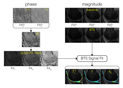 |
Novel 3D Quantitative Magnetization Transfer Imaging of The
Whole Knee Cartilage
Albert Jang1,2,
Zachary Stewart2,
Martin Torriani2,
Miho Tanaka3,
and Fang Liu1,2
1Athinoula A. Martinos Center for Biomedical Imaging, Harvard Medical School, Charlestown, MA, United States, 2Radiology, Massachusetts General Hospital, Boston, MA, United States, 3Orthopedic Surgery, Massachusetts General Hospital, Boston, MA, United States Keywords: Cartilage, Magnetization transfer We propose to characterize cartilage extracellular matrix of the whole knee using a novel quantitative magnetization transfer (MT) imaging technique. This new technique achieves accurate T1 and MT quantification by using a new sequence incorporating off-resonance RF pulses that create two simultaneous effects: 1) Bloch-Siegert phase shift to measure and correct for B1+ inhomogeneity and 2) direct saturation of macromolecules to model MT. This new method, termed BTS (Bloch-Siegert and magnetization Transfer Simultaneously), is presented and validated in both phantom and in-vivo knee imaging experiments. |
| 09:43 |
1120.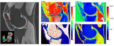 |
MR Fingerprinting in the Knee Cartilage Compared to Conventional
Methods in Combination with Automated Cartilage Segmentation
Diana Bencikova1,
Martin A. Cloos2,
Veronika Janacova1,
Siegfried Trattnig1,3,4,5,
and Vladimir Juras1
1Department of Biomedical Imaging and Image-Guided Therapy, Medical University Vienna, Vienna, Austria, 2University of Queensland, Queensland, Austria, 3CD Laboratory for MR Imaging Biomarkers (BIOMAK), Vienna, Austria, 4Austrian Cluster for Tissue Regeneration, Ludwig Boltzmann Institute for Experimental and Clinical Traumatology, Vienna, Austria, 5Karl Landsteiner Society, Institute for Clinical Molecular MRI in the Musculoskeletal System, Vienna, Austria Keywords: Cartilage, MR Fingerprinting, MSK, MR value, Osteoarthritis, Quantitative Imaging, Relaxometry Quantitative MRI has been shown to be sensitive to early stages of cartilage degeneration in osteoarthritis, but conventional MRI techniques can measure only single parameter at a time. This poses time constraints and co-registration challenges. With MR Fingerprinting, multiple parameters can be assessed within single measurement. Here, we evaluated prototype MRF sequence in NIST phantom and healthy volunteers in combination with automatic cartilage segmentation procedure and compared to conventional techniques. We could show that the values provided by MRF sequence agreed with the values provided by conventional techniques. Therefore MRF is accurate and practical diagnosis tool for articular cartilage examination. |
| 09:51 |
1121.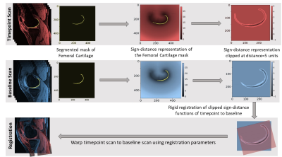 |
3D Cluster Analysis for Cartilage T2 And T1ρ Mapping to Assess
Focal Lesions in ACL-Injured Subjects
Anoosha Pai S1,
Anthony A Gatti2,
Marianne S Black3,
Arjun D Desai4,
Jarrett Rosenberg2,
Katherine A Young2,
Jessica L Asay2,
Seth L Sherman5,
Garry E Gold2,
Feliks Kogan2,
Brian Hargreaves2,
and Akshay S Chaudhari6
1Bioengineering, Stanford University, Palo Alto, CA, United States, 2Radiology, Stanford University, Palo Alto, CA, United States, 3Mechanical Engineering, University of Victoria, Victoria, BC, Canada, 4Electrical Engineering, Stanford University, Palo Alto, CA, United States, 5Orthopaedic Surgery, Stanford University, Palo Alto, CA, United States, 6Radiology/Integrative Biomedical Imaging Informatics, Stanford University, Palo Alto, CA, United States Keywords: Cartilage, Osteoarthritis, ACL-injury, cartilage, knee A 3D pipeline (longitudinal registration followed by cluster analysis) was developed to identify focal-lesions (clusters) of elevated T2 and T1ρ in femoral cartilage over 4-visits (3-weeks, 3, 9, and 18-months) post ACL-reconstruction surgery. Cluster Average (CA) and Cluster Percentage (CP) for T2 were significantly higher for ACL-inured when compared to ACL-contralateral and control-healthy knees. While the CP followed an increasing trend during subsequent visits, CA did not significantly vary across visits. Thus, our method could be effective for identifying and tracking quality (measured by CA) and quantity (measured by CP) of focal lesions in ACL-injured population. |
| 09:59 |
1122.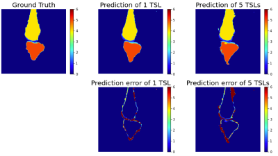 |
Knee Bone and Cartilage Segmentation using Deep Learning Model
Trained with Heterogeneous Data: Preliminary Results
Xiaoxia Zhang1,
Hector L. De Moura1,
Marcelo V. W. Zibetti1,
and Ravinder R. Regatte1
1Grossman School of Medicine, New York University, New York, NY, United States Keywords: Cartilage, Joints In this study, we pre-trained a deep learning model for knee bone and cartilage segmentation with open dataset (MICCAI grand challenge "K2S 2022"), and then fine-tuned it with a small size of locally acquired data for customized task, in which we explored different contrasts as well. We can benefit from the large dataset size to increase the segmentation accuracy and generalization capabilities while reducing the labor and time of manual segmentation for training data. The preliminary results of a small dataset with 10 subjects using a simple 2D U-net are promising for single contrast and multi contrast images. |
| 10:07 |
1123.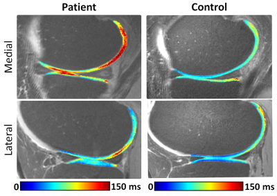 |
Quantitative T2 Mapping of Articular Cartilage in Patients with
Posterior Horn Root Tears of Medial Meniscus
Abdul Wahed Kajabi1,2,
Stefan Zbyn1,2,3,
Jesse Smith1,2,
Morgan Homan4,
Hasan Abbasguliyev5,
Ariel N. Rodriguez4,
Gregory J. Metzger1,
Robert F. LaPrade4,
and Jutta M. Ellermann1,2
1Center for Magnetic Resonance Research, University of Minnesota, Minneapolis, MN, University of Minnesota, Minneapolis, MN, United States, 2Department of Radiology, University of Minnesota, Minneapolis, MN, United States, 3Department of Biomedical Engineering, Lerner Research Institute, Cleveland, OH, United States, 4Twin Cities Orthopedics, Edina, MN, United States, 5Department of Diagnostic and Interventional Radiology, Ataturk University Research Hospital, Erzurum, Turkey Keywords: Cartilage, Quantitative Imaging The aim of this study was to evaluate degenerative changes in articular cartilage of patients with posterior root tear of the medial meniscus. Multi-echo quantitative T2 mapping was acquired on a 7T clinical scanner. For reference, age- and gender-matched healthy controls were scanned using the same 7T protocol. Arthroscopic evaluation of articular cartilage and menisci was performed in patients during the repair of posterior medial root tears. Significantly higher T2 values were found in the lateral and medial femoral cartilage of the patients compared to healthy controls. |
 Back to Meeting Home
 Back to the Program-at-a-Glance
The International Society for Magnetic Resonance in Medicine is accredited by the Accreditation Council for Continuing Medical Education to provide continuing medical education for physicians.
 Back to Meeting Home
 Back to the Program-at-a-Glance
 View Presentation Video