Oral Session
Technical Developments of MSK Imaging
ISMRM & ISMRT Annual Meeting & Exhibition • 03-08 June 2023 • Toronto, ON, Canada

Oral Session
Technical Developments of MSK Imaging
| 15:45 |
0624.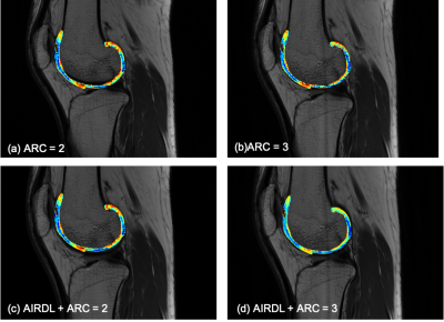 |
High-resolution and rapidly-acquired T2 mapping using
deep-learning reconstruction and parallel imaging in evaluation
of knee cartilage
Weiyin Vivian Liu1,
Xiaxia Wu2,
and Yunfei Zha2 1GE Healthcare, Beijing, China, 2Department of Radiology, Renmin Hospital of Wuhan University, Wuhan, China Keywords: MSK, Quantitative Imaging T2 mapping reflects biochemical composition of tissues such as cartilage, tendon, ligament, meniscus without requirement for a contrast injection or special MRI imaging hardware, and there are numerous available techniques for post-processing of T2 images. However, scan time and signal-to-noise ratio are critical factors in clinical implementation. With the development of parallel imaging in combination of deep-learning based reconstruction algorithm, high-resolution and reproducible T2 mapping was potentially achieved to identify cartilage injury or degeneration in spite that T2 relaxation time cannot directly refer to previous studies but showed high consistency with the same acceleration factor for T2 mapping. |
| 15:53 |
0625.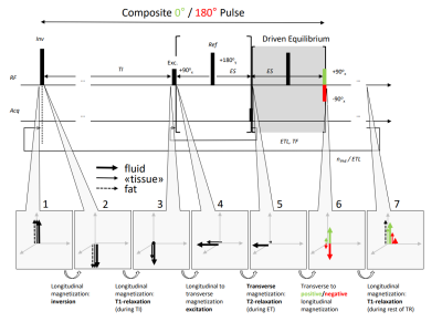 |
Enhancing fluid signal in driven-equilibrium short-tau
inversion-recovery (STIR) imaging with short TR times.
Constantin von Deuster1,2 and
Daniel Nanz2,3
1Advanced Clinical Imaging Technology, Siemens Healthineers International AG, Zurich, Switzerland, 2Swiss Center for Musculoskeletal Imaging, Balgrist Campus AG, Zurich, Switzerland, 3University of Zurich, Zurich, Switzerland Keywords: MSK, Contrast Mechanisms, Driven Equilibrium Fluid-sensitive Turbo-Spin-Echo (TSE) imaging in combination with Short-Tau Inversion Recovery for fat suppression (STIR) is commonly employed in musculoskeletal MRI. A Driven Equilibrium (DE) module following the imaging train can be used to reduce the Repetition Time (TR) without significant SNR loss. However, due to the STIR preparation, a conventional DE element rotates the fluid magnetization onto the negative z-axis instead of the positive z-axis, which delays longitudinal relaxation and attenuates the fluid signal. We implemented a modified DE module that increased fluid-signal intensity in STIR-TSE images of the spine. |
| 16:01 |
0626.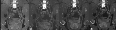 |
Improved 3D DESS MR Neurography of the Lumbosacral Plexus with
Deep Learning and Geometric Image Combination
Yenpo Lin1,2,
Ek T. Tan1,
Gracyn Campbell1,
Philip G. Colucci1,
Sumedha Singh1,
Yan Wen3,
Qian Li1,
and Darryl B. Sneag1 1Hospital for Special Surgery, New York, NY, United States, 2Chang Gung Memorial Hospital, Taipei, Taiwan, 3GE Healthcare, Waukesha, WI, United States Keywords: Neurography, Neurography Geometric image combination (GIC) and deep learning (DL) reconstructions were together applied to improve nerve visualization in lumbosacral plexus MR neurography (MRN) using the 3D dual echo steady-state free precession (DESS) sequence. Qualitative comparisons were made against standard image reconstruction of the 2nd echo of the DESS sequence. While standard-of-care (SOC) reconstructions of 3D DESS images provided effective vascular suppression and good nerve conspicuity, the DL-GIC reconstructed images demonstrated similar or improved nerve conspicuity. |
| 16:09 |
0627.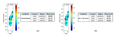 |
Evaluation of an accelerated Deep Learning-reconstructed T2
mapping technique through knee cartilage regional analysis using
DOSMA framework
Laura Carretero Gómez1,2,
Maggie Fung3,
Bruno Nunes4,
Valentina Pedoia5,
Sharmila Majumdar5,
Akshay Chaudhari 6,
Arjun Divyang Desai6,7,
Anthony Andrea Gatti6,
Feliks Kogan6,
and Mario Padron8
1GE Healthcare, Munich, Germany, 2LAIMBIO, Rey Juan Carlos University, Madrid, Spain, 3GE Healthcare, New York, NY, United States, 4GE Healthcare, San Ramon, CA, United States, 5University of California, San Francisco, San Francisco, CA, United States, 6Radiology, Stanford University, Stanford, CA, United States, 7Electrical Engineering, Stanford University, Stanford, CA, United States, 8Clinica CEMTRO, Madrid, Spain Keywords: Cartilage, Quantitative Imaging The clinical translation of MRI Quantitative Imaging is still hampered by the high variability and suboptimal reproducibility of the cartilage biomarkers. The purpose of this work is to validate the consistency of a novel accelerated DL reconstructed T2 mapping technique compared to conventional reconstructed acquisition, on knee patient population. To access both femoral cartilage T2 maps, we propose a semi-automatic workflow through AI-based cartilage segmentation and regional quantification using DOSMA framework. Relaxometry analysis showed no difference between both T2 mapping techniques, implying a great step into an extensive clinical adoption. |
| 16:17 |
0628.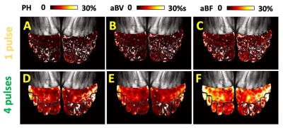 |
Non-Contrast MRI of Micro-Vascularity of the Feet and Toes
Won Bae1,2,
Vadim Malis1,
Asako Yamamoto3,
Yoshimori Kassai4,
Jun Isogai5,
Katsumi Nakamura6,
John Lane7,
and Mitsue Miyazaki1
1Radiology, University of California, San Diego, La Jolla, CA, United States, 2VA San Diego Healthcare System, San Diego, CA, United States, 3Radiology, Teikyo University, Tokyo, Japan, 4Canon Medical Systems Corp, Otawara, Japan, 5Radiology, Asahi General Hospital, Chiba, Japan, 6Kyoritsu Tobata Hospital, Kitakyusyu, Japan, 7University of California, San Diego, La Jolla, CA, United States Keywords: MSK, Arterial spin labelling, Bilateral feet perfusion, ASL, Feet and Toes Non-contrast MR perfusion techniques were developed to assess micro-vascularity of the foot in humans. Using flow-out spin labeling with 3D SSFSE acquisition at multiple Tis, we obtained perfusion signal into distal feet and toes. Signal vs. TI data was fit to determine perfusion metrics including peak height (PH), apparent blood volume (aBV) and apparent blood flow (aBF). Compared to 1-tag, when 4-tag pulses were used PH, aBV, and aBF were significantly greater, suggesting increased signal afforded by the 4-tag pulses. This will be useful for subjects with low blood flow, such as those with peripheral artery disease. |
| 16:25 |
0629.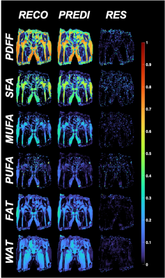 |
Unraveling Bone Marrow Adipose Tissue Composition Through 3T
Deep Learning Chemical Shift Encoded-MRI
Dimitri Martel1,
Benjamin Leporq2,
Anmol Monga1,
Stephen Honig3,
and Gregory Chang1
1Radiology, NYU Langone, New York, NY, United States, 2Université de Lyon; CREATIS CNRS UMR 5220, Inserm U1206, INSA-Lyon, Villeurbanne, France, 3Osteoporosis Center, Hospital for Joint Diseases, NYU Langone, New York, NY, United States Keywords: Bone, Fat Chemical Shift Encoded (CSE) MRI method has been used to highlight subregional differences in the femoral bone marrow in terms of fatty acids composition in proximal femur, suggesting an important role of the marrow in bone quality. This method requires a certain number of echoes (3 for fat/water separation and >8 for fatty acids mapping). Deep Learning (DL) has been recently applied to accelerate, improve the quality and efficiency for fat/water separation in CSE-MRI. Our aim was to develop a DL-CSE method and evaluate it for fatty acids composition mapping; using different echo numbers. |
| 16:33 |
0630.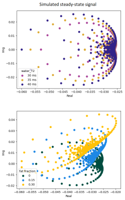 |
Quantification of muscle fat fraction and water T2 via RF
phase-modulated 3D gradient-echo imaging
Eléonore Vermeulen1,
Pierre-Yves Baudin1,
Marc Lapert2,
and Benjamin Marty1
1NMR Laboratory, Neuromuscular Investigation Center, Institute of Myology, Paris, France, 2Siemens Healthcare SAS, Saint-Denis, France Keywords: Muscle, Quantitative Imaging Intramuscular fat fraction (FF) is a frequently used biomarker of neuromuscular disease severity while water-T2 has been identified as a biomarker of disease activity. In this feasibility study, we explored the possibility to exploit RF phase-modulated 3D gradient-echo imaging to obtain multi-parametric mapping adapted to the study of skeletal muscles. Monte Carlo simulations were conducted to evaluate the robustness to noise of this proposed approach. An in vivo proof of concept on healthy volunteers was performed. |
| 16:41 |
0631.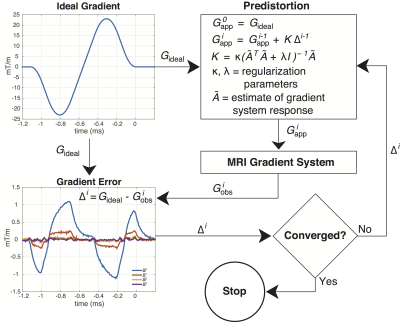 |
Application of fast, oblique 2D-UTE to musculoskeletal imaging
with a library of predistorted slice select gradient waveforms
Kevin D Harkins1,2,3,
Nicholson S Chadwick1,
and Mark D Does2,3
1Radiology and Radiological Sciences, Vanderbilt University Medical Center, Nashville, TN, United States, 2Institute of Imaging Science, Vanderbilt University Medical Center, Nashville, TN, United States, 3Biomedical Engineering, Vanderbilt University, Nashville, TN, United States Keywords: MSK, Pulse Sequence Design The adoption of 2D UTE for general musculoskeletal imaging has been slowed by technical limitations—especially gradient errors that cause slice profile distortions. In this work, a library of predistorted gradient waveforms were used to interpolate slice select gradient waveforms for oblique oriented half-pulse 2D UTE. Example multi-echo and multi-slice acquired 2D UTE images are shown in the tibia, ankle, & knee. Positive contrast in collagen rich tissues like bone and tendon could provide a new source of information to potentially diagnose MSK injuries. |
| 16:49 |
0632. |
Volume isotropic 3D bone Imaging with broadband IR-prepared
FLORET UTE and Fibonacci interleaved trajectory ordering
Masami Yoneyama1,
Minako Azuma2,
Masahiro Enzaki3,
Guruprasad Krishnamoorthy4,5,
Ryan Robison4,
Iain Ball6,
Hiroshi Hamano1,
and Marc Van Cauteren7
1Philips Japan, Tokyo, Japan, 2Department of Radiology, Faculty of Medicine, University of Miyazaki, Miyazaki, Japan, 3Division of Radiology, Miyazaki University Hospital, Miyazaki, Japan, 4Philips Healthcare, Rochester, MN, United States, 5Department of Radiology, Mayo Clinic College of Medicine and Science, Rochester, MN, United States, 6Philips Australia & New Zealand, North Ryde, Australia, 7Philips Healthcare, Best, Netherlands Keywords: Bone, Bone MR bone imaging has gained more attention for detecting and assessing bone pathology. In this study, we proposed a new technique consisting of broadband inversion recovery preparation with FLORET UTE with Fibonacci interleaved trajectory ordering (FLORET BoneVIEW) to obtain 3D isotropic bone images within a clinically feasible scan time. FLORET BoneVIEW has great potential to help a more accurate assessment of bone pathology as an alternative to CT bone imaging. |
| 16:57 |
0633.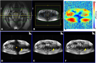 |
Application of Direct Signal Control with Variable Excitation
and Refocusing for T2-w TSE Musculoskeletal Imaging at 7 Tesla
Oliver Kraff1,
Jenni Schulz2,
Markus W May1,3,
Tom WJ Scheenen1,2,
Thomas B Meurs4,
Sebastian Schmitter5,
Jens M Theysohn6,
and Harald H Quick1,3
1Erwin L Hahn Institute for MRI, University Duisburg-Essen, Essen, Germany, 2Medical Imaging, Radboud UMC, Nijmegen, Netherlands, 3High-Field and Hybrid MR Imaging, University Hospital Essen, Essen, Germany, 4Tesla Dynamic Coils, Zaltbommel, Netherlands, 5Physikalisch-Technische Bundesanstalt (PTB), Braunschweig und Berlin, Germany, 6Department of Diagnostic and Interventional Radiology and Neuroradiology, University Hospital Essen, Essen, Germany Keywords: Joints, RF Pulse Design & Fields, UHF First experiences with direct signal control with variable excitation and refocusing (DiSCoVER) for T2-weighted TSE imaging in 7 Tesla musculoskeletal (MSK) applications of hip, shoulder, and ankle are presented. Images were compared to static RF shimming aiming at a homogeneous excitation by using the MR system’s framework as well as optimizing for a B1+-efficient shim in an offline calculation. DiSCoVER yielded satisfying results within the ROI defined for signal optimization but showed more pronounced signal dropouts outside the ROI compared to static RF shimming. Workflow improvements were particularly noted for DiSCoVER as it calculates pTx scale factors under SAR constraints. |
| 17:05 |
0634.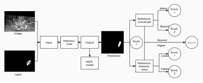 |
Deep-learning-based scoring for inflammatory lesions using
SPARCC system
Yingying Lin1,
Yin Chung Ho2,
Shirley Chui Wai Chan3,
Kam Ho Lee4,
and Peng Cao3
1the university of HongKong, Hongkong, Hong Kong, 2Chiron Medical, HongKong, Hong Kong, 3the university of HongKong, HongKong, Hong Kong, 4Queen Mary Hospital, HongKong, Hong Kong Keywords: Joints, Joints, deep learning, MRI, Ankylosing spondylitis Our work proposed a work pipeline to automatic score the inflammatory lesion based on the SPARCC scoring system. This pipeline included the reference vessel intensity set-up, template-based registration, and lesion detection with scoring. With this pipeline, a time-saving and objective scoring method based on SPARCC scoring system could be achieved and widely applied to AS patient management. Our result showed reasonable performance for score-2 and score-3 lesions demonstrating the deep learning initial capacity of performing the SPARCC scoring. |
| 17:13 |
0635.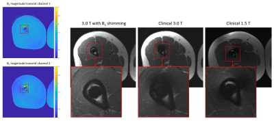 |
Local $$$B_1^+$$$ shimming improves visualization of the
bone-metal interface in patients with orthopedic hardware
Iman Khodarahmi1,
Mahesh B Keerthivasan2,
and Jan Fritz1 1NYU Langone School of Medicine, New York, NY, United States, 2Siemens Medical Solutions USA Inc., Malvern, PA, United States Keywords: Bone, Bone $$$B_1^+$$$ field inhomogeneity is a source of metal artifacts in patients with orthopedic hardware. Local $$$B_1^+$$$ shimming can potentially decrease these artifacts and improve visualization of the bone-metal interface. Our proposed turbo-spin echo-based $$$B_1^+$$$ mapping technique enables accurate estimation of the $$$B_1^+$$$ field near the metal hardware. After optimization for in-vivo applications, the technique was successfully employed on a clinical 3.0 T parallel-transmit system aiming at $$$B_1^+$$$ shimming near the orthopedic hardware. Our results demonstrate significant improvement in visualization of the bone-metal interface compared to standard 1.5 and 3.0 T acquisitions. |
| 17:21 |
0636.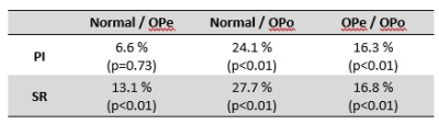 |
MRI-based porosity index and suppression ratio in the tibial
cortex: significant differences in normal, osteopenic, and
osteoporotic subjects
Saeed Jerban1,2,3,
Yajun Ma1,2,
Dina Moazamian1,
Jiyo Athertya1,
Sophia Dwek1,
Hyungseok Jang1,2,
Gina Woods4,
Christine B Chung1,2,
Eric Y Chang1,2,
and Jiang Du1,2
1Department of Radiology, University of California, San Diego, La Jolla, CA, USA, San Diego, CA, United States, 2Radiology Service, Veterans Affairs San Diego Healthcare System, San Diego, La Jolla, CA, USA, San Diego, CA, United States, 3Department of Orthopedic Surgery, University of California, San Diego, La Jolla, CA, USA, San Diego, CA, United States, 4Department of Medicine, University of California, San Diego, La Jolla, CA, USA, San Diego, CA, United States Keywords: Bone, Bone, Tibia The porosity index (PI) and the suppression ratio (SR) are two rapid MRI-based techniques developed using ultrashort echo time (UTE) sequences to evaluate the cortical bone microstructure. We have investigated the performance of PI and SR in detecting tibial bone quality differences between osteoporosis (OPo) patients, osteopenia (OPe) patients, and healthy volunteers with normal bone (Normal). We also investigated the correlations of PI and SR with DEXA T-score performed at the hip in patients. PI and SR were significantly higher in the OPo group compared with the Normal and OPe groups. DEXA T-score was significantly correlated with PI and SR. |
| 17:29 |
0637.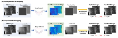 |
Self-supervised T1 Mapping from Two Variable Flip Angle Images
without Requiring Ground Truth T1 Maps
Yan Wu1,
Yajun Ma2,
Zhitao Li1,
Jiang Du2,
John Pauly1,
and Shreyas Vasanawala1 1Radiology, Stanford University, Stanford, CA, United States, 2Radiology, University of California San Diego, San Diego, CA, United States Keywords: Cartilage, Cartilage, supervised learning We propose a self-supervised learning method that derives T1 map from a reduced number of variable flip angle images without requiring ground truth maps, aimed at minimizing data acquisition efforts for obtaining training and testing data. |
| 17:37 |
0638. |
De-noising of 4D real-time joint motion images using a
convolutional neural network trained on static data
Laurel Hales1,
Arjun Desai1,
Valentina Mazzoli 1,
Akshay Chaudhari1,
and Feliks Kogan1
1Radiology, Stanford University, Stanford, CA, United States Keywords: Joints, Image Reconstruction, kinematic We demonstrated the potential applying a noise removal machine learning network trained on images without motion, on a set of similar real-time images acquired with motion. |
 Back to Meeting Home
 Back to the Program-at-a-Glance
The International Society for Magnetic Resonance in Medicine is accredited by the Accreditation Council for Continuing Medical Education to provide continuing medical education for physicians.
 Back to Meeting Home
 Back to the Program-at-a-Glance
 View Presentation Video