Oral Session
Potpourri of Clinical MSK Imaging
ISMRM & ISMRT Annual Meeting & Exhibition • 03-08 June 2023 • Toronto, ON, Canada

| 16:00 |
1437.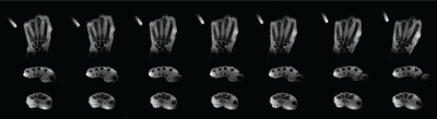 |
Volumetric Dynamic Imaging for Functional Kinematic Assessment
of the Wrist
Ruoxun Zi1,
Bili Wang1,
Jerzy Walczyk1,
Ryan Brown1,
Catherine Petchprapa1,
James Fishbaugh2,
Guido Gerig2,
Kai Tobias Block1,
and Riccardo Lattanzi1
1The Bernard and Irene Schwartz Center for Biomedical Imaging, Department of Radiology, New York University Grossman School of Medicine, New York, NY, United States, 2Department of Computer Science and Engineering, New York University Tandon School of Engineerin, Brooklyn, NY, United States Keywords: Joints, MSK, Wrist Dynamic MRI can be useful for evaluation of wrist instability. However, most available real-time MRI methods are either limited due to their 2D nature or provide only low temporal resolution and insufficient image quality. Here, we propose a novel approach for volumetric dynamic wrist examination by assembling 2D real-time data into 3D snapshots using MRI-visible markers. The method has been demonstrated for ulnar-radial deviation using a flexible wrist coil and 3D-printed support platform for guiding motion. Future work will use a high-resolution static MRI as morphological prior to segment bones on the dynamic volumes and allow for quantitative kinematic assessment. |
| 16:08 |
1438.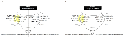 |
Alteration of sacroiliac fatty acids composition in axial
spondyloarthritis: analysis using 3.0 T chemical shift-encoded
MRI
Min Chen1,2,
Chuanli Cheng1,
Chao Zou1,
and Guanxun Cheng2
1Paul C. Lauterbur Research Center for Biomedical Imaging, Shenzhen Institutes of Advanced Technology, Chinese Academy of Sciences, Shenzhen, China, 2Radiology Department, Peking University Shenzhen Hospital, Shenzhen, China Keywords: Joints, Quantitative Imaging, axial spondyloarthritis; bone marrow; fatty acids Changes of bone marrow fatty acids (FAs) composition have been detected in several inflammatory and metabolic diseases by MRI. However, the alteration of sacroiliac FAs composition in axial spondyloarthritis (axSpA) remains unknown. We observed changes of bone marrow FAs composition of the sacroiliac joint in patients with axSpA compared to controls using chemical shift-encoded MRI (CSE-MRI). The changes differed between areas with fat metaplasia and areas without fat metaplasia in axSpA. Our results indicated bone marrow FAs including saturated FA, mono-unsaturated FA and poly-unsaturated FA may play different roles in the pathogenesis of axSpA. |
| 16:16 |
1439.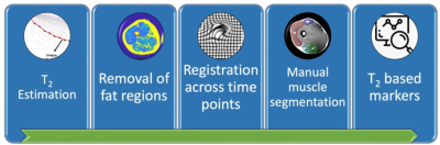 |
Probing muscle recovery following downhill running using precise
mapping of MRI’s T2 relaxation times
Maria Holodov1,
Irit Markus2,
Chen Solomon1,
Shimon Shahar3,
Tamar Blumenfeld-Katzir1,
Yftach Gepner2,
and Noam Ben-Eliezer1,4,5
1Department of Biomedical Engineering, Tel Aviv University, Tel Aviv, Israel, 2Department of Epidemiology and Preventive Medicine, School of Public Health and Sylvan Adams Sports Institute, Tel Aviv University, Tel Aviv, Israel, 3Center of AI and Data Science (TAD), Tel Aviv University, Tel Aviv, Israel, 4Sagol School of Neuroscience, Tel Aviv University, Tel Aviv, Israel, 5Center for Advanced Imaging Innovation and Research (CAI2R), New-York University Langone Medical Center, New York, NY, United States Keywords: Muscle, Skeletal Post-exercise recovery rate is vital for planning training protocols, maintaining high-level performance, and preventing injuries. Notwithstanding the advancement of noninvasive imaging, there is still a lack in efficient tools for monitoring muscle state and post-exercise due to the relatively small changes in the muscles’ microarchitecture, and the high variability in recovery rate between and within participants. Here we utilized the echo-modulation-curve (EMC) algorithm, a highly accurate and precise tool for mapping T2 relaxation times, to track muscle damage and recovery following exercise. Results show that this approach provides new insights into the microstructural processes that occur following exercise. |
| 16:24 |
1440.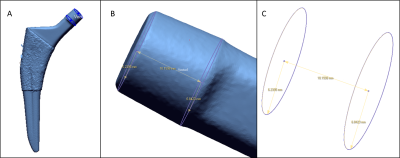 |
Flexural Rigidity of THAs is Associated with MRI and
Histological findings
Sara E Sacher1,
Elexis Padgett1,
John P Neri1,
Timothy Wright1,
Thomas W Bauer1,
Michael Parides1,
Douglas Padgett1,
Hollis G Potter1,
and Matthew F Koff1
1Hospital for Special Surgery, New York, NY, United States Keywords: MSK, MSK, Total hip arthroplasty Micromotion at interfaces in modular total hip arthroplasties (THAs) induces corrosion and generation of adverse local tissue reactions (ALTRs), potentially leading to premature failure. The flexural rigidities (FR) of THA femoral stem trunnions retrieved from revision surgery were correlated with MRI metrics and histopathological outcomes. Increased FR was associated with osteolysis and a reduced synovial response, while a lower FR was associated with granulomas, different synovitis types, and tissue necrosis. These results indicate that MRI can serve as a useful biomarker of the local tissue response around THA. |
| 16:32 |
1441.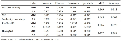 |
Differentiation between myelodysplastic syndrome and aplastic
anemia using transfer learning of vision transformer
Miyuki Takasu1,
Yasutaka Baba2,
Konagi Takeda1,
Saki Kawai1,
Hiroaki Sakane1,
Nobuko Tanitame1,
Akihisa Tamura1,
Makoto Iida1,
and Kazuo Awai3
1Diagnostic Radiology, Hiroshima City Hiroshima Citizens Hospital, Hiroshima, Japan, 2Diagnostic Radiology, International Medical Center, Saitama Medical University, Hidaka, Japan, 3Hiroshima University Hospital, Hiroshima, Japan Keywords: Bone, Machine Learning/Artificial Intelligence We examined the use of a vision transformer (ViT)-based deep learning model for the task of differentiation between aplastic anemia and myelodysplastic syndrome using lumbar T1-weighted images. Three sagittal images per patient were obtained and made square using zero-padding and were resized (224 × 224). The overall accuracy and area under the curve of the pre-trained ViT model were higher than those of ViT without pre-training, ResNet-110, and BinaryNet at the optimum hyperparameters. We utilized Grad-CAM images to highlight the information that is important for decision-making. ViT combined with Grad-CAM successfully recognized variability in the distribution of bone marrow components. |
| 16:40 |
1442.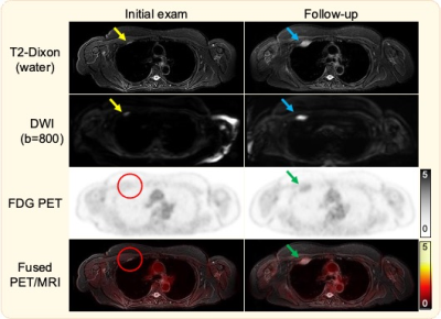 |
Diagnostic performance of Rapid Whole-body MRI with uniformly
fat-suppressed T2-weighted imaging for multiple myeloma
Rianne A van der Heijden1,
Timothy M Schmidt2,
Scott B Reeder1,3,4,5,6,
and Ali Pirasteh1,3
1Department of Radiology, University of Wisconsin-Madison, Madison, WI, United States, 2Carbone Cancer Center, University of Wisconsin-Madison, Madison, WI, United States, 3Department of Medical Physics, University of Wisconsin-Madison, Madison, WI, United States, 4Department of Biomedical Engineering, University of Wisconsin-Madison, Madison, WI, United States, 5Department of Medicine, University of Wisconsin-Madison, Madison, WI, United States, 6Department of Emergency Medical Physics, University of Wisconsin-Madison, Madison, WI, United States Keywords: Skeletal, PET/MR, Multiple Myeloma In this study we evaluated the diagnostic performance of whole-body T2-weighted imaging with uniform 2-point Dixon fat-suppression for the detection of multiple myeloma. Furthermore, we evaluated the added sensitivity, change in disease stage, and change in treatment plan with the sequential addition of DWI and FDG PET. T2-Dixon demonstrated higher sensitivity than both DWI and PET. Adding DWI did not change stage or treatment plan. However, the addition of PET did change the treatment plan in one patient with extra-osseous disease. Whole-body PET-MRI with T2-Dixon is a promising and efficient tool for assessment of multiple myeloma. |
| 16:48 |
1443. |
Inspiratory dilatory tongue movement, as measured with tagged
MRI, and intramuscular neural drive in obstructive sleep apnoea
patients
Lauriane Jugé1,2,
Angela Liao1,2,
Jade Yeung1,
Fiona Knapman1,2,
Christopher Bull1,2,
Peter Burke1,3,
Elizabeth Brown1,4,
Simon Gandevia1,2,
Danny Eckert1,5,
Jane Butler1,2,
and Lynne Bilston1,2
1Neuroscience Research Australia, Sydney, Australia, 2University of New South Wales, Sydney, Australia, 3Macquarie Medical School, Macquarie University, Sydney, Australia, 4Prince of Wales Hospital, Sydney, Australia, 5Flinders Health and Medical Research Institute, Flinders University, Adelaide, Australia Keywords: Muscle, Muscle, electromyography, sleep, tagged MRI As measured by tagged MRI, inspiratory tongue dilatory movement might be useful to shed new light on mechanisms controlling upper airway dilation in obstructive sleep apnoea (OSA). Nine healthy controls and 37 untreated OSA patients underwent an upper airway MRI scan and tongue intramuscular electromyography (EMG) assessment. Results identified two opposing relationships between inspiratory tongue movement and phasic EMG with variable impacts on upper airway function for controls and OSA patients. These results suggest that there are complex, and unexpected, relationships between neural drive and anterior tongue movement that suggest upper airway function cannot be predicted from EMG alone. |
| 16:56 |
1444.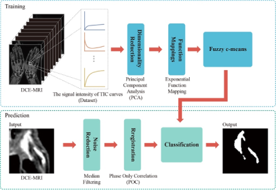 |
Segmentation and quantification of hand synovitis in rheumatoid
arthritis on DCE-MRI-based clustering
Wanxuan Fang1,
Yijun Mao2,
Yujie An1,
Hiroyuki Sugimori2,
Shinji Kiuch3,
and Tamotsu Kamishima2
1Graduate School of Health Sciences, Hokkaido University, Sapporo, Japan, 2Faculty of Health Sciences, Hokkaido University, Sapporo, Japan, 3AIC Yaesu Clinic, Tokyo, Japan Keywords: Rheumatoid Arthritis, DSC & DCE Perfusion Volumetry of enhancing pannus can be used as a marker for disease activity in synovial proliferative disorders such as RA. Enhancing pannus quantified by manual outlining as well as semiquantitative visual assessment of synovitis is too time-consuming to be justified in busy clinical practice. Our study showed great potential for automated quantitative segmentation and quantification of enhancing pannus, which would allow for accurate estimation of disease aggressiveness and thus prompt early therapeutic intervention for patients and assessment of treatment efficacy without increasing the burden on physicians. |
| 17:04 |
1445.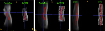 |
Intelligent automatic slice prescription of scout scans of MSK
MRI imaging using surface coil sensitivities
Kavitha Manickam1,
Muhan Shao2,
Dawei Gui1,
Chitresh Bhushan2,
and Dattesh D Shanbhag3
1GE HealthCare, Waukesha, WI, United States, 2GE Global Research, Niskayuna, NY, United States, 3GE Healthcare, Bangalore, India Keywords: MSK, Machine Learning/Artificial Intelligence, Automatic prescription, coil sensitivity map Large-FOV, low-resolution 3D surface coil sensitivities maps are acquired as part of pre-scan which is typically used to calibrate the hardware of MRI system. In this abstract, we propose to use the coil sensitivity maps to automatically find the region of interest of spine anatomy. This information can be used to position the scout images accurately and automatically and thereby reduce any repeat of the scout scans and increase throughput. Our proposal does not need any additional scan and utilizes the existing information available in the MRI scanner. |
| 17:12 |
1446.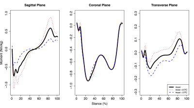 |
Lower-limb walking kinetics associated with acetabular and
femoral bone remodeling in hip OA: a PET-MR study
Koren Roach1,2,
Radhika Tibrewala2,3,
Valentina Pedoia2,
Emma Bahroos2,
Sharmila Majumdar2,
and Richard Souza2 1University of Calgary, Calgary, AB, Canada, 2University of California, San Francisco, San Francisco, CA, United States, 3New York University, New York, NY, United States Keywords: Osteoarthritis, MSK Early hip osteoarthritis is clinically hypothesized to include alterations in joint motion and bone remodeling. This study investigated the relationships between lower-limb joint kinetics and bone remodeling, measured via positron emission tomography. Our results suggest that greater hip flexion and internal rotation loading and more uniform knee abduction loading are associated with bone remodeling and may serve as early biomechanical biomarkers for hip OA. |
| 17:20 |
1447.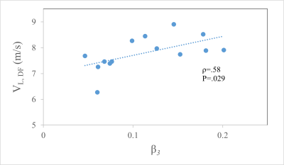 |
Multiexponential UTE Relaxation in Achilles Tendon of Ballet
Dancers Reveals Matrix Proton Pool Associated with Tendon Shear
Stiffness
Anna M Horner1,
Felix M Gonzalez2,
Courtney N Gleason1,
Amanda Blackmon3,4,
Emma Faulkner3,5,
and David A Reiter1
1Emory University School of Medicine, Atlanta, GA, United States, 2Radiology, AdventHealth, Orlando, FL, United States, 3Atlanta Dance Medicine, Atlanta, GA, United States, 4Physical Therapy, Mercer University, College of Health Professions, Macon, GA, United States, 5Dance, Emory University, Atlanta, GA, United States Keywords: Tendon/Ligament, Low-Field MRI, UTE MRI, SWE, ballet dancers Multi-echo ultrashort echo time (UTE) MRI provides quantitative structural information on both tendon matrix constituents and water distribution, which influence the mechanical function of tendons. UTE images of professional ballet dancers' Achilles tendons (AT) were combined with Shear Wave Elastography (SWE) Ultrasound (US) measurements of their AT to provide insight on the structure-function. The signal fraction corresponding to the off-resonance relaxation tendon components were found to positively correlate with the long-axis SWE velocity in this dancer cohort. |
| 17:28 |
1448.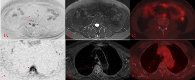 |
Whole-Body Diffusion-weighted MRI, non-diffusion MRI, and PET-CT
in detection of Spinal Lesions in Multiple Myeloma Patients
Kai Ye1,
Huishu Yuan1,
Weifang Zhang2,
Xianchang Zhang3,
and Lihua Zhang1
1Department of Radiology, Peking University Third Hospital, Beijing, China, 2Peking University Third Hospital, Beijing, China, 3MR Collaboration, Siemens Healthineers Ltd, Beijing, China Keywords: Bone, Diffusion/other diffusion imaging techniques, Multiple Myeloma This study investigated the performance of whole-body diffusion-weighted imaging (WBDWI), non-diffusion MRI, and 18F-FDG PET-CT in detecting spinal lesions in patients with an initial diagnosis of multiple myeloma (MM). The results showed that WBDWI discovered more lesions compared with both non-diffusion MRI and 18F-PET-CT. And the detection of spinal lesions in WBDWI was not significantly decreased even with the decreased degree of marrow infiltration in patients with MM. This suggests that WBDWI should be the preferentially recommended examination for patients with an initial diagnosis of MM. |
| 17:36 |
1449.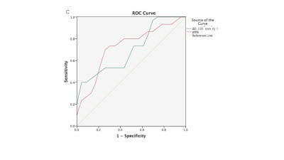 |
Intravoxel Incoherent Motion Diffusion-weighted MR Imaging and
IDEAL-IQ for Assessment of Treatment Response in Multiple
Myeloma
Yanhua Tang1,
Li Wang2,
and Yichen Ma3 1Radiology, Beijing Chao-Yang Hospital, Capital Medical Universit, Beijing, China, 2Beijing Chao-Yang Hospital, Capital Medical Universit, Beijing, China, 3Beijing Chao-Yang Hospital, Capital Medical University, Beijing, China Keywords: Bone, Diffusion/other diffusion imaging techniques, intravoxel incoherent motion, diffusion-weighted imaging, iterative decomposition of water and fat with echo asymmetry and least-squares estimation quantitation sequence (IDEAL-IQ), multiple myeloma, treatment response This study explored the feasibility of IVIM DWI and IDEAL-IQ parameters in evaluating the treatment response of patients with multiple myeloma (MM) who received systemic treatment within 6 months. The results showed that within 6 months after treatment, the changes of D and f values of IVIM DWI in deep-responders before and after treatment were significantly different from those in non-deep responders.This indicates that the changes of D and f values of IVIM DWI may be helpful to evaluate the short-term (within 6 months) therapeutic response of myeloma, while the evaluation of IDEAL-IQ on short-term therapeutic response needs further study. |
| 17:44 |
1450.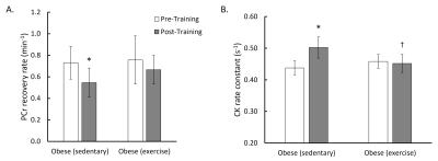 |
The Effect of Long-term Exercise Training on Metabolic Responses
in Obese Zucker Fatty Diabetic Rats using Phosphorous-31 MRS
Kihwan Kim1,
Yuran Zhu1,
Raodatullah Abodunrin1,
Sonia Kumar 1,
Jessica Meng1,
Luchen Yu1,
Allison McKenzie 1,
and Xin Yu1,2
1Biomedical Engineering, Case Western Reserve University, Cleveland, OH, United States, 2Case Center for Imaging Research, Case Western Reserve University, Cleveland, OH, United States Keywords: Muscle, Diabetes In this study, metabolic responses to 10-week treadmill exercise on obese Zucker rats were examined using 31P magnetic resonance spectroscopy. Exercise resulted in improvement in glucose tolerance and aerobic capacity (VO2max) while retaining mitochondrial oxidative capacity (MOC) and creatine kinase (CK) activity. Sedentary rats, however, showed poor glucose tolerance and reduced VO2max, which were also marked by significant changes in their MOC and CK activity. |
| 17:52 |
1451.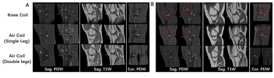 |
An initial evaluation of image quality for double knee imaging
with air coil as compared with traditional knee coils
Chao Han1,
Feifei Zhang1,
Chengbin Tian1,
Wenwen Kang1,
Weimin Yu1,
and Jinxia Guo2
1General Hospital of Pingmei Shenma Group, Pingdingshan, China, 2GE Healthcare, MR Research, Beijing, China Keywords: Joints, Joints This study investigated the feasibility and potential advantages of adaptive imaging receiver (AIR) coil for knee imaging. The results show that the SNR and CNR of air coil images were inferior to that of traditional knee coil, but the visual image qualities were comparable between two coils. The air coil can be feasible for knee imaging, especially for patients with large size and double knee imaging. |
 Back to Meeting Home
 Back to the Program-at-a-Glance
The International Society for Magnetic Resonance in Medicine is accredited by the Accreditation Council for Continuing Medical Education to provide continuing medical education for physicians.
 Back to Meeting Home
 Back to the Program-at-a-Glance
 View Presentation Video