Oral Session
Novel MR Techniques & Clinical Applications in Neurofluids
ISMRM & ISMRT Annual Meeting & Exhibition • 03-08 June 2023 • Toronto, ON, Canada

Oral Session
Novel MR Techniques & Clinical Applications in Neurofluids
| 16:00 |
1452.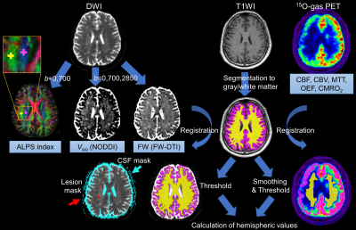 |
Do adult patients with moyamoya disease have glymphatic system
dysfunction? - evaluation using diffusion along perivascular
space
Shoko Hara1,2,3,
Junko Kikuta2,
Kaito Takabayashi2,
Koji Kamagata2,
Shihori Hayashi1,4,
Motoki Inaji1,3,
Yoji Tanaka1,
Masaaki Hori2,
Kenji Ishii4,
Tadashi Nariai1,4,
Toshiaki Taoka5,
Shinji Naganawa6,
Shigeki Aoki2,
and Takeoshi Maehara1
1Neurosurgery, Tokyo Medical and Dental University, Tokyo, Japan, 2Radiology, Juntendo University, Tokyo, Japan, 3Research Team of Neuroimaging, Tokyo Metropolitan Institute of Gerontology, Tokyo, Japan, 4Research Team for Neuroimaging, Tokyo Metropolitan Institute of Gerontology, Tokyo, Japan, 5Innovative Biomedical Visualization, Nagoya University Graduate School of Medicine, Nagoya, Japan, 6Radiology, Nagoya University, Nagoya, Japan Keywords: Neurofluids, Diffusion/other diffusion imaging techniques, glymphatic system We aimed to evaluate the glymphatic system of adult moyamoya disease (MMD) by measuring diffusion along the perivascular space (DTI-ALPS index). We evaluated 46 patients using diffusion MRI, perfusion parameters of 15O-gas PET, and cognitive tests, and 34 age-sex-matched normal controls. Compared to normal controls, patients with MMD showed significantly lower DTI-ALPS index. DTI-ALPS index in MMD revealed the correlation between perfusion and freewater parameters, and executive dysfunction, and suggested that dysfunction of the glymphatic system may exist, correlate with the degree of hemodynamic disturbance, lead to increased parenchymal free water, and relate to cognitive dysfunction in adult MMD. |
| 16:08 |
1453.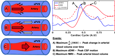 |
Multimodal MR imaging approach to evaluate the interaction
between cardiac pulsation and perivascular CSF motion
Adam M. Wright1,2,
Yunjie Tong1,
Yu-Chien Wu2,
and Qiuting Wen2
1Weldon School of Biomedical Engineering, Purdue University, West Lafayette, IN, United States, 2School of Medicine, Indiana University, Indianapolis, IN, United States Keywords: Neurofluids, Neurofluids We designed a non-invasive multimodal MRI approach to evaluate the relationship between cardiac pulsation and perivascular cerebrospinal fluid (pCSF) dynamics in humans. We utilized cardiac-aligned resting-state functional MRI and dynamic diffusion-weighted imaging to describe cerebral vascular events and pCSF motion, respectively. This approach revealed that changes in cerebral blood volume preceded an increase in pCSF motion, demonstrating the cardiac cycle’s effect on pCSF dynamics. Our results parallel preclinical two-photon imaging findings of perivascular CSF dynamics in mice. Our in-vivo assessment of cardiac pulsations influencing pCSF motion provides a non-invasive look into a crucial part of the human brain’s glymphatic system. |
| 16:16 |
1454.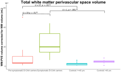 |
TOTAL WHITE MATTER PERIVASCULAR SPACE VOLUME: AN EARLY MARKER
FOR DUTCH-TYPE CEREBRAL AMYLOID ANGIOPATHY
Manon Roxanne Schipper1,
Arie-Tjerk Razoux-Schultz2,
Thijs W. van Harten1,
Jeroen van der Grond1,
Mark van Buchem1,
Steven M. Greenberg3,
Marieke J.H. Wermer4,
Matthias J.P. van Osch1,
Marianne A.A. van Walderveen1,
and Sanneke van Rooden1
1Radiology, Leiden University Medical Center, Leiden, Netherlands, 2Leiden University Medical Center, Leiden, Netherlands, 3Neurology, Mass General Research Institute, Boston, MA, United States, 4Neurology, Leiden University Medical Center, Leiden, Netherlands Keywords: Neurofluids, Neurofluids, Perivascular spaces In this study we semi-automatically quantified MRI-visible perivascular space volume in the white matter (WM-PVS) and compared these volumes between (pre-)symptomatic Dutch-type hereditary Cerebral Amyloid Angiopathy (D-CAA) and matched controls. Main findings show that the WM-PVS volume was significantly higher in symptomatic D-CAA (median=19.9mL) compared with both presymptomatic D-CAA (median=3.5mL; U=19, p=.01) and matched controls (median=4.9mL; U=1, p<.001). Presymptomatic D-CAA carriers had a higher WM-PVS volume than the matched controls (median=2.3mL; U=27, p=.02). This indicates that WM-PVS volume, in contrast to the PVS-CSO visual rating scales, may be an early marker for D-CAA. |
16:24 |
1455.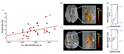 |
Higher blood flow velocity pulsatility relates to increased
interstitial fluid diffusivity - a potential proxy of high
perivascular fluid flow
Merel M. van der Thiel1,2,3,
Marieke van den Kerkhof1,2,
Alida A. Postma1,2,
Walter H. Backes1,2,4,
and Jacobus F.A. Jansen1,2,5
1Department of Radiology & Nuclear Medicine, Maastricht University Medical Center, Maastricht, Netherlands, 2School for Mental Health and Neuroscience, Maastricht University, Maastricht, Netherlands, 3Department of Psychiatry & Neuropsychology, Maastricht University, Maastricht, Netherlands, 4Cardiovascular Research Institute Maastricht, Maastricht University, Maastricht, Netherlands, 5Department of Electrical Engineering, Eindhoven University of Technology, Eindhoven, Netherlands Keywords: Neurofluids, Aging, Diffusion, pulsatility, waste clearance Impaired cerebral waste clearance occurs in healthy ageing and various neurodegenerative diseases and is theorized to be due to compromised arterial pulsatility. Profiting from high-resolution 7 Tesla MRI, the current study investigated the association between pulsatility characteristics of a small artery in the basal ganglia (BG) with interstitial fluid (ISF) characteristics of the BG - as derived with intravoxel incoherent motion. This study found that an increased small vessel velocity pulsatility was related to higher ISF-diffusivity in the BG of an elderly sample. This increased ISF-diffusivity might represent increased perivascular fluid diffusivity, influencing the waste fluid transport out of these spaces. |
| 16:32 |
1456.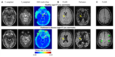 |
DWI with dynamic b-value cycling reveals evidence of reduced
suprasellar cistern neurofluid motion in Parkinson’s disease
Gabriela Pierobon Mays1,
Kilian Hett1,
Jarrod Eisma1,
Colin D McKnight1,
Jason Elenberger1,
Alexander K Song1,
Ciaran Considine1,
Caleb Han1,
Daniel O Claassen1,
and Manus J Donahue1 1Vanderbilt University, Nashville, TN, United States Keywords: Neurofluids, Parkinson's Disease, Glymphatic The goal of this study was to use a novel diffusion weighted imaging (DWI) protocol with dynamic cycling of low b-values to test fundamental hypotheses regarding neurofluid movement along inflow and egress pathways of the suprasellar cisterns in older adults with and without Parkinson’s disease (PD). Contrast consistent with reduced neurofluid motion was observed in PD relative to healthy participants, with reduced fluid movement corresponding to choroid plexus hyperemia. Modeling of DWI signal decay as a function of b-value provides quantitative neurofluid kinetics and may present a candidate non-tracer technology for quantifying neurofluid flow non-invasively in vivo in humans. |
| 16:40 |
1457.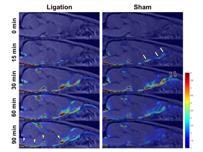 |
Blockage of CSF Outflow via Nasal Lymphatic Pathway in Rats
after Deep Cervical Lymph Node Ligation Observed Using
Intrathecal Gd-based MR Imaging
Naoya Kinota1,2,
Hiroyuki Kameda3,4,
Kazuyuki Minowa3,
and Kohsuke Kudo1,5
1Department of Diagnostic Imaging, Faculty of Medicine and Graduate School of Medicine, Hokkaido University, Sapporo, Japan, 2Department of Dental Radiology, Hokkaido University Hospital, Sapporo, Japan, 3Department of Radiology, Faculty of Dental Medicine, Hokkaido University, Sapporo, Japan, 4Department of Diagnostic and Interventional Radiology, Hokkaido University Hospital, Sapporo, Japan, 5Global Center for Biomedical Science and Engineering, Faculty of Medicine, Hokkaido University, Sapporo, Japan Keywords: Neurofluids, Neurofluids, Glymphatic System Previous reports showed that deep cervical lymph nodes (DCLNs) receive cerebrospinal fluid (CSF) outflow. DCLN ligation resulted in intracranial accumulation of waste proteins due to impaired CSF outflow; however, changes in extracranial outflow after DCLN ligation have not been directly observed. We examined extracranial CSF outflow in rats after DCLN ligation by using intrathecal gadolinium (Gd)-based dynamic contrast-enhanced MRI. DCLN ligation blocked CSF- tracer outflow into the nasal cavity. A weak trend towards CSF-tracer retention in the ventral cistern was also observed. These results suggest that DCLN ligation affects CSF outflow in rodents. |
| 16:48 |
1458.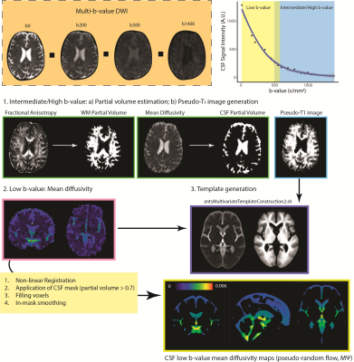 |
CSF pseudo-diffusion spatial statistics for whole-brain
voxel-wise analysis of CSF flow
Arash Nazeri1,
Taher Dehkharghanian2,
Pamela J. LaMontagne1,
Tammie L.S. Benzinger1,
and Aristeidis Sotiras1,3
1Mallinckrodt Institute of Radiology, Washington University in St. Louis, St. Louis, MO, United States, 2Faculty of Health Science, McMaster University, Hamilton, ON, Canada, 3Institute for Informatics, Washington University in St. Louis, St. Louis, MO, United States Keywords: Neurofluids, Diffusion/other diffusion imaging techniques, cerebrospinal fluid Alterations in CSF flow patterns have been implicated in various brain disorders. At low b-values, diffusion-weighted imaging is sensitive to pseudorandom CSF flow. Here, we present CSF pseudo-diffusion spatial statistics (CΨSS), a whole-brain multi-subject voxel-wise analysis framework that exploits low-b-value diffusion-weighted imaging to study regional patterns of CSF flow. Using this technique, we show how brain atrophy and ventricular volumes affect regional CSF pseudorandom flow. In conclusion, CΨSS is a simple and effective approach for characterizing determinants of regional CSF pseudorandom flow. |
| 16:56 |
1459.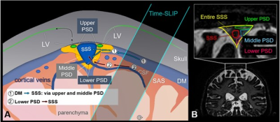 |
Physical exercise activates intrinsic CSF outflow metrics in
healthy humans
Mitsue Miyazaki1,
Vadim Malis1,
Asako Yamamoto2,
Jirach Kungsamutr3,
Marin McDonald1,
Linda McEvoy1,
and Won Bae1,4
1Radiology, University of California, San Diego, La Jolla, CA, United States, 2Radiology, Teikyo University, Tokyo, Japan, 3Bioengineer, University of California, San Diego, La Jolla, CA, United States, 4VA San Diego Healthcare System, San Diego, CA, United States Keywords: Neurofluids, Neurofluids, CSF, glymphatic egress pathways, active and sedentary Possible two intrinsic CSF egress pathways of dura mater and the lower parasagittal dura (PSD) are observed using non-contrast spin-labeling MRI. Intrinsic CSF outflow metrics increase in the adults with an active lifestyle than adults with sedentary lifestyle. However, after 3 weeks of increased physical activity, the sedentary group showed improved CSF outflow metrics. This improvement was notable at the lower PSD, where outflow metrics were highest among the active group. These quantitative CSF results indicate a new pathway of CSF flow from the lower PSD to the superior sagittal sinus that is most evident in physically active individuals. |
| 17:04 |
1460.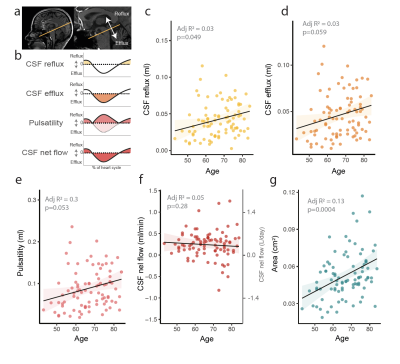 |
Cerebrospinal fluid pathways are perturbed in aging and
prodromal Alzheimer’s disease
Tekla Maria Kylkilahti1,2,
Max Wictor1,2,
David Berron3,
Johannes Töger4,
Karin Markenroth Bloch5,
Niklas Mattson-Carlgren3,
Oskar Hansson3,6,
and Iben Lundgaard1,2
1Experimental medical science, Lund University, Lund, Sweden, 2Wallenberg Centre for Molecular Medicine, Lund University, Lund, Sweden, 3Clinical Memory Research Unit, Department of Clinical Sciences Malmö, Lund University, Malmö, Sweden, 4Skåne University Hospital, Department of Clinical Sciences Lund, Diagnostic Radiology, Lund University, Lund, Sweden, 5Lund University Bioimaging Center, Lund University, Lund, Sweden, 6Neuropsychiatric Clinic, Malmö University Hospital, Malmö, Sweden Keywords: Neurofluids, Velocity & Flow Cerebrospinal fluid (CSF) circulation through the parenchyma by the glymphatic system has been suggested to play a role in the clearance of metabolic waste from the brain, including Amyloid β. In this 7T MRI study of CSF flow in the cerebral aqueduct, we find that ageing, cognitive status and amyloid status influence CSF dynamics and morphology of the aqueduct. These changes appear to precede major cognitive changes in Alzheimer’s disease. Disruption of flow dynamics and anatomy of CSF flow pathways may reflect dysfunctional CSF circulation in downstream compartments of the glymphatic system, and contribute to amyloid accumulation in the brain. |
| 17:12 |
1461.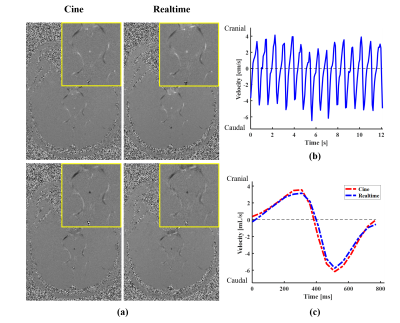 |
Real-Time Imaging of Cerebrospinal Fluid Flow with Low-Rank and
Subspace Modeling
Aiqi Sun1,
Lekang Yin2,
Bingyi Wang1,
Hengfa Lu3,
Peng Wu4,
and Bo Zhao3,5
1Institute of Science and Technology for Brain-Inspired Intelligence, Fudan University, Shanghai, China, 2Department of Radiology, Zhongshan Hospital, Fudan University, Shanghai, China, 3Department of Biomedical Engineering, University of Texas at Austin, Austin, TX, United States, 4Philips Healthcare, Shanghai, China, 5Oden Institute for ComputationalEngineering and Sciences, University of Texas at Austin, Austin, TX, United States Keywords: Neurofluids, Velocity & Flow Quantification of cerebrospinal fluid (CSF) flow is critical for studying the physiological and pathological mechanisms of CSF dynamics and related neurological diseases. Conventional cine phase-contrast MRI provides an effective tool to quantify CSF flow, however, this method is not suitable for evaluating beat-by-beat flow variabilities associated with cardiac arrhythmia and/or respiratory regulation. This work presents a real-time flow MRI method at a spatial resolution of 0.5 mm and temporal resolution of 52 ms for assessment of CSF flow in cerebral aqueduct at 3T, which can well resolve beat-by-beat CSF flow variations. Its feasibility has been demonstrated in multiple healthy subjects. |
| 17:20 |
1462.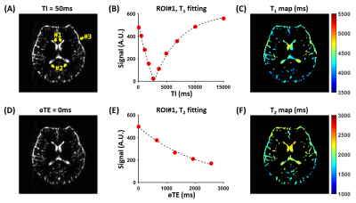 |
Quantification of CSF T1 and T2 in the Subarachnoid Space:
Implication for Brain-CSF Exchange
Dengrong Jiang1,
Yifan Gou2,
Zhiliang Wei1,
Xirui Hou1,
Vivek Yedavalli1,
and Hanzhang Lu1
1Department of Radiology, Johns Hopkins University School of Medicine, Baltimore, MD, United States, 2Department of Biomedical Engineering, Johns Hopkins University School of Engineering, Baltimore, MD, United States Keywords: Neurofluids, Neurofluids It has been suggested that the brain’s waste clearance system involves water exchange between brain tissue and CSF in perivascular spaces, which are part of the subarachnoid space (SAS). In this work, we found CSF T1 and T2 in SAS were shorter than corresponding values in the frontal-horns of lateral ventricle, which have minimal exchange. Numerical simulations showed that a higher brain-CSF water exchange rate was associated with lower apparent relaxation time in SAS, providing a potential means to estimate brain-CSF water exchange rate in vivo. |
| 17:28 |
1463.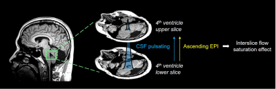 |
Measurement of CSF pulsation in Parkinson’s disease patients
using EPI-based fMRI data
Jun-Hee Kim1,
Suhong Kim1,
Jae-Geun Im1,
Seok Jong Chung2,
Phil Hyu Lee2,
Yong Jeong1,
and Sung-Hong Park1
1Korea Advanced Institute of Science and Technology, Daejeon, Korea, Republic of, 2Yonsei University Colledge of Medicine, Seoul, Korea, Republic of Keywords: Neurofluids, Parkinson's Disease The fMRI dataset for Parkinson’s disease with mild cognitive impairment (PD-MCI) was studied using a recently-proposed method of simultaneous CSF pulsation and BOLD activity imaging. The PD group was classified to dementia (PDD) high-risk and low-risk groups. The CSF pulsation of both PDD high-risk and low-risk groups was higher than that of healthy control. Opposite to the tendency of CSF pulsation, coefficient of variation of CSF pulsation was reduced in PD group compared to healthy control. These results indicate association of CSF pulsation with brain waste clearance in these patients, which requires further investigations for elucidation. |
| 17:36 |
1464.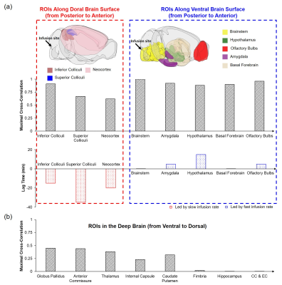 |
The impact of infusion rate on Gd-DTPA transport in the
glymphatic system in mouse brain
Yuran Zhu1,
Guanhua Wang2,
Kihwan Kim1,
Chris A. Flask1,3,4,
and Xin Yu1,3,5
1Department of Biomedical Engineering, Case Western Reserve University, Cleveland, OH, United States, 2Department of Biomedical Engineering, University of Michigan, Ann Arbor, MI, United States, 3Department of Radiology, Case Western Reserve University, Cleveland, OH, United States, 4Department of Pediatrics, Case Western Reserve University, Cleveland, OH, United States, 5Department of Physiology and Biophysics, Case Western Reserve University, Cleveland, OH, United States Keywords: Neurofluids, Contrast Agent, Glymphatic system This study evaluated the impact of infusion rate on tracer transport in mouse glymphatic system. Gd-DTPA was administered via cisterna magna at a rate either comparable to the CSF production rate in mouse brain (0.33 μL/min) or three-fold higher (1 μL/min). The kinetics and distribution of Gd-DTPA were assessed by DCE-MRI for 2 hours. Our results show a significantly increased transport along the dorsal brain regions and penetration into the deep brain regions at a slow infusion rate, suggesting that the infusion rate may alter the pressure gradient and affect tracer transport and distribution in the brain. |
17:44 |
1465.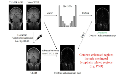 |
Synthesizing contrast-enhancement map from non-contrasted
black-blood images for brain lymphatic imaging using a deep
neural network
Jun-Hee Kim1,
Roh-Eul Yoo2,3,
Seung-Hong Choi2,3,
and Sung-Hong Park1
1Korea Advanced Institute of Science and Technology, Daejeon, Korea, Republic of, 2Department of Radiology, Seoul National University College of Medicine, Seoul, Korea, Republic of, 3Seoul National University Hospital, Seoul, Korea, Republic of Keywords: Neurofluids, Machine Learning/Artificial Intelligence In this study, we proposed non-invasive brain lymphatic region mapping by synthesizing contrast-enhancement maps (CEM) from non-contrast enhanced black blood imaging (non-CEBB). T1 images were used as secondary input along with non-CEBB, which helped the network to better distinguish lymphatic regions from blood vessels. From the reconstructed 3D CEM segmentation, enhancement was mainly distributed in dorsal parasagittal dura, parasagittal regions, brain basal region and around choroid plexus, consistent with previous studies. This study could be applied to the segmentation of the brain lymphatic region with less ambiguity and may help automatic segmentation rather than intensity-based segmentation by adapting self-supervised learning. |
| 17:52 |
1466.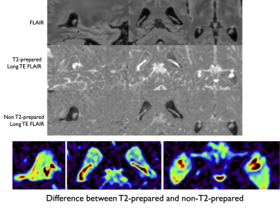 |
Imaging water exchange in the choroid plexus using T2-prepared
long TE Fluid-Attenuated Inversion Recovery
Manuel Taso1 and
David C Alsop1 1Division of MRI Research, department of Radiology, Beth Israel Deaconess Medical Center, Harvard Medical School, Boston, MA, United States Keywords: Neurofluids, Brain The choroid plexus (CP) plays a key role in brain homeostasis and waste clearance as the main source of CSF production in the brain. However, this small structure sitting in the lateral ventricles is not well characterized, including its dysfunction or impairment in several pathologies or just normal aging. Water exchange measurement using various methods has been proposed to evaluate structural but also functional properties of the CP such as ASL or other spin labeling strategies mainly in rodents. In this work, we propose a new look into water exchange between CP and CSF using T2-prepared, long TE Fluid-Attenuated-Inversion-Recovery (FLAIR). |
 Back to Meeting Home
 Back to the Program-at-a-Glance
The International Society for Magnetic Resonance in Medicine is accredited by the Accreditation Council for Continuing Medical Education to provide continuing medical education for physicians.
 Back to Meeting Home
 Back to the Program-at-a-Glance
 View Presentation Video