Oral Session
Novel Biomarkers for Alzheimer's Disease & Dementia
ISMRM & ISMRT Annual Meeting & Exhibition • 03-08 June 2023 • Toronto, ON, Canada

Oral Session
Novel Biomarkers for Alzheimer's Disease & Dementia
08:15 |
0406.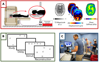 |
Brain Stiffness, Aerobic Fitness, and Memory Performance
Differences Between Older Adults with and without Mild Cognitive
Impairment
Mary K Kramer1,
Peyton L Delgorio1,
Alexa M Diano1,
Olivia M Bailey1,
Grace McIlvain1,
Kyra E Twohy2,
Matthew Overstreet3,
David G Edwards3,
Christopher R Martens3,
and Curtis L Johnson1,2
1Biomedical Engineering, University of Delaware, Newark, DE, United States, 2Mechanical Engineering, University of Delaware, Newark, DE, United States, 3Kinesiology and Applied Physiology, University of Delaware, Newark, DE, United States Keywords: Alzheimer's Disease, Elastography Magnetic resonance elastography (MRE) is a robust and sensitive tool used to measure brain mechanical properties that can accurately detect improvements in brain health and function. Exercise and aerobic fitness levels are strongly tied to these brain mechanical properties and their related functionality. In healthy older adults, greater fitness is associated with better memory and increased mechanical integrity of the brain. In a population of older adults with mild cognitive impairment, there is a notable decrease in memory function and aerobic fitness compared to healthy controls, and this decrease is measurable in the mechanical properties of the brain using MRE. |
| 08:23 |
0407.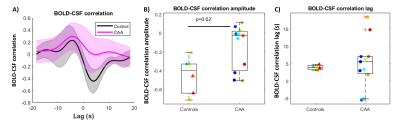 |
Coupling between low-frequency hemodynamic oscillations and
cerebrospinal fluid flow is altered in patients with cerebral
amyloid angiopathy
Lydiane Hirschler1,2,
Maria Clara Zanon Zotin1,
Laura D Lewis3,4,
Mitchell J Horn1,
M. Edip Gurol1,
Anand Viswanathan1,
Jonathan R. Polimeni4,
Matthias JP van Osch2,
Susanne J van Veluw1,2,
and Steven M Greenberg1 1Neurology, Massachusetts General Hospital, Harvard Medical School, Boston, MA, United States, 2C.J. Gorter MRI Center, Department of Radiology, Leiden University Medical Center, Leiden, Netherlands, 3Biomedical Engineering, Boston University, Boston, MA, United States, 4Athinoula A. Martinos Center for Biomedical Imaging, Department of Radiology, Harvard Medical School, Massachusetts General Hospital, Charlestown, MA, United States Keywords: Dementia, Neurofluids, Neurovascular, small vessel disease Cerebral amyloid angiopathy (CAA) is characterized by the accumulation of amyloid-β in the vessel walls. Prominent manifestations of CAA include enlarged perivascular spaces (ePVS) and lower amyloid-β concentrations in CSF, which may be the result of impaired brain clearance. CAA patients also demonstrate impaired evoked vascular reactivity. Whether vascular dysfunction is associated with impaired fluid movement in the human brain remains unclear. Here we show that BOLD-CSF coupling is reduced in CAA patients compared to elderly controls, which is a first demonstration of a cerebral small vessel disease affecting CSF motion. |
| 08:31 |
0408.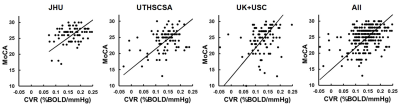 |
Cerebrovascular reactivity (CVR) MRI as a biomarker for small
vessel disease related cognitive decline: validation in the
MarkVCID Consortium
Peiying Liu1,2,
Zixuan Lin1,
Kaisha Hazel1,
George Pottanat1,
Cuimei Xu1,
Dengrong Jiang1,
Emma Lucke1,
Christopher E. Bauer3,
Brian T. Gold3,
Steven M. Greenberg4,
Karl G. Helmer5,
Kay Jann6,
Gregory A. Jicha3,
Joel Kramer7,
Pauline Maillard8,
Rachel Mulavelil9,
Pavel Rodriguez9,
Claudia L. Satizabal9,
Sudha Seshadri9,
Herpreet Singh5,
Angel G. Velarde9,
Danny J.J. Wang6,
Rita R. Kalyani1,
Abhay Moghekar 1,
Paul B. Rosenberg1,
Sevil Yasar1,
Marilyn Albert1,
and Hanzhang Lu1
1Johns Hopkins University School of Medicine, Baltimore, MD, United States, 2University of Maryland School of Medicine, Baltimore, MD, United States, 3University of Kentucky, Lexington, KY, United States, 4Harvard Medical School, Boston, MA, United States, 5Massachusetts General Hospital, Boston, MA, United States, 6University of Southern California, Los Angeles, CA, United States, 7University of California, San Francisco, San Francisco, CA, United States, 8University of California, Davis, Sacramento, CA, United States, 9UT Health San Antonio, San Antonio, TX, United States Keywords: Dementia, Blood vessels Small vessel disease (SVD) related vascular contributions to cognitive impairment and dementia (VCID) represent a major factor in cognitive decline in older adults. However, there has not been a validated biomarker for the diagnosis and treatment monitoring of this condition. Recently, the US National Institute on Neurological Disorders and Stroke (NINDS), a branch of NIH, funded a MarkVCID consortium, the goal of which is to identify and validate clinical-trial-ready biomarkers for VCID. Cerebrovascular reactivity (CVR) MRI was one of the selected biomarkers that underwent multi-site testing. The present work reports the relationship between CVR and cognitive function, and examines whether the pre-specified hypothesis can be reproduced at each of the sites. |
| 08:39 |
0409.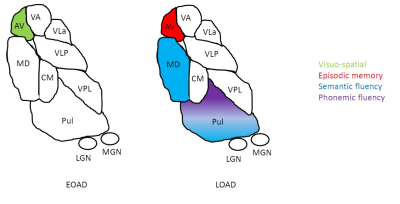 |
Thalamic nuclei changes in early vs. late onset Alzheimer’s
Disease
Gonzalo Forno1,2,
Michael Hornberger3,
Albert Lladó1,
and Manojkumar Saranathan4 1Alzheimer’s Disease and Other Cognitive Disorders Unit, Neurology Service, Hospital Clínic of Barcelona, Institut d’Investigacions Biomèdiques August Pi i Sunyer (IDIBAPS), Universitat de Barcelona, Barcelona, Spain, 2Escuela de Psicología, Universidad de los Andes, Santiago, Chile, 3University of East Anglia, Norwich, United Kingdom, 4Radiology, University of Massachusetts Chan Medical School, Worcester, MA, United States Keywords: Alzheimer's Disease, Segmentation Age at symptom onset distinctly affects thalamic nuclei alongside AD. While Early Onset AD showed significant anteroventral (AV) nucleus changes associated with increasing tau pathology, Late onset AD AV nucleus was associated with Aβ42 levels but no significant volume changes were found. |
08:47 |
0410.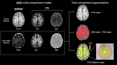 |
Alzheimer's disease neuropathology contributes to perivascular
and parenchymal free water diffusion characteristics
Kirsten Mary Lynch1,
Arthur W Toga1,
and Jeiran Choupan1,2
1USC Mark and Mary Stevens Institute for Neuroimaging and Informatics, Keck School of Medicine of USC, Los Angeles, CA, United States, 2Research, NeuroScope Inc., Scarsdale, NY, United States Keywords: Alzheimer's Disease, Neurofluids Disruption of perivascular and interstitial fluid transport in the brain may contribute to the accumulation of toxic metabolic deposits observed in neurodegeneration. However, the relationship between AD neuropathology and free water fluid dynamics are not well understood. Using multi-compartment diffusion models, we assessed the relationship between PET tau and Aβ uptake and free water diffusion properties. We found tau deposition was associated with reduced free water anisotropy in brain regions that undergo neurodegeneration in pre-clinical AD. These findings provide a preliminary link between AD pathology and fluid transfer that may indicate waste clearance functional alterations. |
| 08:55 |
0411.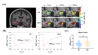 |
Vascular-water-exchange imaging to detect blood-brain barrier
breakdown in Alzheimer’s disease without contrast agent
Yifan Zhang1,2,
Yue Wang3,4,
Zhaoqing Li1,2,
Shiping Li3,4,
Yi-Cheng Hsu5,
Jiong Shi3,4,
Binbin Sui3,
and Ruiliang Bai1,2
1College of Biomedical Engineering and Instrument Science, Zhejiang University, Hangzhou, China, 2School of Medicine, Zhejiang University, Hangzhou, China, 3National Clinical Research Center for Neurological Diseases, Beijing Tiantan Hospital, Capital Medical University, Beijing, China, 4Department of Neurology, Beijing Tiantan Hospital, Capital Medical University, Beijing, China, 5MR Collaboration, Siemens Healthcare, Shanghai, China Keywords: Alzheimer's Disease, Alzheimer's Disease, Blood-brain barrier Blood-brain-barrier (BBB) impairment is an important pathophysiological process in Alzheimer’s disease (AD). However, most neuroimaging methods assessing BBB function require contrast agent injection, limiting the methods’ application. Vascular-water-exchange imaging (VEXI), a version of filter-exchange imaging (FEXI), is a contrast-agent-free method assessing BBB permeability to water. We quantitatively measured BBB permeability using VEXI in normal subjects, mild cognitive impairment patients, and AD patients and found BBB breakdown occurred specifically in the hippocampus, worsening with disease progression. In addition, BBB permeability to water showed a significant correlation with cognitive impairment. Therefore, VEXI might be a potential contrast-agent-free neuroimaging method. |
| 09:03 |
0412.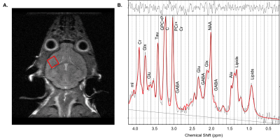 |
Investigating the impact of ketogenic diet in Alzheimer’s
disease using 1H MRS
Chaitali Anand1,
Lydia Le Page1,
Marina Radoul1,
and Myriam M Chaumeil1
1UCSF, San Francisco, CA, United States Keywords: Alzheimer's Disease, Spectroscopy, metabolism Ketogenic diet (KD) may be used to treat cognitive dysfunction in Alzheimer’s disease (AD). In hAPPJ20 AD mice fed KD, improved spatial learning has been noted, but mechanisms driving this cognitive change remains unclear. Here we used 1H MRS to probe the effect of KD on brain metabolism in hAPPJ20 mice fed either a control diet or KD. Increases in prefrontal creatine and Glx in KD-fed mice and increased Tau in male KD-fed mice were observed. Male KD-fed mice also demonstrated better spatial learning on the Morris water maze. Thus, KD may improve brain energetics and neurotransmission supporting cognitive function. |
| 09:11 |
0413.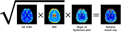 |
Increased vessel size accompanies decreased cerebral blood flow
in the hippocampus in Alzheimer’s disease
Hansol Lee1,2,
Kyla Gaudet1,
Annie G. Bryant3,
Rachel E. Bennett3,
David H. Salat1,
Yi-Fen Yen1,
and Susie Y. Huang1
1Department of Radiology, Athinoula A. Martinos Center for Biomedical Imaging, Massachusetts General Hospital, Charlestown, MA, United States, 2Department of Biomedical Engineering, Ulsan National Institute of Science and Technology, Ulsan, Korea, Republic of, 3Department of Neurology, Massachusetts General Hospital, Charlestown, MA, United States Keywords: Alzheimer's Disease, Perfusion The purpose of this study was to probe alterations in vessel size and their contributions to perfusion deficits observed in the early stages of Alzheimer’s disease (AD). AD and mild cognitive impairment (MCI) patients along with older and young adults underwent perfusion and diffusion magnetic resonance imaging (MRI). Increased vessel size was observed in conjunction with decreased cerebral blood flow in the hippocampus of individuals with AD and MCI. These results highlight potential pathologic mechanisms of abnormal vascular supply to the hippocampus in AD. Further investigations into vessel density and the relationship between cerebrovascular structure to AD pathology are warranted. |
| 09:19 |
0414.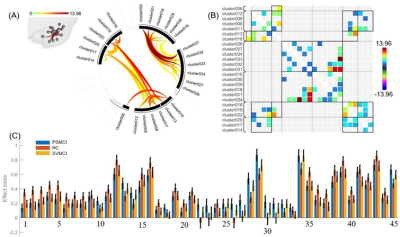 |
Functional Abnormalities of Cerebellum in Vascular Cognitive
Impairment
Zhao Ruan1,
Weiyin Vivian Liu2,
and Haibo Xu3
1Department of Radiology, Zhongnan Hospital of Wuhan University, Wuhan, China, 2GE Healthcare, MR Research China, Beijing, Beijing, China, 3Zhongnan Hospital of Wuhan University, Wuhan, China Keywords: Dementia, Brain Connectivity, vascular cognitive impairment The potential prevalence of vascular cognitive impairment (VCI) is high; early diagnosis and treatment is essential for disease prognosis. In this study, compared to healthy controls (HCs), there were differences of functional connectivity (FC) between the cerebellum and cerebral regions of VCI patients, mainly involving default mode network (DMN), sensorimotor network (SMN) and frontoparietal network (FPN). Furthermore, FC between multiple sub-regions in the cerebellum reduced in patients with VCI compared to HCs. These findings may expand our understanding of the neural mechanisms of VCI in the perspective of neurovascular coupling. |
| 09:27 |
0415.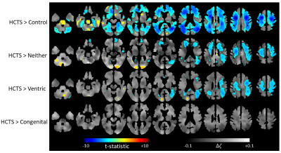 |
Unique brain stiffness and damping ratio patterns associated
with morphological phenotypes of normal pressure hydrocephalus
Pragalv Karki1,
Matthew C Murphy1,
Petrice M Cogswell1,
Matthew L Senjem1,
Jonathan Graff-Radford2,
Clifford R Jack Jr1,
Richard L Ehman1,
and John Huston III1
1Department of Radiology, Mayo Clinic, Rochester, MN, United States, 2Department of Neurology, Mayo Clinic, Rochester, MN, United States Keywords: Dementia, Brain, Normal pressure hydrocephalus Normal pressure hydrocephalus (NPH) is a brain disorder that is often misdiagnosed as Alzheimer’s or Parkinson’s disease due to overlapping symptoms. However, unlike such neurodegenerative diseases, NPH can be surgically treated by shunt placement with a success rate of around 80%, depending on phenotype. Therefore, correct diagnosis and categorization of NPH is important. As a step towards that goal, we demonstrate that analyzing the different morphologic phenotypes of NPH using magnetic resonance elastography can help establish unique viscoelastic signatures associated with each phenotype providing distinguishing biomarkers. |
|
09:35 |
0416.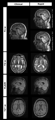 |
An ultra-fast dementia diagnosis MRI protocol enabled by
Wave-CAIPI
Haroon R. Chughtai1,2,
David L. Thomas3,4,
Miguel Rosa-Grilo3,
Eoin Mulroy3,
Millie Beament3,
Will Coath3,
Lloyd Prosser3,
Ian Malone3,
Danny Alexander5,
Frederik Barkhof 3,
Catherine J. Mummery3,
Nick C. Fox3,
and Geoff J. M. Parker1,6
1Centre for Medical Image Computing (CMIC), Medical Physics & Biomedical Engineering, UCL, London, United Kingdom, 2Advanced Research Computing (ARC) Centre, UCL, London, United Kingdom, 3Dementia Research Centre (DRC), UCL Queen Square Institute of Neurology, UCL, London, United Kingdom, 4Department of Brain Repair and Rehabilitation, UCL Queen Square Institute of Neurology, UCL, London, United Kingdom, 5Department of Computer Science, Centre for Medical Image Computing (CMIC), University College London, London, United Kingdom, 6Bioxydyn Limited, Manchester, United Kingdom Keywords: Data Acquisition, Dementia Structural brain imaging is vital in the diagnostic pathway for cognitive disorders and dementias, including the identification of Alzheimer’s disease. MRI is the recommended modality for this but is often not used due to long scan times and lower availability relative to CT. Reduced scan times are needed to enable more widespread adoption of MRI as a first-line modality for cognitive disorders. Our ultra-fast protocol enabled by Wave-CAIPI shows promise in reducing the diagnostic scan time for dementias from around 18 minutes to under 6 minutes whilst retaining clinical utility across several contrasts. |
|
09:43 |
0417.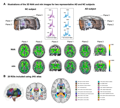 |
White Matter Neurometabolite Vulnerability Predicts Cognitive
Decline in Alzheimer’s Disease: A High-Resolution 3D 1H-MRSI
Study
Danni Wang1,
Miao Zhang2,
Yibo Zhao3,4,
Yudu Li3,5,
Wen Jin3,4,
Jialin Hu1,
Yaoyu Zhang1,
Biao Li2,
Jun Liu6,
Binyin Li6,
Zhi-Pei Liang3,4,
and Yao Li1
1School of Biomedical Engineering, Shanghai Jiao Tong University, Shanghai, China, 2Department of Nuclear Medicine, Ruijin Hospital, Shanghai Jiao Tong University of Medicine, Shanghai, China, 3Beckman Institute for Advanced Science and Technology, University of Illinois at Urbana-Champaign, Urbana, IL, United States, 4Department of Electrical and Computer Engineering, University of Illinois at Urbana-Champaign, Urbana, IL, United States, 5National Center for Supercomputing Applications, University of Illinois at Urbana-Champaign, Urbana, IL, United States, 6Department of Neurology and Institute of Neurology, Ruijin Hospital, Shanghai Jiao Tong University of Medicine, Shanghai, China Keywords: Alzheimer's Disease, Spectroscopy White matter (WM) damage plays an important role in AD and different mechanisms have been suggested for different brain areas from postmortem studies. Understanding the spatial patterns of pathological changes in WM is of great importance in AD diagnosis. Using a high-resolution 3D MRSI technique, we investigated the spatial patterns of neurometabolic changes in WM regions. We firstly derived neurometabolite vulnerability maps in AD, showing spatially varying patterns of NAA reduction and mIn elevation in WM regions, in distinct association with gray matter volume or Aβ deposition, respectively. The neurometabolic biomarkers showed improved prediction of cognitive decline of AD patients. |
|
09:51 |
0418.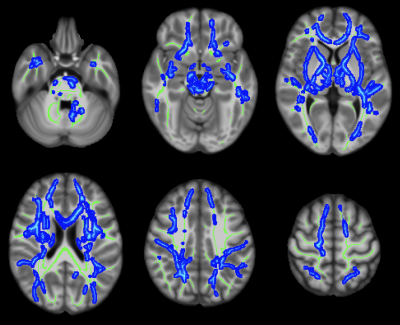 |
Diffusion abnormalities linked to brain arteriolosclerosis: An
in-vivo MRI and pathology study in community-based older adults
Ana Tomash1,
Mahir Tazwar1,
Md Tahmid Yasar1,
Shengwei Zhang2,
Arnold M Evia2,
David A Bennett2,
Julie A Schneider2,
and Konstantinos Arfanakis1,2
1Department of Biomedical Engineering, Illinois Institute of Technology, Chicago, IL, United States, 2Rush Alzheimer’s Disease Center, Rush University Medical Center, Chicago, IL, United States Keywords: Dementia, Vessels, Alzheimer's disease, Aging, Hypertension Brain arteriolosclerosis is one of the main pathologies of cerebral small vessel disease, is common in older adults, and is associated with lower cognitive and motor function and higher odds of dementia. This work combined in-vivo MRI and pathology in a community-cohort of older adults to investigate the independent association of brain arteriolosclerosis with diffusion abnormalities in white matter. Brain arteriolosclerosis was shown to be associated with lower FA and higher trace of the diffusion tensor that extended throughout white matter, and these associations were independent of the effects of other neuropathologies or white matter hyperintensities. |
| 09:59 |
0419.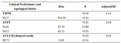 |
Topological properties of individual gray matter morphological
network effectively identify the preclinical stages of
Alzheimer’s disease
Zhihao Wang1,
Hongyuan Ding2,
Yin Tang3,
Yao Tang4,5,
Ming Qi6,
Weiqiang Dou7,
Long Qian7,
Yaxin Gao8,9,
Tong Wang10,
Qian Zhong11,
Xi Yang10,
Huifang Tian12,
Ling Zhang2,
and Yi Zhu10
1School of Biological Science & Medical Engineering, Southeast University, Nanjing, China, 2Department of Radiology, The First Affiliated Hospital of Nanjing Medical University, Nanjing, China, 3Department of Medical imaging, Jingjiang People's Hospital, Jingjiang, China, 4School of Rehabilitation Medicine, Nanjing Medical University, Nanjing, China, 5Rehabilitation Medicine Department, Geriatric Hospital of Nanjing Medical University, Nanjing, China, 6Department of Radiology, The First Affiliated Hospital of Nanjing Medical University, Naning, China, 7MR Research China, GE Healthcare, Beijing, China, 8Department of Rehabilitation, The Affiliated Suzhou Hospital of Nanjing Medical University, Suzhou Municipal Hospital, Suzhou, China, 9Gusu School, Nanjing Medical University, Suzhou, China, 10Rehabilitation Medicine Center, The First Affiliated Hospital of Nanjing Medical University, Nanjing, China, 11Department of Rehabilitation, Nanjing Drum Tower Hospital Clinical College of Nanjing Medical University, Nanjing, China, 12Clinical Medicine Research Institution, The First Affiliated Hospital of Nanjing Medical University, Nanjing, China Keywords: Alzheimer's Disease, Neurodegeneration In this study, the topology of normalized individual morphological network (IMN) has been investigated for patients with subjective cognitive decline (SCD), mild cognitive impairment (MCI) and healthy controls (HCs), respectively. Significantly different topology has been revealed among SCD and MCI patients and HCs, both at global and regional level. Moreover, the alterations of the regional topological metrics, including degree centrality and nodal efficiency, at the caudate nucleus exhibited significant correlations with all clinical scales and statistically predicted the cognitive performances. Therefore, the alterations of IMN can be regarded as an effective biomarker in early detection of SCD and MCI. |
| 10:07 |
0420.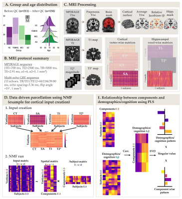 |
Cortical morphometry and hippocampal microstructure predict
aging and Alzheimer’s disease progression.
Aurélie Bussy1,
Raihaan Patel2,
Olivier Parent1,
Alyssa Salaciak1,
Sarah Farzin1,
Stephanie Tullo1,
Sylvia Villeneuve1,
Judes Poirier1,
John CS Breitner1,
Gabriel A. Devenyi1,
Christine L. Tardif1,
and M. Mallar Chakravarty1 1McGill University, Montréal, QC, Canada, 2University of Oxford, Oxford, United Kingdom Keywords: Alzheimer's Disease, Quantitative Imaging Morphometric and quantitative MRI metrics have rarely been used simultaneously to characterize healthy aging and Alzheimer’s disease (AD) progression. Here, cortical vertex-wise and hippocampal voxel-wise metrics were extracted to infer atrophy progression (using cortical thickness, surface area or relative Jacobians), myelin and iron contents (T1 and T2* respectively). A data-driven approach was used to parcellate the cortex and the hippocampus. A multivariate statistical technique was used to link hippocampal and cortical metrics to demographics and cognitive scores of interest. Our results suggest that AD-related neural risk is associated with neurodegeneration and microstructural changes in the cortex and hippocampus, respectively. |
 Back to Meeting Home
 Back to the Program-at-a-Glance
The International Society for Magnetic Resonance in Medicine is accredited by the Accreditation Council for Continuing Medical Education to provide continuing medical education for physicians.
 Back to Meeting Home
 Back to the Program-at-a-Glance
 View Presentation Video