Oral Session
Brain Tumors: Acquisition
ISMRM & ISMRT Annual Meeting & Exhibition • 03-08 June 2023 • Toronto, ON, Canada

| 13:45 |
0139.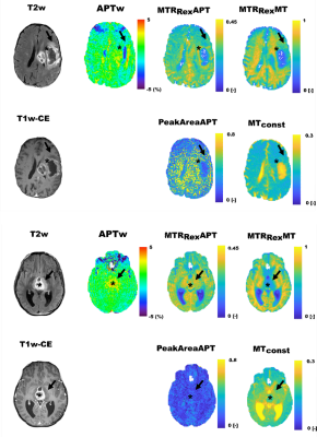 |
CEST Imaging of the APT and ssMT predict the overall survival of
patients with glioma at the first follow-up after completion of
radiotherapy at 3T
Nikolaus von Knebel Doeberitz1,
Florian Kroh2,3,
Johannes Breitling 4,
Laila König5,
Srdjan Maksimovic1,
Svenja Grass1,
Jürgen Debus5,6,7,
Peter Bachert3,4,
Heinz-Peter Schlemmer1,7,
Mark E. Ladd3,4,7,
Andreas Korzowski4,
Steffen Goerke4,
and Daniel Paech1,8 1Division of Radiology, German Cancer Research Center, Heidelberg, Germany, 2Divion of Medical Physics in Radiology, German Cancer Research Center, Heidelberg, Germany, 3Department of Physics and Astronomy, University of Heidelberg, Heidelberg, Germany, 4Division of Medical Physics in Radiology, German Cancer Research Center, Heidelberg, Germany, 5Department of Radiation Oncology, University Hospital Heidelberg, Heidelberg, Germany, 6Clinical Cooperation Unit Radiation Oncology, German Cancer Research Center, Heidelberg, Germany, 7Faculty of Medicine, University of Heidelberg, Heidelberg, Germany, 8Department of Neuroradiology, University Hospital Bonn, Bonn, Germany Keywords: CEST & MT, Treatment, Glioma In this prospective clinical study we compared the ability of asymmetry-based amide proton transfer-weighted (APTw) imaging with Lorentzian-fit-based (PeakAreaAPT and MTconst) and relaxation-compensated (MTRRexAPT and MTRRexMT) CEST-MRI of the amide proton transfer (APT) and semisolid magnetization transfer (ssMT) at 3T for the prediction of the overall survival of patients with glioma at the first follow-up after completion of radiotherapy. The APTw (HR=4.66, p<0.001) was more strongly associated with survival compared to the MTRRexAPT (HR=2.44, p=0.056) and MTconst (HR=2.54, p=0.044). The MTRRexMT and PeakAreaAPT did not display any association with survival. |
13:53 |
0140.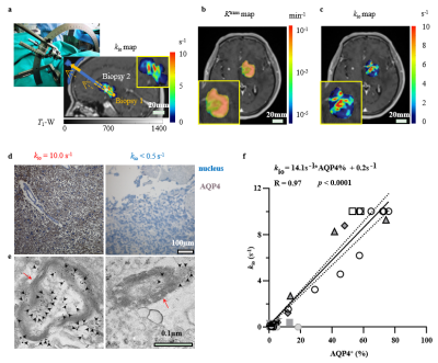 |
Transmembrane water-efflux rate measured by water exchange DCE-MRI:
a sensitive and specific biomarker of Aquaporin-4 in gliomas
Yinhang Jia1,2,
Guangxu Han1,
Zejun Wang1,
Yi-Cheng Hsu3,
Yi Sun3,
Bao Wang4,
Yingchao Liu5,6,
and Ruiliang Bai2,7
1Key Laboratory of Biomedical Engineering of Ministry of Education, College of Biomedical Engineering and Instrument Science, Zhejiang University, Hangzhou, China, 2School of Medicine, Zhejiang University, Hangzhou, China, 3MR Collaboration, Siemens Healthcare, Shanghai, China, 4Department of Radiology, Qilu Hospital of Shandong University, Jinan, China, 5Department of Neurosurgery, Provincial Hospital Affiliated to Shandong First Medical University, Jinan, China, 6Shandong National Center for Applied Mathematics, Shandong University, Jinan, China, 7College of Biomedical Engineering and Instrument Science, Zhejiang University, Hangzhou, China Keywords: Tumors, Brain, Glioma, Aquaporins-4, transmembrane water-efflux rate, therapy-resistant. The water-selective channel aquaporin-4 (AQP4) contributes to the migration and proliferation of gliomas, and to their resistance to therapy. Here, we show, in glioma animal models, and in glioma patients, that transmembrane water-efflux rate is a sensitive and specific biomarker of AQP4 expression and can be measured via dynamic-contrast-enhanced magnetic resonance imaging. Water-efflux rates correlated with changes in the heterogeneity of intratumoural and intertumoural AQP4 in human gliomas and following treatment with the AQP4 inhibitor TGN020. Regions with low water-efflux rates contained higher fractions of stem-like slow-cycling cells and therapy-resistant cells, suggesting that maps of water-efflux rates could be used to identify gliomas that are resistant to therapies. |
|
14:01 |
0141.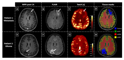 |
Non-invasive blood brain barrier integrity mapping in patients
with high-grade glioma and metastasis by time-encoded arterial
spin labelling
Gabriel Hoffmann1,2,
Christine Preibisch1,2,
Matthias Günther3,4,5,
Amnah Mahroo3,
Matthias JP van Osch6,7,
Lena Václavů6,
Lena Schmitzer1,
Claus Zimmer1,2,
Benedikt Wiestler1,
and Stephan Kaczmarz1,2,8
1Department of Diagnostic and Interventional Neuroradiology, Klinikum rechts der Isar, Technical University of Munich, Munich, Germany, 2TUM-Neuroimaging Center, Klinikum rechts der Isar, Technical University of Munich, Munich, Germany, 3MR Physics, Fraunhofer Institute for Digital Medicine MEVIS, Bremen, Germany, 4MR-Imaging and Spectroscopy, University of Bremen, Bremen, Germany, 5mediri GmbH, Heidelberg, Germany, 6C.J. Gorter MRI Center, Department of Radiology, Leiden University Medical Center, Leiden, Netherlands, 7Leiden Institute of Brain and Cognition, Leiden University, Leiden, Netherlands, 8Philips GmbH Market DACH, Hamburg, Germany Keywords: Tumors, Permeability, Blood Brain Barrier High-grade glioma are known to cause blood brain barrier (BBB) disruption, facilitating molecular leakage from inside the vessels into tissue. Current methods to probe BBB integrity are either not quantitative or, by using Gadolinium, insensitive to subtle changes. Here, we present first data of arterial spin labelling (ASL)-based BBB mapping of water exchange time (Texch) in brain tumour patients. Results show faster exchange (shorter Texch) in tumorous and even normal-appearing tissue compared to healthy subjects. This highlights the potential of ASL-based water exchange mapping as a proxy measure for BBB integrity, and its potential sensitivity to even subtle impairments. |
| 14:09 |
0142.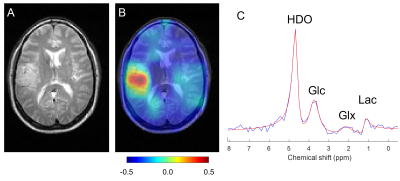 |
Imaging metabolism of deuterated glucose in patients with
primary brain tumors
Zachary A. Corbin1,
Yanning Liu2,
Robert K. Fulbright3,
Serena Thaw-Poon1,
Joachim M. Baehring1,
Nicholas Blondin1,
Peter Kim1,
Antonio Omuro1,
Veronica L. Chiang4,
Jennifer Moliterno4,
Sacit B. Omay4,
Joseph M. Piepmeier4,
Douglas L. Rothman3,
Robin A. de Graaf3,
and Henk M. De Feyter3
1Department of Neurology, Yale University, New Haven, CT, United States, 2Department of Biomedical Engineering, Yale University, New Haven, CT, United States, 3Department of Radiology and Biomedical Imaging, Yale University, New Haven, CT, United States, 4Department of Neurosurgery, Yale University, New Haven, CT, United States Keywords: Tumors, Metabolism, deuterium Deuterium metabolic imaging (DMI), a combination of 2H MRSI with administration of a deuterated substrate, was used to map regional metabolism of [6,6’-2H2]-glucose in 24 patients with multiple types of brain tumors. DMI data were acquired 70-90 minutes after oral intake of the deuterated glucose and revealed strong tumor-to-brain image contrast in high-grade tumors. Our metric, based on the labeling of specific glucose metabolites, reflects the canonical glucose metabolism in aggressive tumors – the Warburg Effect. The Warburg Effect appears higher in high-grade tumors and showed potential as a biomarker of treatment effect. |
| 14:17 |
0143.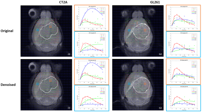 |
Probing Tumor Heterogeneity in Mouse Glioblastoma with Dynamic
Glucose-Enhanced Deuterium Metabolic Imaging
Rui Vasco Simoes1,2,
Rafael N Henriques1,
Jonas L Olesen3,4,
Beatriz M Cardoso1,
Francisca F Fernandes1,
Tania Carvalho1,
Sune N Jespersen3,4,
and Noam Shemesh1 1Champalimaud Research, Champalimaud Foundation, Lisbon, Portugal, 2(Present Address) Preclinical MRI, Institute for Research & Innovation in Health (i3S), Porto, Portugal, 3Center of Functionally Integrative Neuroscience (CFIN) and MINDLab, Department of Clinical Medicine, Aarhus University, Aarhus, Denmark, 4Department of Physics and Astronomy, Aarhus University, Aarhus, Denmark Keywords: Tumors, Deuterium, glioma Dynamic glucose-enhanced deuterium MRS (DGE 2H-MRS) coupled with Marchenko-Pastur PCA (MPPCA) denoising has been recently applied to immunocompetent mouse glioblastoma subtypes (GL261 and CT2A), demonstrating the ability to measure glucose metabolism through glycolysis and mitochondrial oxidation non-invasively, and its association with tumor proliferation. Here, we extend this approach to DGE Deuterium Metabolic Imaging (DGE-DMI) coupled with tensor MPPCA (tMPPCA) denoising, to map glucose fluxes in the same mouse models of glioblastoma. Our results demonstrate a strong association between glycolytic rates and MRI and histologic features of inter/intra-tumor heterogeneity. |
| 14:25 |
0144.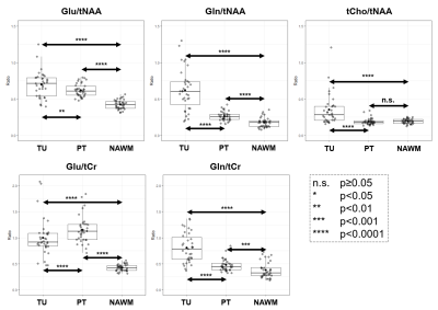 |
An exploration of peritumoral glutamate and glutamine in diffuse
gliomas using 7T MRSI
Gilbert Hangel1,2,3,
Philipp Lazen1,2,
Sukrit Sharma2,
Cornelius Cadrien1,2,
Thomas Roetzer-Pejrimovsky4,
Eva Niess2,
Lukas Hingerl2,
Stephan Gruber2,
Bernhard Strasser2,
Barbara Kiesel1,
Adelheid Woehrer4,
Matthias Preusser5,
Julia Furtner6,7,
Wolfgang Bogner2,
Siegfried Trattnig2,
Karl Rössler1,
and Georg Widhalm1
1Department of Neurosurgery, Medical University of Vienna, Vienna, Austria, 2High-field MR Center, Department of Biomedical Imaging and Image-guided Therapy, Medical University of Vienna, Vienna, Austria, 3Christian Doppler Laboratory for MR Imaging Biomarkers, Vienna, Austria, 4Division of Neuropathology and Neurochemistry, Department of Neurology, Medical University of Vienna, Vienna, Austria, 5Division of Oncology, Department of Internal Medicine I, Medical University of Vienna, Vienna, Austria, 6Division of Neuroradiology and Musculoskeletal Radiology, Department of Biomedical Imaging and Image-guided Therapy, Medical University of Vienna, Vienna, Austria, 7Medical Image Analysis und AI, Danube Private University, Krems, Austria Keywords: Tumors, Spectroscopy Research indicates that glutamate (Glu) and glutamine (Gln) play a role in the infiltrative properties of diffuse gliomas. Overcoming limitations of previous MRSI techniques, our 7T MRSI approach allows high-resolution Glu imaging. We investigated intra- and peritumoral Glu and Gln in a cohort of 36 patients and found significant increases in peritumoral Glu/tNAA, Gln/tNAA, Glu/tCr, and Gln/tCr compared to a NAWM control region. We established peritumoral Dice similarity coefficients of 0.67 for Glu/tNAA and Gln/tNAA compared to 0.31 for tCho/tNAA. Our results that Glu/Gln imaging could investigate the metabolism of infiltrative gliomas. |
| 14:33 |
0145.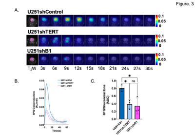 |
Hyperpolarized [1-13C] Gluconolactone Can Visualize TERT
Silencing Directly or via the Upstream Transcriptional Factor
GABPB1 in Glioblastoma
Noriaki Minami1,
Donghyun Hong1,
Celine Taglang1,
Georgios Batsios1,
Anne Marie Gillespie1,
Pavithra Viswanath1,
Nicholas Stevers2,
Joseph F Costello2,
and Sabrina M Ronen1
1Radiology and Biomedical Imaging, UCSF, San Francisco, CA, United States, 2Department of Neurological Surgery, UCSF, San Francisco, CA, United States Keywords: Tumors, Brain, Glioblastoma, GBM, TERT, GABP, imaging, MRI, MRS, NMR TERT promoter mutation is a genetic hallmark of glioblastoma, and targeting TERT or its upstream transcriptional factor GABPB1 are considered ideal therapeutic targets. Accordingly, establishing reliable imaging modalities that will enable monitoring of TERT silencing are needed as early indicators of target engagement and response to these molecular targeting therapies. Here, we demonstrate that using 13C MRS to monitor the metabolism of hyperpolarized δ-[1-13C] gluconolactone via the pentose phosphate pathway to 6-phosopho-[1-13C]gluconate (6PG) provides such a noninvasive imaging readout of TERT or GABPB1 silencing in glioblastoma. |
| 14:41 |
0146.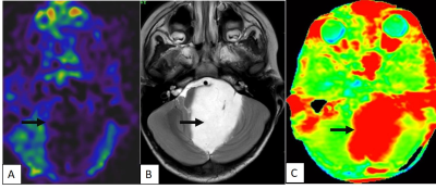 |
AMIDE PROTON TRANSFER IMAGING-ARTERIAL SPIN LABELING MISMATCH –
A NEW IMAGING BIOMARKER FOR PILOCYTIC ASTROCYTOMA
Adhithyan Rajendran1,
Chidambaranathan Natesan2,
Lavanya Yegnaraman2,
Rakesh Jalali3,
Rashmi Rao4,
Manikandan Mariyapillai5,
Prashant Jawahar1,
Roopesh Kumar6,
Srinivas Chilukuri7,
and Sushma Patil8
1Radiology, Apollo Proton Cancer Centre, Chennai, India, 2Radiology, Apollo Hospitals Greams Road, Chennai, India, 3Radiation Oncology, Apollo Proton Cancer Centre, chennai, India, 4Radiology, Philips India Ltd, Bangalore, India, 5Philips India Ltd, Chennai, India, 6Neurosurgery, Apollo Proton Cancer Centre, Chennai, India, 7Radiation Oncology, Apollo Proton Cancer Centre, Chennai, India, 8Pathology, Apollo Proton Cancer Centre, Chennai, India Keywords: Tumors, CEST & MT, Amide Proton Transfer Imaging Atypical features of Pilocytic astrocytoma (PA) on conventional MR imaging are often challenging and can be mistaken as High-grade glioma. Advanced Novel techniques of Amide Proton Transfer (APT) and Arterial spin labelling (ASL) have shown promising results in the grading of gliomas. This study to explore the added value and utility in characterizing the atypical form of PA, if these two non-contrast techniques are combined in the clinical practice. Mismatch between APT and ASL signals, was seen in all the cases of PA and this unique imaging appearance of APT ASL imaging mismatch could be a biomarker for pilocytic astrocytoma. |
| 14:49 |
0147.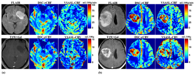 |
Tumor Blood Volume Measurement in Brain Gliomas Using
Velocity-Selective Arterial Spin Labeling
Yaoming Qu1,
Qin Qin2,3,
and Zhibo Wen1
1Radiology, Zhujiang Hospital of Southern Medical University, Guangzhou, China, 2The Russell H. Morgan Department of Radiology and Radiological Science, Division of MR Research, Johns Hopkins University School of Medicine, Baltimore, MD, United States, 3F.M. Kirby Research Center for Functional Brain Imaging, Kennedy Krieger Institute, Baltimore, MD, USA, Baltimore, MD, United States Keywords: Tumors, Perfusion Velocity selective arterial spin labeling (VSASL) has shown comparable CBF measurements in brain gliomas with DSC-PWI. As CBV derived from DSC-PWI is the most widely adopted perfusion marker of brain tumor angiogenesis, the clinical utility for neurooncology imaging of VSASL based CBV quantification worth investigation. This study on preoperative patients with brain gliomas demonstrated that VSASL provided highly correlated quantifications of relative tumor blood volume (R2=0.83) compared to DSC-PWI, and further improved diagnostic performance than VSASL derived relative tumor blood flow measurements (ROC AUC=0.94 vs. 0.89), indicating its potential as a viable non-contrast alternative to DSC-PWI for brain tumor applications. |
| 14:57 |
0148.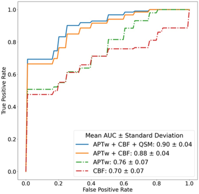 |
Improved diagnostic performance of APTw MRI to multiparametric
non-contrast-enhanced MRIs in patients with post-treatment
high-grade gliomas
Qianqi Huang1,2,
Jingpu Wu1,3,
Yiqing Shen1,2,
Nhat Le1,2,
Pengfei Guo1,2,
Karisa Schreck4,
David Kamson4,
Lindsay Blair4,
Hye Young Heo1,
Xu Li1,5,
Wenbo Li1,5,
Haris Sair1,
Jaishri Blakeley4,
John Laterra4,
Matthias Holdhoff6,
Stuart Grossman6,
Debraj Mukherjee7,
Chetan Bettegowda 7,
Peter van Zijl 1,5,
Jinyuan Zhou1,
and Shanshan Jiang1
1Department of Radiology, Johns Hopkins University, Baltimore, MD, United States, 2Department of Computer science, Johns Hopkins Univeristy, Baltimore, MD, United States, 3Department of Applied Mathematics and Statistics, Johns Hopkins Univeristy, Baltimore, MD, United States, 4Department of Neurology, Johns Hopkins University, Baltimore, MD, United States, 5Department of Radiology, Kennedy Krieger Institute, Baltimore, MD, United States, 6Department of Oncology, Johns Hopkins University, Baltimore, MD, United States, 7Department of Neurosurgery, Johns Hopkins University, Baltimore, MD, United States Keywords: Tumors, Treatment We explored non-contrast-enhanced MRI performances of APTw, DWI, SWI, and pCASL at 3 Tesla in glioma patients post-treatment. APTw, ADC, QSM, and CBF histogram parameters from volumetric ROIs were recorded. Multivariable logistics regression with principal component analysis (PCA) was built for differentiating treatment effect from tumor recurrence. Results showed that the regression model trained on the combination of APTw, CBF, and QSM achieved the highest classification performance, with an AUC of 0.90. |
| 15:05 |
0149.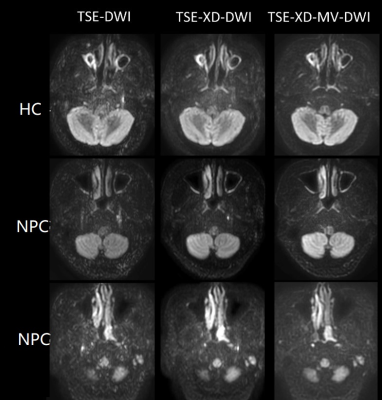 |
Feasibility study of diusion weighted imaging of nasopharynx
using split-echo TSE-DWI combined with MultiVane acquisition
Kun Wang1,
Yujie Yu1,
Maoxue Wang1,
Ming Li1,
Xiance Zhao2,
Peng Wu2,
and Kan Deng3
1The Affiliated Drum Tower Hospital of Nanjing University Medical School, Nanjing, China, 2Philips Healthcare, Shanghai, China, 3Philips Healthcare, Guangzhou, China Keywords: Tumors, Brain Diusion weighted imaging (DWI) has high sensitivity in the dierential diagnosis of nasopharyngeal carcinoma. However,the DWI based on SE-EPI has poor imaging quality in the nasopharynx. A combination of Multi-Vane (MV) and SPLICE(MV-SPLICE) were developed to reduce ELT, as well as improve SNR and the imaging quality of DWI. This study compares theDWI images of three acquisition sequences in nasopharynx,including DWI TSE without SPLICE(TSE), TSE with SPLICE(TSE-XD), and TSE with MV-SPICE(TSE-XD-MV) and proves that TSE with MV-SPLICE DWI based TSE with MV-SPLICE provides anexcellence imaging scheme of DWI for the clinical diagnosis of nasopharyngeal diseases. |
| 15:13 |
0150.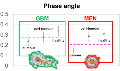 |
Tumour virtual histology with Magnetic Resonance Elastography
Giacomo Annio1,2,
Robin Bugge3,
Siri Fløgstad Svensson 3,
Omar Darwish4,
Giorgio Seano5,
Donata Biernat 6,
Karoline Skogen 6,
Jon Ramm-Pettersen 7,
Einar Vik-Mo 7,
Katharina Schregel 8,
Kyrre Eeg Emblem3,
and Ralph Sinkus1
1INSERM - King's College, Paris, France, 2School of Biomedical Engineering and Imaging Sciences, King's College London, London, United Kingdom, 3Department of Diagnostic Physics, Division of Radiology and Nuclear Medicine, Oslo University Hospital, Oslo, Norway, 4Department of Biomedical Engineering, King's College London, London, United Kingdom, 5U1021 INSERM, Institut Curie, Paris, France, 6Department of Radiology Ullevål, Division of Radiology and Nuclear Medicine,, Oslo University Hospital, Oslo, Norway, 7Department of Neurosurgery, Division of Clinical Neuroscience,, Oslo University Hospital, Oslo, Norway, 8Department of Neuroradiology, Heidelberg University Hospital, Heidelberg, Germany Keywords: Tumors, Elastography The reciprocal interaction between cancer cells and the surrounding creates a tumorigenic feedback loop capable of shifting the anti-neoplastic feature of the microenvironment towards a tumour growth-promoting one. Unfortunately infiltrating tumours, like GBMs, do not have discrete boundaries and intra-axial metastases are not visible on conventional MR images. However they are expected to change the tissue biomechanics. Here we explored tumour microenvironment biomechanics non-invasively using MRE in a cohort of 23 patients with brain tumours - 13 meningiomas, 10 glioblastomas. We show how MRE provides a comprehensive characterization of the tumour microenvironment, explaining histological features and tumour invasive features. |
| 15:21 |
0151.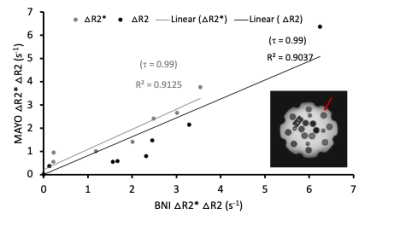 |
Repeatability and reproducibility of a multi-vendor spin and
gradient echo (SAGE) pulse sequence for dynamic susceptibility
contrast MRI
Poonam Choudhary1,
YuXiang Zhou2,
Sudarshan Ragunathan3,
Ethan Mathew1,
Aliya Anil1,
Natenael Semmineh4,
Belinda Gutierrez1,
John P Karis5,
Leland S. Hu2,
Kathleen M. Schmainda6,
Ashley M. Stokes1,
and C. Chad Quarles4
1Division of Neuroimaging Research and Barrow Neuroimaging Innovation Center, Barrow Neurological Institute, Phoenix, AZ, United States, 2Department of Radiology, Mayo Clinic College of Medicine, Phoenix, AZ, United States, 3Hyperfine Inc., Guilford, CT, United States, 4The University of Texas MD Anderson Cancer Center, Houston, TX, United States, 5Neuroradiology, Southwest Neuroimaging at Barrow Neurological Institute, Phoenix, AZ, United States, 6Department of Biophysics Medical College of Wisconsin, Milwaukee, WI, United States Keywords: Tumors, DSC & DCE Perfusion, sequence Single-echo pulse sequences are commonly employed for DSC-MRI. The multi-echo spin and gradient echo (SAGE) sequence can be useful in multi-contrast and multi-scale imaging of morphologic and functional features of brain tumor vasculature. The goal of this study is to establish harmonized SAGE sequence and analysis protocol across primary MRI vendors to facilitate multi-site clinical trials. In this work, we present initial results of SAGE sequence multi-site reproducibility and repeatability in phantom, healthy volunteers and high-grade glioma patients. |
| 15:29 |
0152.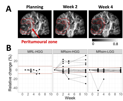 |
Diffusion tensor imaging on a 1.5T MR-Linac and comparison to a
3T diagnostic scanner in glioma patients
Liam S. P. Lawrence1,
Rachel W. Chan1,
James Stewart2,
Mark Ruschin2,
Aimee Theriault2,
Sten Myrehaug2,
Jay Detsky2,
Pejman J. Maralani3,
Chia-Lin Tseng2,
Hany Soliman2,
Mary Jane Lim-Fat4,
Sunit Das5,
Greg J. Stanisz1,6,
Arjun Sahgal2,
and Angus Z. Lau1
1Physical Sciences, Sunnybrook Research Institute, Toronto, ON, Canada, 2Department of Radiation Oncology, Sunnybrook Health Sciences Centre, Toronto, ON, Canada, 3Medical Imaging, Sunnybrook Health Sciences Centre, Toronto, ON, Canada, 4Division of Neurology, Department of Medicine, Sunnybrook Health Sciences Centre, Toronto, ON, Canada, 5Department of Surgery, St. Michael's Hospital, Toronto, ON, Canada, 6Department of Neurosurgery and Paediatric Neurosurgery, Medical University, Lublin, Poland Keywords: Tumors, Diffusion Tensor Imaging Diffusion tensor imaging was implemented on a 1.5T MR-Linac and used to scan ten glioma patients and four healthy volunteers. Mean diffusivity and fractional anisotropy (FA) measurements were compared to those at 3T in 35 glioma patients. FA values surrounding the tumour and in white matter structures (genu, splenium, and body of corpus callosum) were investigated for the detection of tumour infiltration and radiation-induced damage, respectively. There was no evidence of dose-dependent white matter changes, in contrast to previous literature. Peritumoural FA changes occurred more frequently for high-grade compared to low-grade gliomas. |
| 15:37 |
0153.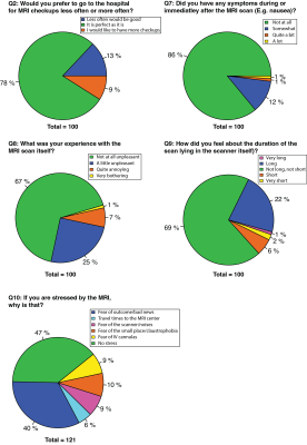 |
The Patients’ Experience on Neuroimaging of Gliomas: A
Cross-Sectional Survey Study
Ivar J.H.G. Wamelink1,2,
Hugo Hempel1,
Elsmarieke van de Giessen1,3,
Mark Vries1,
Philip de Witt Hamer2,4,
Frederik Barkhof1,5,
and Vera C. Keil1,2,3
1Radiology & Nucleaire Medicine, Amsterdam UMC location Vrije Universiteit Amsterdam, Amsterdam, Netherlands, 2Brain Tumor Center Amsterdam, Cancer Center Amsterdam, Amsterdam, Netherlands, 3Neuroscience, Amsterdam UMC location Vrije Universiteit Amsterdam, Amsterdam, Netherlands, 4Neurosurgery, Amsterdam UMC location Vrije Universiteit Amsterdam, Amsterdam, Netherlands, 5UCL institutes of Neurology and Healthcare Engineering, London, United Kingdom Keywords: Tumors, Challenges, Patient experience Glioma patient experience of MRI scans, repeated follow-up, and gadolinium-based contrast agents (GBCA) are unknown, despite the vulnerability of this patient group and relevant implications for clinics and science. This cross-sectional survey questioned 100 patients with primary brain tumors and found generally positive experiences with neuro-oncological scans. Age, diagnosis, and number of previous scans had little impact on the patient experience. However, women found MRI and intravenous injection significantly more uncomfortable. Also, patients would prefer GBCA-free MRIs if diagnostically equivalent. Patient knowledge towards GBCAs was limited and better information regarding GBCA and the MRI itself is needed. |
 Back to Meeting Home
 Back to the Program-at-a-Glance
The International Society for Magnetic Resonance in Medicine is accredited by the Accreditation Council for Continuing Medical Education to provide continuing medical education for physicians.
 Back to Meeting Home
 Back to the Program-at-a-Glance
 View Presentation Video