Oral Session
Brain Tumors: Analysis
ISMRM & ISMRT Annual Meeting & Exhibition • 03-08 June 2023 • Toronto, ON, Canada

16:00 |
0254.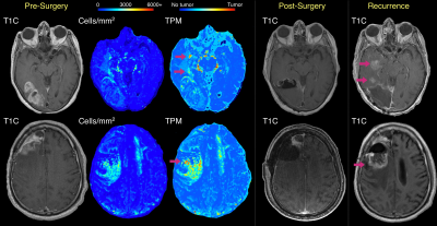 |
Comparison of radio-pathomic maps of tumor probability to 5-ALA guided surgical resection cavities in glioblastoma patients
Samuel A Bobholz1, Allison K Lowman1, Savannah R Duenweg2, Aleksandra Winiarz2, Margaret Stebbins2, Fitzgerald Kyereme1, Jennifer Connelly3, Dylan Coss4, Wade M Mueller5, Mohit Agarwal1, Anjishnu Banerjee6, Max Krucoff5, and Peter S LaViolette1,7 1Radiology, Medical College of Wisconsin, Milwaukee, WI, United States, 2Biophysics, Medical College of Wisconsin, Milwaukee, WI, United States, 3Neurology, Medical College of Wisconsin, Milwaukee, WI, United States, 4Pathology, Medical College of Wisconsin, Milwaukee, WI, United States, 5Neurosurgery, Medical College of Wisconsin, Milwaukee, WI, United States, 6Biostatistics, Medical College of Wisconsin, Milwaukee, WI, United States, 7Biomedical Engineering, Medical College of Wisconsin, Milwaukee, WI, United States Keywords: Tumors, Brain This study sought to compare previously published autopsy-based radio-pathomic maps of pre-surgical cellularity and tumor probability to beyond-contrast 5-ALA guided resection margins to assess concordance in identifying non-enhancing tumor between the techniques. In a series of 10 cases, radio-pathomic maps were able to identify areas of non-enhancing tumor prior to surgery that coincided with either 5-ALA guided resection margins or areas of future recurrence in 7 of 10 cases, suggesting that highlighted areas on these maps indicate active, progressive tumor. |
| 16:08 | 0255.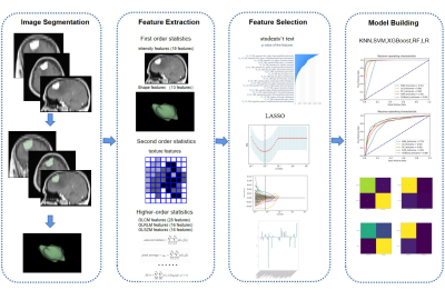 |
Preoperative Prediction of Brain Invasion in Meningiomas: A Radiomics Model based on Multimodal MRI and Quantitative Clinical Factors
Jinli Li1, Xianchang Zhang2, Ruijun Yang1, Quanzhi Feng1, Dong Yuan1, Jiajia Zhang1, and Tong Han1
1Tianjin Huanhu Hospital, No.6 Jizhao Road, Jinnan District, Tianjin 300350, China., Tianjin, China, 2MR Collaboration, Siemens Healthineers Ltd., Beijing, China., Beijing, China Keywords: Tumors, Radiomics, brain invasion This study aimed to construct a radiomic model based on a large patient cohort to predict brain invasion (BI) in meningioma. By analyzing 97 patients with BI and 935 patients without BI, we found that the clinical risk factors for BI were male sex, tumor located at the skull base, and peritumoral edema volume. A binary logistic regression model combining these risk factors and multimodal MRI radiological characteristics was established. The constructed model achieved an excellent performance (AUC: 0.928) in terms of BI classification with an accuracy of 91.77%, which may be helpful for personalized treatment plan in meningioma. |
| 16:16 | 0256.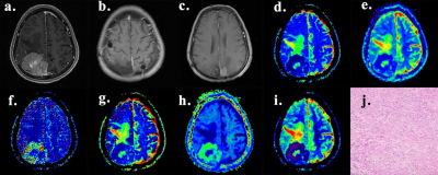 |
The Comparison of Diffusion Weighted Imaging and Advanced Diffusion Techniques in Predicting Postoperative Recurrence of Meningioma
Hui Zheng1, Lingmin Zheng1, Zongmeng Wang1, Rufei Zhang1, Yang Song2, and Lin Lin1
1Fujian Medical University Union Hospital, Fuzhou, China, 2MR Scientific Marketing, Siemens, Healthineers Ltd., Shanghai, China Keywords: Tumors, Diffusion/other diffusion imaging techniques Prior investigators have found that diffusion weighted imaging (DWI) contributes to improving the prediction of postoperative recurrence of meningioma. This study evaluated the feasibility of intravoxel incoherent motion (IVIM) and diffusional kurtosis imaging (DKI) in predicting postoperative recurrence of meningioma and compared them with DWI. Histogram metrics derived from DWI, IVIM, and DKI parameters were compared between the recurrent group and non-recurrent group. It was found that the lower ADC, D, f value and the higher D*, K value, the higher risk of postoperative recurrence of meningioma. However, IVIM and DKI derived parameters had similar diagnostic performance compared with DWI. |
| 16:24 | 0257.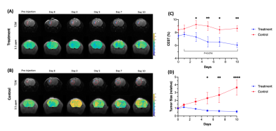 |
Early detection of brain tumor response during local hydrogel based treatment using CEST MRI
Se Weon Park1,2, Joseph H.C Lai1, Xiongqi Han3, Jianpan Huang1, Vivian W.M Leung1, Peng Xiao1, and Kannie W.Y Chan1,2,4,5 1Department of Biomedical Engineering, City University of Hong Kong, Hong Kong, Hong Kong, 2Hong Kong Centre for Cerebro-Cardiovascular Health Engineering (COCHE), Hong Kong, Hong Kong, 3SiBionics, Shenzhen, China, 4Russell H. Morgan Department of Radiology and Radiological Science, The Johns Hopkins University School of Medicine, Baltimore, MD, United States, 5City University of Hong Kong Shenzhen Research Institute, Shenzhen, China Keywords: Tumors, Treatment CEST MRI is a non-invasive molecular imaging approach, while APT has been applied to reveal molecular changes in brain tumors. Based on our theranostic hydrogel studies, here, we demonstrated the therapeutic effect on brain tumor with a continuous decrease in tumor size over 10 days. Importantly, the APT signal showed significant decrease on day 3 after treatment when tumor size did not have a significant change. Moreover, a strong correlation between APT signal and Ki-67 staining was shown. This could be a potential application to detect early tumor responses toward local hydrogel based treatment. |
| 16:32 | 0258.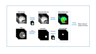 |
Differentiation of subtypes and genotypes in adult-type diffuse gliomas by APT imaging
Tongling Jiang1, Minghao Wu2, Cong Xie2, Xianchang Zhang3, Yaou Liu2, and Yi Zhang1
1Key Laboratory for Biomedical Engineering of Ministry of Education, Department of Biomedical Engineering, College of Biomedical Engineering & Instrument Science, Zhejiang University, Hangzhou, China, 2Department of Radiology, Beijing Tiantan Hospital, Capital Medical University, Beijing, China, 3MR Collaboration, Siemens Healthineers Ltd., Beijing, China Keywords: Tumors, Brain APT-related metric maps were applied to differentiate subtypes and genotypes of adult-type diffuse gliomas following the latest 2021 WHO guidelines. One hundred and twenty-nine patients were imaged on a 3T scanner. Regions of interest were obtained with an automatic segmentation algorithm on conventional anatomical images, which were then resampled and matched with the CEST images. The mean CEST metric values were calculated in the automatically-defined regions of interest, and the receiver operating characteristic analysis was implemented, achieving generally successful performance in identifying the IDH status and part of the subtypes. |
| 16:40 | 0259.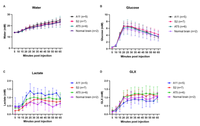 |
Deuterium metabolic imaging distinguishes metabolic subtypes and detects early treatment response to current standard of care in glioblastoma
Jacob Chen Ming Low1, Jianbo Cao1, Friederike Hesse1, Alan Wright1, and Kevin Brindle1
1CRUK Cambridge Institute, Cambridge, United Kingdom Keywords: Tumors, Deuterium The potential for DMI measurements of deuterated glucose metabolism to differentiate between metabolic subtypes in GBM has been demonstrated in patient-derived xenografts in mice. The glycolytic subtype showed increased lactate labelling whereas two oxidative subtypes showed increased glutamate/glutamine (Glx) labelling. There was decreased lactate labelling in a glycolytic subtype and decreased lactate and Glx labelling in an oxidative subtype within 24 h after the completion of standard-of-care chemoradiotherapy, demonstrating that the technique can also be used to detect early treatment response in this tumor type. |
| 16:48 | 0260.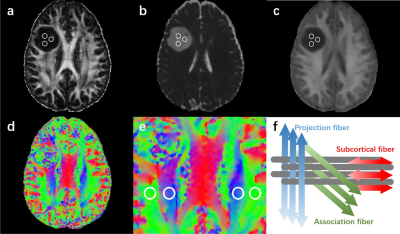 |
An application of diffusion tensor image analysis along perivascular space (DTI-ALPS) in grading gliomas and predicting IDH1 mutation status
Hongquan Zhu1, Yuanhao Li1, Li Li1, Weiyin Vivian Liu2, and WenZhen Zhu1
1Department of Radiology, Tongji Hospital, Tongji Medical College, Huazhong University of Science and Technology, Wuhan, China, 2GE Healthcare, MR Research China, Beijing, China Keywords: Tumors, Brain, Glioma Glioma could impair the glymphatic function with reduced CSF outflow. We aimed to use DTI-ALPS method to investigate the pathological change of glymphatic function in gliomas and evaluate its value in grading gliomas and predicting IDH1 mutation status. The lower-grade gliomas (LrGGs) showed higher ALPS Index than glioblastomas (GBMs). IDH1 wild-type group showed decreased ALPS Index compared with the IDH1 mutant group in LrGGs. But there was no difference between the IDH1 subtypes in GBMs. The ALPS Index showed good efficiency in grading gliomas and predicting IDH1 mutation status, it may be a potential imaging biomarker for diagnosing gliomas. |
| 16:56 | 0261.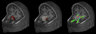 |
Fixel-based and tensor analysis of tractography-reconstructed anterior optic pathway in patients with sellar/parasellar tumor
Giovanni Sighinolfi1, Laura Ludovica Gramegna1,2, Alessandro Carrozzi1, Cristiana Fiscone1, Matteo Zoli1,2, Diego Mazzatenta1,2, Claudia Testa2,3, Raffaele Lodi1,2, Caterina Tonon1,2, and David Neil Manners1
1Department of Biomedical and Neuromotor Sciences, University of Bologna, Bologna, Italy, 2IRCCS Istituto delle Scienze Neurologiche di Bologna, Bologna, Italy, 3Department of Physics and Astronomy, University of Bologna, Bologna, Italy Keywords: Tumors, Tractography & Fibre Modelling, Anterior Optic Pathway Tumors of the sellar/paraellar region are likely to displace the optic chiasm, making it valuable to study the entire course and microstructural properties of the anterior optic pathway (AOP, including optic nerves, chiasm and optic tracts). We present the first fully automated tractography pipeline to entirely reconstruct the AOP. The pipeline was applied on a group of patients with sellar/parasellar tumors displacing the chiasm, achieving excellent reliability. We also estimated mean diffusivity, fractional anisotropy and fiber density along the AOP, demonstrating alterations in patients compared to controls, which we explain based on chiasmal compression. |
| 17:04 | 0262.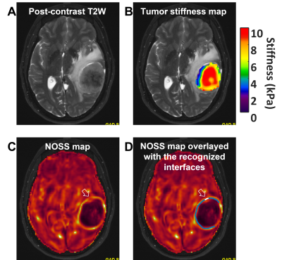 |
MRE-assessed meningioma stiffness and peripheral adhesion characteristics predict the extent of resectability and recurrence probability
Keni Zheng1, Matthew C. Murphy 1, Emanuele Camerucci 1, Xiang Shan1, Yi Sui1, Armando Manduca 2, Jamie J. Van Gompel3, Richard L. Ehman 1, John III Huston1, and Ziying Yin1
1Radiology, Mayo Clinic, Rochester, MN, United States, 2Physiology and Biomedical Engineering, Mayo Clinic, Rochester, MN, United States, 3Neurosurgery, Mayo Clinic, Rochester, MN, United States Keywords: Tumors, Brain, Meningiomas, tumor stiffness, tumor adhesion, slip interface imaging, tumor recurrence, tumor resection This work explored the value of MR elastography (MRE)-measured tumor stiffness and slip interface imaging (SII)-assessed adhesion metrics in predicting the extent of tumor resection and the probability of meningioma recurrence in 52 patients with meningiomas. Tumor adhesion percentage was assessed based on pattern recognition of the normalized octahedral shear strain (NOSS) map. Tumor stiffness was calculated using a neural network-based inversion (NNI). We found that stiffness correlates with the extent of resection (EOR) that was possible at surgery, while the extent of tumor adhesion showed good correlation with tumor recurrence among aggressive tumors with atypical features. |
| 17:12 | 0263.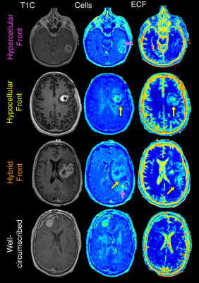 |
Radio-pathomic maps of glioblastoma identify phenotypes of non-enhancing infiltration associated with bevacizumab treatment response
Samuel A Bobholz1, Alisha Hoefs1, Jordyn Hamburger1, Allison K Lowman1, Savannah R Duenweg2, Aleksandra Winiarz2, Margaret Stebbins2, Fitzgerald Kyereme1, Jennifer Connelly3, Dylan Coss4, Wade M Mueller5, Mohit Agarwal1, Anjishnu Banerjee6, and Peter S LaViolette1,7 1Radiology, Medical College of Wisconsin, Milwaukee, WI, United States, 2Biophysics, Medical College of Wisconsin, Milwaukee, WI, United States, 3Neurology, Medical College of Wisconsin, Milwaukee, WI, United States, 4Pathology, Medical College of Wisconsin, Milwaukee, WI, United States, 5Neurosurgery, Medical College of Wisconsin, Milwaukee, WI, United States, 6Biostatistics, Medical College of Wisconsin, Milwaukee, WI, United States, 7Biomedical Engineering, Medical College of Wisconsin, Milwaukee, WI, United States Keywords: Tumors, Tumor We tested the hypothesis that autopsy-based radio-pathomic maps of glioblastoma pathology reveal distinct phenotypes (hypercellular, hypocellular, hybrid, and well-circumscribed fronts) that differ in patient survival and bevacizumab treatment response. Patients with tumor invasion beyond contrast showed worse survival outcomes compared to patients with well-circumscribed tumors. Additionally, patients with hypocellular components of the non-enhancing front selectively benefit from bevacizumab treatment, with an observable reduction in the hypocellular volume over the course of bevacizumab use. |
| 17:20 | 0264.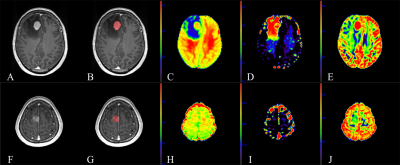 |
Neurite orientation dispersion and density MR imaging: discriminating atypical high-grade glioma from primary central nervous system lymphoma
Eryuan Gao1, Guohua Zhao1, Huiting Zhang2, Peipei Wang1, Xiaoyue MA1, Jie Bai1, Xu Yan2, Guang Yang3, and Jingliang Cheng1
1Department of Magnetic Resonance, the First Affliated Hospital of Zhengzhou University, Zhengzhou, China, 2MR Scientific Marketing, Siemens Healthcare, Shanghai, China, 3Shanghai Key Laboratory of Magnetic Resonance, East China Normal University, Shanghai, China Keywords: Tumors, Neuro, atypical high-grade glioma, primary central nervous system lymphoma Preoperative differentiation of atypical high-grade glioma (HGG) (with no or little necrosis) and primary central nervous system lymphoma (PCNSL) may help to develop treatment plans. However, they share similar appearance in routine MR images. As a new diffusion model, neurite orientation dispersion and density imaging (NODDI) reflects the microstructure of tissue by measuring different components within it. Through quantitative analysis, we found that all parameters of NODDI performed excellent in distinguishing between atypical HGG and PCNSL. |
| 17:28 | 0265.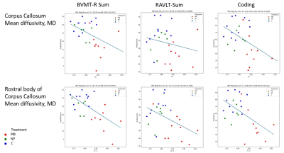 |
Exploring neuroimaging biomarkers and their correlations to neurocognitive function in adult childhood cancer survivors
Maria Ljungberg1,2, Oscar Jalnefjord1,2, Isabelle Rydén3, Erik Fernström4, Linnea Andersson1,2, Justin Schneiderman3, Malin Blomstrand4, Marie Kalm5, Isabella Björkman-Burtscher6, and Marianne Jarfelt4
1Department of Medical Radiation Sciences, Institute of Clinical Sciences, Sahlgrenska Academy, University of Gothenburg, Gothenburg, Sweden, 2Department of Medical Physics and Biomedical Engineering, Sahlgrenska University Hospital, Gothenburg, Sweden, 3Department of Clinical Neuroscience, Institute of Neuroscience and Physiology, Sahlgrenska Academy, University of Gothenburg, Gothenburg, Sweden, 4Department of Oncology, Institute of Clinical Sciences, Sahlgrenska Academy, University of Gothenburg, Gothenburg, Sweden, 5Department of Pharmacology, Institute of Neuroscience and Physiology , Sahlgrenska Academy, University of Gothenburg, Gothenburg, Sweden, 6Department of Radiology, Institute of Clinical Sciences, Sahlgrenska Academy, University of Gothenburg, Gothenburg, Sweden Keywords: Tumors, Radiotherapy, DTI, MR spectroscopy Cranial radiotherapy (CRT) is effective; however, survivors of childhood cancer often suffer from cognitive deficits. Two adult patient groups that received different CRT exposures as children were included, as well as healthy volunteers. The MR-examination comprised of anatomical imaging, DTI and MR spectroscopy of the hippocampus. Differences were found between the group that received highest CRT dose and the control group in hippocampal volume, tNAA/tCr-ratio of hippocampus and several DTI-measures in e.g., corpus callosum. Correlations was found between DTI-measures (MD/RD/AD) in some of the studied white matter tracts and several of the used neuropsychological tests, e.g., BVMT-R Sum and Coding. |
| 17:36 | 0266.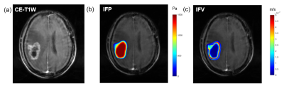 |
Predicting IDH1 Expression using Computational Modeling of Interstitial Fluid Pressure in Glioblastoma
Jianan Zhou1,2,3, Wentao Hu4, Zhengyang Zhu5, Xin Zhang1,2,3, Yongming Dai4, and Bing Zhang2,3,5,6,7
1Department of Radiology,Nanjing Drum Tower Hospital Clinical College of Nanjing Medical University, Nanjing,Jiangsu Province,China, China, 2Institute of Medical Imaging and Artificial Intelligence, Nanjing University, Nanjing, China, 3Medical Imaging Center, Affiliated Drum Tower Hospital, Medical School of Nanjing University, Nanjing, China, 4MR Collaboration, Central Research Institute, United Imaging Healthcare, Shanghai, China, Shanghai, China, 5Department of Radiology, The Affiliated Drum Tower Hospital of Nanjing University Medical School, Nanjing,Jiangsu Province,China, China, 6Jiangsu Key Laboratory of Molecular Medicine, Nanjing, China, 7Institute of brain Science, Nanjing University, Nanjing, China Keywords: Tumors, Contrast Agent The immunohistochemistry features of glioblastoma have important influence on its occurrence and prognosis. Understanding the noninvasive detection of interstitial fluid pressure (IFP) and velocity (IFV) based on dynamic contrast enhanced (DCE)-MRI to explore the correlation between IFP and IFV with immunohistochemical markers of glioblastoma. The investigation demonstrates that IFP indicators have significant correlation with isocitrate dehydrogenase 1 (IDH1) expression and Ki-67 level of glioblastoma, and could help diagnose IDH1-mutation patients. Keywords Glioblastoma,Interstitial Fluid Pressure and Velocity,IDH1,Ki-67 |
| 17:44 | 0267.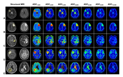 |
Identification of IDH and tumor subtype of adult-type diffuse gliomas based on WHO CNS5 using standard, high and ultra-high b value DWI
Xueqin Wang1, Xinru Shu2, Pingping He3, Yang Song4, and Rifeng Jiang5
1School of Medical Imaging, Fujian Medical University, Fujian, China, 2School of Basic Medical Sciences, Fujian Medical University, Fuzhou, Fujian, China, 3School of Medical Imaging, Fujian Medical University, Fuzhou, Fujian, China, 4SIEMENS Healthcare, Diagnostic Imaging, Shanghai, China, 5Radiology, Fujian Medical University Union Hospital, Fuzhou, Fujian, China Keywords: Tumors, Nervous system To date, few studies have investigated the association between ADC values and tumor subtypes of adult-type diffuse gliomas based on WHO CNS5. By comparing standard, high and ultra-high b value DWI, our research found that all ADC values can differentiate IDH genotypes with ADC8,000 having the highest AUC of 0.827, and mostly ADC values can differentiate the tumor subtypes of gliomas, with ADC8,000 and ADC4,000 having the highest AUC of 0.780 and 0.902, respectively. High and ultra-high b value DWI, is a potent approach in identifying IDH genotype and tumor subtype of adult-type diffuse gliomas based on WHO CNS5.
|
17:52 |
0268.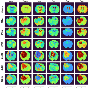 |
Soma and Neurite Density Imaging (SANDI) characterizes microscopic differences between two glioma mouse model subtypes
Andrada Ianus1, Marco Palombo2,3, Rui V. Simoes1,4, Rafael N. Henriques1, Tania Carvalho1, and Noam Shemesh1
1Champalimaud Research, Champalimaud Foundation, Lisbon, Portugal, 2Cardiff University Brain Research Imaging Centre, School of Psychology, Cardiff University, Cardiff, United Kingdom, 3School of Computer Science and Informatics, Cardiff University, Cardiff, United Kingdom, 4Institute for Research & Innovation in Health (i3S), University of Porto, Porto, Portugal Keywords: Tumors, Diffusion/other diffusion imaging techniques Diffusion MRI (dMRI) provides an indirect characterisation of tissue features at the microscopic-scale. The Soma and Neurite Density Imaging (SANDI) methodology was initially proposed to quantity parameters of cell soma and neurite in brain white and gray matter. Here, we investigate the potential of employing SANDI as a general representation of isotropic, restricted, highly anisotropic and isotropic Gaussian components in two mouse models of glioma, derived from two cell lines with distinct microstructure (CT2A and GL261). The results reveal differences across the two tumour subtypes which are consistent with qualitative histological assessment. |
 Back to Meeting Home
 Back to the Program-at-a-Glance
The International Society for Magnetic Resonance in Medicine is accredited by the Accreditation Council for Continuing Medical Education to provide continuing medical education for physicians.
 Back to Meeting Home
 Back to the Program-at-a-Glance
 View Presentation Video