Oral Session
Aging Brain
ISMRM & ISMRT Annual Meeting & Exhibition • 03-08 June 2023 • Toronto, ON, Canada

13:45 |
1298.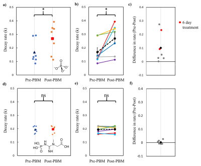 |
A ray of light against age-related Neurodegenerative disease: A
31P Magnetisation Transfer MRS study
Elizabeth Jane Fear1,
Frida Torkelsen2,
Heidi A Baseler3,
and Aneurin James Kennerley4
1Biomolecular Sciences, University of Urbino Carlo Bo, Urbino, Italy, 2Chemistry, University of York, York, United Kingdom, 3Psychology, University of York, York, United Kingdom, 4Sport and Exercise Science, Institute of Science, Manchester Metropolitan University, Manchester, United Kingdom Keywords: Neurodegeneration, Magnetization transfer, 31P Through combined theoretical Monte Carlo stimulation and practical 31P Magnetisation Transfer Magnetic Resonance Spectroscopy we quantify the effects of 670 nm photobiomodulation treatment on healthy aging brains. Mitochondrial function declines with age and many pathological processes of neurodegenerative diseases stem from this mitochondrial dysfunction when they fail to produce the necessary energy required. Therefore, an aging population coupled with associated increases in cases of neurological conditions amplifies the need to develop safe, inexpensive treatments to restore mitochondrial function and offer neuronal protection as we grow old. Evidence shows that non-invasive transcranial red/infrared photobiomodulation (PBM) therapy can offer such neuroprotective benefits. |
|
13:53 |
1299.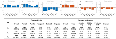 |
Age-related alterations in soma and neurite fraction obtained
from high-gradient diffusion MRI data across the lifespan
Hansol Lee1,
Hong-Hsi Lee1,
Laleh Eskandarian1,
Kyla Gaudet1,
Qiyuan Tian1,
Eva A. Krijnen2,3,
Eric C. Klawiter2,
and Susie Y. Huang1
1Department of Radiology, Athinoula A. Martinos Center for Biomedical Imaging, Massachusetts General Hospital, Charlestown, MA, United States, 2Department of Neurology, Massachusetts General Hospital, Harvard Medical School, Boston, MA, United States, 3MS Center Amsterdam, Anatomy and Neurosciences, Amsterdam Neuroscience, Amsterdam UMC location VUmc, Amsterdam, Netherlands Keywords: Neurodegeneration, Aging We studied alterations in soma and neurite signal fractions with age based on Soma And Neurite Density imaging (SANDI) in 43 healthy adults across the lifespan using multi-shell dMRI measurements acquired on the MGH Connectome scanner. We observed decreases in soma fraction with age in all cortical lobes, especially in the frontal lobe. The neurite fraction predominantly decreased with age in the genu of corpus callous. These results suggest a regionally selective aging effect on changes in compartmental composition within the brain, potentially reflecting alterations in microstructure associated with neurodegeneration. |
| 14:01 |
1300.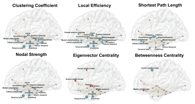 |
Structural network integrity in limbic-predominant age-related
TDP-43 encephalopathy neuropathological change (LATE-NC)
Mahir Tazwar1,
Arnold M Evia2,
Abdur Raquib Ridwan2,
David A Bennett2,
Julie A Schneider2,
and Konstantinos Arfanakis1,2
1Department of Biomedical Engineering, Illinois Institute of Technology, Chicago, IL, United States, 2Rush Alzheimer’s Disease Center, Rush University Medical Center, Chicago, IL, United States Keywords: Neurodegeneration, White Matter, LATE-NC, TDP-43, neuropathology, aging Limbic-predominant age-related TDP-43 encephalopathy neuropathological change (LATE-NC) is a common pathological finding in the brain of older adults, but its impact on structural network integrity remains unknown. In this work, we studied structural connectivity network abnormalities associated with LATE-NC using graph theory. Our results demonstrated that severity of LATE-NC was independently associated with weaker network integration and segregation, and increased vulnerability in a network of brain regions typically affected by LATE-NC. |
| 14:09 |
1301.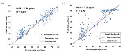 |
Brain Age Prediction Based on Quantitative Susceptibility
Mapping
Mingxing Chen1,
Yuting Shi1,
Yuyao Zhang2,
and Hongjiang Wei1
1School of Biomedical Engineering, Shanghai Jiao Tong University, Shanghai, China, 2School of Information and Science and Technology, ShanghaiTech University, Shanghai, China Keywords: Neurodegeneration, Quantitative Susceptibility mapping The aging process of the human brain is known to be complex, resulting in considerable structural and functional changes in the brain. In this study, a brain age prediction method based on QSM was proposed and then applied to predict the brain age of PD patients. The model achieved a high prediction accuracy with the MAE of 4.40 years and the R2 of 0.91 in healthy subjects. The PADs of the PD patients were significantly higher than HC subjects. The results show that brain age prediction based on QSM can provide a new biomarker to explore iron-related brain age changes. |
| 14:17 |
1302.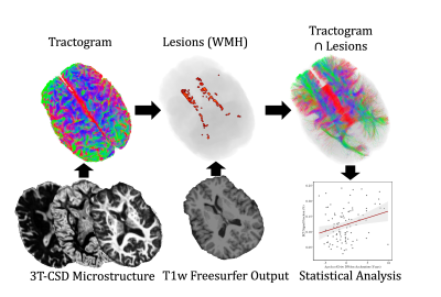 |
Epigenetic age acceleration predicts subject-specific white
matter degeneration in the human brain.
Benjamin T Newman1,2,
Joshua S Danoff2,
Morgan E Lynch3,
Stephanie N Giamberardino4,
Simon G Gregory4,5,
Jessica J Connelly2,
T Jason Druzgal1,
and James P Morris2
1Radiology and Medical Imaging, University of Virginia, Charlottesville, VA, United States, 2Department of Psychology, University of Virginia, Charlottesville, VA, United States, 3Department of Psychology, Univeristy of Southern California, Los Angeles, CA, United States, 4Duke Molecular Physiology Institute, Duke University, Durham, NC, United States, 5Department of Neurology, Duke University, Durham, NC, United States Keywords: Neurodegeneration, Neurodegeneration Epigenetic clocks provide powerful tools for estimating health and lifespan but their ability to predict brain degeneration and neuronal damage during the aging process is unknown. This study uses the epigenetic clock GrimAge to longitudinally investigate brain cellular microstructure in axonal white matter from a healthy aging cohort. We reconstructed subject-specific axonal networks damaged by white matter hyperintensities, a visible neurological manifestation of small vessel disease. A chronological age-adjusted version of GrimAge was significantly correlated with longitudinal markers of neuronal decline. This study is the first to establish a relationship between accelerated epigenetic GrimAge and brain cellular microstructure in humans. |
| 14:25 |
1303.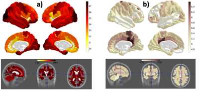 |
Pronounced gender and lifestyle effects in the oxygen extraction
fraction (OEF) of the ageing brain
Ana-Maria Oros-Peusquens1,
Luis Hau1,
Junghun Cho2,
Nora Bittner3,4,
Svenja Caspers3,5,
Yi Wang2,
and N. Jon Shah1,6,7,8,9 1INM-4, Research Centre Juelich, Juelich, Germany, 2Department of Radiology, Weill Cornell Medical College, New York, NY, United States, 3INM-1, Research Centre Juelich, Juelich, Germany, 44Institute for Anatomy I, Medical Faculty & University Hospital Düsseldorf, Heinrich Heine University, Duesseldorf, Germany, 5Institute for Anatomy I, Medical Faculty & University Hospital Düsseldorf, Heinrich Heine University, Duesseldorf, Germany, 6RWTH Aachen University, Aachen, Germany, 7INM-11, JARA, Forschungszentrum Jülich, Juelich, Germany, 8JARA - BRAIN - Translational Medicine, Aachen, Germany, 9Department of Neurology, RWTH Aachen University, Aachen, Germany Keywords: Neurodegeneration, Aging, oxygen extraction fraction, OEF, gender differences, menopause Regionally-resolved oxygen extraction fraction was investigated in an elderly cohort. OEF was obtained from a single 3D mGRE scan using a novel integrated model of QSM phase signal and quantitative blood oxygenation level dependent magnitude signal. Whereas age showed little influence on this metabolic parameter, a pronounced gender effect was observed. A lifestyle index reflecting physical and social activity as well as alcohol and nicotine consumption showed strong correlations with OEF. Venous blood volume fraction and tissue R2*, but not tissue QSM, also reflected lifestyle influence, showing that brain age is more than number of years. |
| 14:33 |
1304.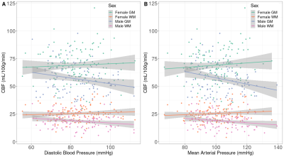 |
Cardiovascular and cerebrovascular health in 70-year-olds: a
population-based ASL study
Mathijs B.J. Dijsselhof1,2,
Sarah-Naomi James3,
Luigi Lorenzini1,2,
Lyduine Collij1,2,
David L. Thomas4,5,
Catherine Scott5,
Emily Manning5,
Tamás I. Józsa1,2,
Dave Cash5,
Insight 46 study team5,
Carole Sudre3,
Alun D. Hughes3,
Marcus Richards3,
Frederik Barkhof1,2,6,
Jonathan Schott5,
Jan Petr1,2,7,
and Henk J.M.M. Mutsaerts1,2 1Radiology & Nuclear Medicine, Amsterdam University Medical Centers, Vrije Universiteit, Amsterdam, Netherlands, 2Amsterdam Neuroscience, Brain Imaging, Amsterdam, Netherlands, 3MRC Unit of Lifelong Health at Ageing at UCL, University College London, London, United Kingdom, 4Neuroradiological Academic Unit, Department of Brain Repair and Rehabilitation, UCL Queen Square Institute of Neurology, London, United Kingdom, 5Dementia Research Centre, UCL Queen Square Institute of Neurology, London, United Kingdom, 6Queen Square Institute of Neurology and Centre for Medical Image Computing, University College London, London, United Kingdom, 7Helmholtz-Zentrum Dresden-Rossendorf, Institute of Radiopharmaceutical Cancer Research, Dresden, Germany Keywords: Neurodegeneration, Arterial spin labelling While mid-life cardiovascular pathology may lead to late-life cognitive decline, our understanding of the role of cerebrovascular health as an intermediate biomarker is limited. We explored the association between cardiovascular health biomarkers and cross-sectional and longitudinal cerebrovascular health assessed by ASL MRI in Insight46, a well-characterised cognitively normal population-based sample. We found several cross-sectional and longitudinal associations between blood pressure and CBF. These findings suggest that the effects of BP on cerebrovascular health can be imaged with ASL perfusion MRI, possibly offering opportunities to prevent or intervene before cognitive decline sets in. |
| 14:41 |
1305.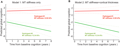 |
Medial Temporal Brain Stiffness Predicts Cognition Decline in
Aging and Alzheimer's Disease
KowsalyaDevi Pavuluri1,
John Huston III1,
Richard L. Ehman1,
Armando Manduca1,2,
Prashanthi Vemuri1,
Clifford R. Jack Jr1,
Matthew L. Senjem3,
and Matthew C. Murphy1
1Department of Radiology, Mayo Clinic, Rochester, MN, United States, 2Department of Physiology and Biomedical Engineering, Mayo Clinic College of Medicine, Rochetser, MN, United States, 3Department of Information Technology, Mayo Clinic, Rochester, MN, United States Keywords: Neurodegeneration, Aging, Stiffness and Cognition Aging is associated with neurodegeneration, cognitive function decline, and increased risk of dementia. Objective methods for the longitudinal prediction of cognitive trajectories are needed for design of comprehensive prevention strategies. We tested the hypothesis that measurements of brain mechanical properties will complement existing biomarkers in predicting future cognitive decline. Using linear mixed effect modelling, we evaluated the role of baseline medial temporal stiffness in predicting future cognitive function in participants along the Alzheimer’s disease spectrum. |
14:49 |
1306.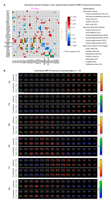 |
Brain-wide fMRI Connectivity and Regional Genetic Modulations
underlying Optogenetically-evoked Spindles in Rescuing Memory
Decline in Aging
Xunda Wang1,2,
Pit Shan Chong3,
Lee-Wei Lim3,
Alex T. L. Leong1,2,
and Ed X. Wu1,2,3 1Laboratory of Biomedical Imaging and Signal Processing, The University of Hong Kong, Hong Kong SAR, China, 2Department of Electrical and Electronic Engineering, The University of Hong Kong, Hong Kong SAR, China, 3School of Biomedical Sciences, The University of Hong Kong, Hong Kong SAR, China Keywords: Neurodegeneration, Aging Memory consolidation, the ability to transform newly learned information into long-term memory, declines with age. Our previous study revealed targeted neuromodulation of spindle activities can arrest memory consolidation dysfunction in aging brains through strengthening multi-target memory representations. However, whether and how spindle activities influence memory consolidation via acting on inter-regional information integration remained unclear. Here, we demonstrate in aging animals that optogenetically-evoked spindle activities alleviate memory consolidation dysfunction through modulating brain-wide inter-regional connectivity and regional genetic expression. Our work provides an approach combining fMRI analysis and genetic expression profiling to bridge systems- and molecular-level understandings of memory consolidation. |
| 14:57 |
1307.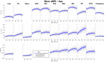 |
Normative trajectories of quantitative MRI parameters in
sub-cortical grey matter on healthy ageing
Kwok-Shing Chan1,2,
Michelle G. Jansen1,
Joukje Oosterman1,
David G. Norris1,3,4,
Christian F. Beckmann1,2,
and José P. Marques1
1Donders Institute for Brain, Cognition and Behaviour, Nijmegen, Netherlands, 2Department of Cognitive Neuroscience, Radboud University Medical Center, Nijmegen, Netherlands, 3The Erwin L. Hahn Institute, Essen, Germany, 4University of Twente, Twente, Netherlands Keywords: Neurodegeneration, Aging The effects of ageing on quantitative MRI, including R1, R2* and tissue susceptibility, are investigated in subcortical grey matter using a healthy cohort. General Linear Regression analysis indicates that ageing has significant impacts on these quantitative measures: the mean R1 has inverted U-shape appearances with time, while the mean R2* and susceptibility are closer to linear. We further studied the spatial variation in these structures and the results show that the spatial gradient of some structures (e.g. caudate and putamen) also changes with age. Normative trajectories of these parameters in subcortical grey matter associated with ageing are also investigated. |
| 15:05 |
1308.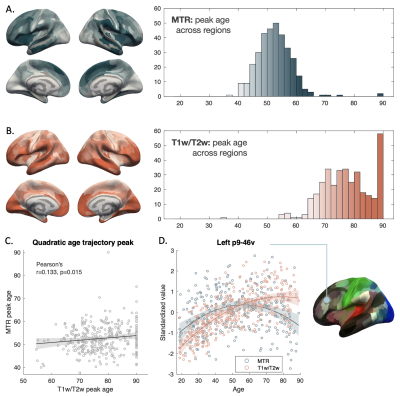 |
Differences in age trajectories of intracortical myelin across
lifespan measured by MTR and T1w/T2w
Yu Veronica Sui1,
Ryn Flaherty1,
Arjun V. Masurkar2,3,4,
Thomas Wisniewski2,4,5,6,
Henry Rusinek1,5,
and Mariana Lazar1
1Department of Radiology, New York University Grossman School of Medicine, New York, NY, United States, 2Department of Neurology, New York University Grossman School of Medicine, New York, NY, United States, 3Department of Neuroscience and Physiology, New York University Grossman School of Medicine, New York, NY, United States, 4Neuroscience Institute, New York University Grossman School of Medicine, New York, NY, United States, 5Department of Psychiatry, New York University Grossman School of Medicine, New York, NY, United States, 6Department of Pathology, New York University Grossman School of Medicine, New York, NY, United States Keywords: Neurodegeneration, Aging In a large cohort of healthy participants (N=349, age range 18-90 years), we characterized intracortical myelin lifespan trajectories using two commonly used myelin proxies, magnetization transfer ratio (MTR) and T1w/T2w, combined with a surface-based image processing method. We showed that both measures exhibit an inverted-U shape trajectory for most cortical regions, with T1w/T2w values peaking later than MTR. A smoothed spline fitting of the trajectories allowed further delineation of onset decline age, a potential aging milestone. Frontal and temporal lobe regions showed consistently later peak ages than parietal and sensory motor regions across MRI metrics and curve fitting methods. |
|
15:13 |
1309.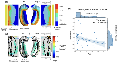 |
Age-related alterations of hippocampal microstructure quantified
using diffusion MRI in a unfolded hippocampal space
Yixin Ma1,2,
Hansol Lee1,2,
Qiyaun Tian1,2,
Susie Y Huang1,2,
and Hong-Hsi Lee1,2
1Radiology, Athinoula A. Martinos Center for Biomedical Imaging, Massachusetts General Hospital, Charlestown, MA, United States, 2Radiology, Harvard Medical School, Boston, MA, United States Keywords: Neurodegeneration, Microstructure The hippocampus plays an important role in cognition and memory. Microstructural alterations can happen in specific sub-regions within the hippocampus in normal aging. Applying more advanced diffusion models and pinpointing changes in specific locations in the hippocampus, though challenging, may offer greater insight into subfield-specific neurodegeneration. Here, we applied advanced biophysical models of dMRI and correlated tissue parameters with age across the unfolded hippocampal coordinates in 43 healthy adults covering a wide age span. |
|
15:21 |
1310.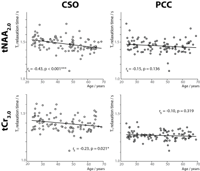 |
Metabolite T1 relaxation times differ across the adult lifespan
Saipavitra Murali-Manohar1,2,
Aaron T. Gudmundson1,2,
Kathleen E. Hupfeld1,2,
Helge J. Zöllner1,2,
Steve C.N. Hui1,2,
Yulu Song1,2,
Christopher W. Davies-Jenkins1,2,
Tao Gong3,4,
Guangbin Wang3,4,
Georg Oeltzschner1,2,
and Richard A.E. Edden1,2
1The Russell H. Morgan Department of Radiology and Radiological Science, Johns Hopkins University School of Medicine, Baltimore, MD, United States, 2F.M. Kirby Research Center for Functional Brain Imaging, Kennedy Krieger Institute, Baltimore, MD, United States, 3Departments of Radiology, Shandong Provincial Hospital Affiliated to Shandong First Medical University, Shandong, China, 4Departments of Radiology, Shandong Provincial Hospital, Shandong University, Shandong, China Keywords: Spectroscopy, Aging, T1 relaxation times, Macromolecules, Human brain This work investigates the age-dependence of metabolite T1 relaxation times at 3T. T1 relaxation times were estimated by modeling the residual metabolite amplitudes in macromolecular spectra, acquired with pre-inversion. Posterior cingulate (PCC) and centrum semiovale (CSO) spectra were acquired in 102 healthy volunteers across five decades of adult life (20 to 69 years). T1 relaxation times of both tNAA2.0 and tCr3.0 significantly negatively correlated with age in CSO, and not in PCC. This has important implications for MRS studies of aging which tend to assume T1 relaxation times are constant as a function of age. |
| 15:29 |
1311. |
White matter changes underlying cortical changes across the
lifespan
Kurt G Schilling1,
Victor Nozais2,
Francois Rheault3,
Derek Archer4,
Muwei Li1,
Leon Y Cai5,
Flavio Dell'Acqua6,
John C Gore1,
and Bennett A Landman5 1Vanderbilt University Medical Center, Nashville, TN, United States, 2Institut des Maladies Neurodégénératives-UMR, Bordeaux, France, 3Sherbrooke University, Sherbrooke, QC, Canada, 4Vanderbilt University School of Medicine, Nashville, TN, United States, 5Vanderbilt University, Nashville, TN, United States, 6King's College London, London, United Kingdom Keywords: White Matter, Aging Brain functions are a property of the interaction between brain areas. While much is known about cortical structure and the structural connections in the white matter, the interaction between the cortex and white matter is underexplored. Here, we aim to investigate and characterize the relationship between white and cortical changes across the lifespan. We use the functionnectome framework to associate white matter to morphological features of the cortex. Biologically, this white matter influences (or is influenced by) these cortical changes. |
| 15:37 |
1312. |
Aging-related morphology and microstructure variations:
Discoveries from the Lifespan Human Connectome Project - Aging
Tyler D. Robinson1,
Yutong L. Sun1,
Paul T. H. Chang1,
and J. Jean Chen1,2,3
1Rotman Research Institute, Baycrest, Toronto, ON, Canada, 2Medical Biophysics, University of Toronto, Toronto, ON, Canada, 3Biomedical Engineering, University of Toronto, Toronto, ON, Canada Keywords: White Matter, Microstructure This study compared age-related differences in white matter morphology and microstructure across ten major tracts of the human brain using diffusion data from 535 participants of the Human Connectome Project in Aging. The results are additionally assessed for agreement with retrogenesis predictions of white matter decline in normal aging. While whole-brain relationships between morphometry and white matter integrity were identified, high variability was also observed between tracts. While our data do not fully support retrogenesis models, we demonstrate patterns that may provide partial support, and highlight the need for tract-specific studies of morphological-microstructural interactions in the aging white matter. |
 Back to Meeting Home
 Back to the Program-at-a-Glance
The International Society for Magnetic Resonance in Medicine is accredited by the Accreditation Council for Continuing Medical Education to provide continuing medical education for physicians.
 Back to Meeting Home
 Back to the Program-at-a-Glance
 View Presentation Video