Oral Session
Epilepsy
ISMRM & ISMRT Annual Meeting & Exhibition • 03-08 June 2023 • Toronto, ON, Canada

15:45 |
0994.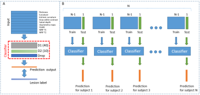 |
Automated Surfaced-based Detection of Focal Cortical Dysplasia
using MR Fingerprinting
Ting-Yu Su1,2,
Siyuan Hu2,
Xiaofeng Wang3,
Sophie Adler4,
Konrad Wagstyl5,
Zheng Ding1,2,
Joon Yul Choi1,
Ken Sakaie6,
Ingmar Blümcke1,7,
Hiroatsu Murakami1,
Stephen Jones6,
Imad Najm1,
Dan Ma2,
and Zhong Irene Wang1
1Epilepsy Center, Neurological Institute, Cleveland Clinic, Cleveland, OH, United States, 2Biomedical Engineering, Case Western Reserve University, Cleveland, OH, United States, 3Quantitative Health Science, Cleveland Clinic, Cleveland, OH, United States, 4University College London Great Ormond Street Institute for Child Health, London, United Kingdom, 5Wellcome Centre for Human Neuroimaging, London, United Kingdom, 6Imaging Institute, Cleveland Clinic, Cleveland, OH, United States, 7Neuropathology, University Hospitals Erlangen, Erlangen, Germany Keywords: Epilepsy, MR Fingerprinting Focal cortical dysplasia (FCD) is a common pathology in medically intractable focal epilepsy and often difficult to detect by visual inspection of conventional MRI. We developed a framework for automatic FCD detection using surface-based processing of conventional MRI and MR fingerprinting data. Thirty-six patients with FCD and 48 healthy controls were included. Improved vertex-wise and cluster-wise performance was seen when MRF and FLAIR features were added to T1w data. A second-stage cluster-wise classifier showed efficacy to reduce false-positive clusters. Interim results of patient-level sensitivity of 76% and low false-positive clusters in controls supported potential clinical applicability of the proposed framework. |
15:53 |
0995.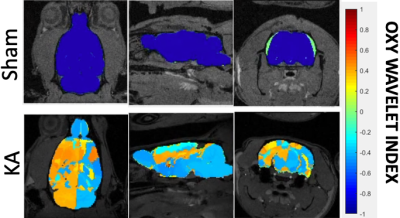 |
Detecting in vivo Mitochondrial Dysfunction with 4D Oxy-wavelet
MRI in Kainic Acid Induced Temporal Lobe Epilepsy
Devin Raine Everaldo Cortes1,2,3,
Margaret C. Stapleton2,3,
Kristina E. Schwab4,5,
Noah W. Coulson2,6,
Dalton R West2,3,
Thomas Becker-Szurszewski4,5,
Sean Hartwick4,5,
Sivakama S. Bharathi7,
Eric Goetzman7,
Kevin M. Kelly8,
Anthony G. Christodoulou9,
and Yijen L Wu1,2,3,7 1Department of Bioengineering, Swanson School of Engineering, University of Pittsburgh, Pittsburgh, PA, United States, 2Department of Developmental Biology, School of Medicine, University of Pittsburgh, Pittsburgh, PA, United States, 3Rangos Research Center Small Animal Imaging Core, Children's Hospital of Pittsburgh of UPMC, Pittsburgh, PA, United States, 4Rangos Research Center Animal Imaging Core, Children's Hospital of Pittsburgh of UPMC, Pittsburgh, PA, United States, 5Department of Pediatrics, University of Pittsburgh, School of Medicine, Pittsburgh, PA, United States, 6Rangos Research Center Small Animal Imaging Core, University of Pittsburgh, Pittsburgh, PA, United States, 7Department of Pediatrics, School of Medicine, University of Pittsburgh, Pittsburgh, PA, United States, 8Allegheny Health Network Research Institue, Allegheny General Hospital, Pittsburgh, PA, United States, 9Biomedical Imaging Research Institue, Cedars-Sinai Medical Center, Los Angeles, CA, United States Keywords: Epilepsy, Metabolism, fMRI, Hypoxia Intractable temporal lobe epilepsy, often acquired after status epilepticus (SE) injury, greatly reduces quality of life. There is an unmet need for a non-invasive method to track progression from SE to epilepsy, which would allow early intervention to prevent irreversible damage to the brain. Mitochondrial dysfunction is becoming a recognized marker of epileptogenesis. Here, we established a novel functional MRI methodology, the 4D Oxy-wavelet MRI, capable of in vivo detection of mitochondrial dysfunction underlying post-SE epileptogenesis in a spatial specific manner. This non-invasive method may aid early detection of subclinical epileptogenesis and serve as a biomarker for therapeutic efficacy. |
| 16:01 |
0996.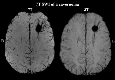 |
A first application of the ILAE consensus protocol for 7T
epilepsy imaging in addition to clinical practice
Gilbert Hangel1,2,3,
Gregor Kasprian4,
Stefanie Chambers1,2,
Lukas Haider4,
Philipp Lazen1,2,
Johannes Koren5,
Robert Diehm6,
Katharina Moser6,
Matthias Tomschik1,
Jonathan Wais1,
Fabian Winter1,
Vitalij Zeiser1,
Stephan Gruber2,
Susanne Aull-Watschinger7,
Tatjana Traub-Weidinger8,
Christoph Baumgartner5,
Martha Feucht6,
Christian Dorfer1,
Wolfgang Bogner2,
Siegfried Trattnig2,
Ekaterina Pataraia7,
and Karl Rössler1
1Department of Neurosurgery, Medical University of Vienna, Vienna, Austria, 2High-field MR Center, Department of Biomedical Imaging and Image-guided Therapy, Medical University of Vienna, Vienna, Austria, 3Christian Doppler Laboratory for MR Imaging Biomarkers, Vienna, Austria, 4Division of Neuroradiology and Musculoskeletal Radiology, Department of Biomedical Imaging and Image-guided Therapy, Medical University of Vienna, Vienna, Austria, 5Department of Neurology, Klinik Hietzing, Vienna, Austria, 6Center for Rare and Complex Childhood Onset Epilepsies, Member of ERN EpiCARE, Department of Pediatrics and, Medical University of Vienna, Vienna, Austria, 7Department of Neurology, Medical University of Vienna, Vienna, Austria, 8Division of Nuclear Medicine, Department of Biomedical Imaging and Image-guided Therapy, Medical University of Vienna, Wien, Austria Keywords: Epilepsy, Brain In a cohort of 38 patients with pharmacoresistant focal epilepsy, our implementation of the 2021 ILAE 7T consensus protocol found lesions in 19% of 3T MR-negative cases and improved delineation of lesions in 88% of 3T MR-positive cases, with statistically significant higher detection confidence at 7T. Our results conform to literature and show the surplus effect of the consensus protocol in addition to clinical standard diagnostics and point at a potential main use of better surgical planning in known lesions. |
| 16:09 |
0997.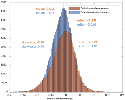 |
7T metabolic MRI in mesial temporal lobe epilepsy
Sarah M Jacobs1,
Zahra Shams1,
Anja G van der Kolk1,2,
Alex Bhogal1,
Jannie P Wijnen1,
Jeroen C.W. Siero1,3,
Pieter van Eijsden4,
Edwin Versteeg1,
Angelika Mühlebner5,6,
Wim Van Hecke5,
Dennis WJ Klomp1,
Maeike Zijlmans4,7,
and Evita C Wiegers1 1Department of Radiology and Nuclear Medicine, University Medical Center Utrecht, Utrecht, Netherlands, 2Department of Medical Imaging, Radboud University Medical Center, Nijmegen, Netherlands, 3Spinoza Centre for Neuroimaging Amsterdam, Amsterdam, Netherlands, 4UMC Utrecht Brain Center, Department of Neurology and Neurosurgery, University Medical Center Utrecht, Utrecht, Netherlands, 5Department of Pathology, University Medical Center Utrecht, Utrecht, Netherlands, 6Department of Pathology, Amsterdam University Medical Center, location AMC, Amsterdam, Netherlands, 7Stichting Epilepsie Instellingen Nederland (SEIN), Heemstede, Netherlands Keywords: Epilepsy, Metabolism We combined quantitative susceptibility mapping (QSM) and single voxel (SV) 1H magnetic resonance spectroscopy (MRS) at 7 tesla (7T) with the aim to characterize mesial temporal lobe epilepsy (mTLE). We discovered in 9 patients that the distribution of quantified susceptibly is negatively skewed in epileptogenic hippocampi compared to contralateral hippocampi, meaning more positive susceptibility values: an indicator for iron deposition. No differences in metabolite ratios could be seen in 7 patients between hippocampi, however our small sample size precludes any final conclusions. |
| 16:17 |
0998.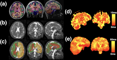 |
Multi-channel 7 Tesla sodium MRI of the brain in children with
epileptogenic SCN1A sodium channel mutations – a pilot study
Jon Orlando Cleary1,
Samuel Rot2,3,
Michael Ayre4,5,6,
Philippa Bridgen6,
Ayse Sila Dokumaci5,6,
Yasmin Blunck7,
Warda Syeda8,
Bhavana S Solanky2,9,
Shaihan J Malik5,6,
Ming Lim4,
Claudia A.M. Gandini Wheeler-Kingshott2,10,11,
Shan-Shan Tang4,
and David W Carmichael5,6
1Department of Radiology, Imperial College Healthcare NHS Trust, London, United Kingdom, 2MR Research Unit, Queen Square MS Centre, Faculty of Brain Sciences, UCL Queen Square Institute of Neurology, London, United Kingdom, 3Department of Medical Physics and Biomedical Engineering, University College London, London, United Kingdom, 4Children's Neurosciences, Evelina London Children's Hospital at Guy's and St Thomas' NHS Foundation Trust, London, United Kingdom, 5Biomedical Engineering Department, School of Biomedical Engineering and Imaging Sciences, King's College London, London, United Kingdom, 6London Collaborative Ultra High Field System (LoCUS), London, United Kingdom, 7Melbourne Brain Centre Imaging Unit, The University of Melbourne, Parkville, Melbourne, Australia, 8Melbourne Neuropsychiatry Centre, The University of Melbourne, Parkville, Melbourne, Australia, 9Quantitative Imaging Group, Department of Medical Physics and Biomedical Engineering, University College London, London, United Kingdom, 10Department of Brain & Behavioural Sciences, University of Pavia, Pavia, Italy, 11Brain Connectivity Centre Research Department, IRCCS Mondino Foundation, Pavia, Italy Keywords: Epilepsy, Non-Proton, brain, sodium MRI, high-field MRI, SCN1A SCN1A gene mutations disrupt sodium channel (NaV1.1) function, causing childhood epilepsy which can be severe. Predicting functional consequences in these children is challenging and new prognostic imaging biomarkers are needed. Sodium MRI directly assesses brain sodium and is a potential in vivo imaging biomarker. Using a multiecho sodium sequence, at 7T, we found children with SCN1A mutations had increased relative sodium concentrations across a large number of brain regions compared to controls. The cause of this change is likely complex, and may reflect an interplay between neuronal/axonal/glial dysfunction – with disruptions in microstructure and physiology – and possible medication effects. |
| 16:25 |
0999.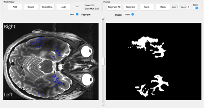 |
Structural changes within the temporal lobe in mesial temporal
lobe epilepsy at 7 Tesla
Mackenzie Langan1,2,
Gauarv Verma2,
Madeline Fields3,
Lara Marcuse3,
Priti Balchandani2,4,5,
and Rebecca Feldman2,6
1Icahn School of Medicine at Mount Sinai, New York, NY, United States, 2Biomedical Engineering and Imaging Institute, Icahn School of Medicine at Mount Sinai, New York, NY, United States, 3Department of Neurology, Mount Sinai Hospital, New York, NY, United States, 4Diagnostic, Molecular and Interventional Radiology, Icahn School of Medicine at Mount Sinai, New York, NY, United States, 5Nash Family Department of Neuroscience, Icahn School of Medicine at Mount Sinai, New York, NY, United States, 6Computer Science, Math, Physics, and Statistics, University of British Columbia, Kelowna, BC, Canada Keywords: Epilepsy, Neuroinflammation, High-Field MRI, perivascular spaces Here we outline a preliminary analysis using a novel method leveraging ultra-high field neuroimaging to measure detectable differences in vasculature within the temporal lobe that may not be detectable at lower field strengths. We provide a tool for detection and quantification of vessels, perivascular spaces, and volumetric changes within the temporal lobe which may be relevant to uncover possible underlying neuroinflammatory processes in mesial temporal lobe epilepsy (MTLE) patients. In our analysis, we found a significant association between volumetric measures of the temporal lobe and vascular and perivascular markers, in patients with lateralized MTLE which may underlie epileptogenic processes. |
|
16:33 |
1000.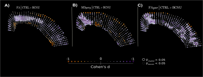 |
Multi-tensor diffusion abnormalities of gray matter in an animal
model of cortical dysplasia.
Paulina Jael Villaseñor1,
Ana Aquiles1,
David Cortes-Servín1,
Aylin Perez-Moriel2,
Hiram Luna-Munguía1,
Alonso Ramirez-Manzanares2,
Jorge Larriva-Sahd1,
and Luis Concha1
1Instituto de Neurobiología, Universidad Nacional Autónoma de México, Querétaro, Mexico, 2Centro de Investigación en Matemáticas, Guanajuato, Mexico Keywords: Epilepsy, Microstructure Focal cortical dysplasias are characterized by abnormal cyto- and myelo- architecture and represent a frequent cause of epilepsy. In some cases these lesions are macroscopically subtle, often going undetected by several conventional imaging techniques, thus warranting the development of alternative imaging methods for their diagnosis. Novel methods for the analysis of diffusion-weighted imaging permit the investigation of the complex architecture of the cortex. Through spatial analysis of diffusion metrics using a multi-tensor approach we demonstrate abnormalities in an animal model of cortical dysplasia that reflect myeloarchitecture disarrangement as seen by histology. |
| 16:41 |
1001.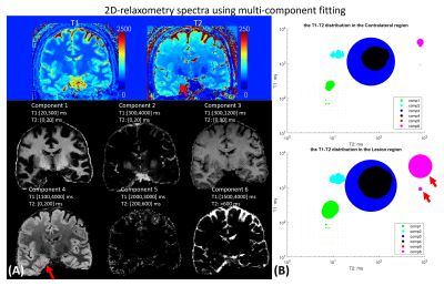 |
A 15-minute 860um whole-brain MR-Fingerprinting and DTI epilepsy
protocol demonstrated on 51 medial temporal lobe epilepsy
patients.
Kang Wang1,
Xiaozhi Cao2,3,
Quan Chen2,3,
Zihan Zhou4,
Dengchang Wu1,
Yunsong Liu5,
Hongjian He4,
Jianhui Zhong4,6,
Kawin Setsompop2,3,
and Congyu Liao2,3
1Department of Neurology, the First Affiliated Hospital, School of Medicine, Zhejiang University, Hangzhou, China, 2Department of Radiology, Stanford University, Stanford, CA, United States, 3Department of Electrical Engineering, Stanford University, Stanford, CA, United States, 4Center for Brain Imaging Science and Technology, College of Biomedical Engineering & Instrument Science, Zhejiang University, Hangzhou, China, 5Signal and Image Processing Institute, University of Southern California, Los Angeles, CA, United States, 6Department of Imaging Sciences, University of Rochester, Rochester, NY, United States Keywords: Epilepsy, Epilepsy In this work, we combined a 5-minute whole-brain 0.86mm-iso 3D-MR fingerprinting (MRF) with a 10-minute whole-brain 0.86mm-iso diffusion MRI protocol, to achieve high-fidelity whole-brain T1/T2/PD and diffusivity maps at sub-millimeter isotropic resolution. This protocol was applied to medial temporal lobe epilepsy (MTLE) patients to enable accurate detection of the hippocampal sclerosis. A multi-parametric analysis was implemented with whole-brain subcortical segmentation. A multi-component 2D-relaxometry spectra was estimated with non-negative joint sparsity for robust suspicious lesion detection. |
| 16:49 |
1002.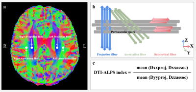 |
Evaluation of glymphatic system function using DTI-ALPS in
patients with temporal lobe epilepsy
Xu Zhao1,
Zhiqiang Zhou1,
and Wenzhen Zhu1
1Tongji hospital of Tongji medical college, Huazhong university of science and technology, Wuhan, China Keywords: Epilepsy, Diffusion Tensor Imaging, glymphatic system, asymmetry The alterations of glymphatic system function in left hemisphere and right hemisphere and the asymmetric features of glymphatic system were not clear in temporal lobe epilepsy (TLE) patients. We investigated the glymphatic system function in TLE patients and evaluated the asymmetric features of glymphatic system by using DTI-ALPS method. Our findings indicated leftward asymmetric tendency of glymphatic system in adult human brain. The abnormality of glymphatic system asymmetry in LTLE was also found. The glymphatic system function was impaired and more severe alterations of glymphatic system in ipsilateral hemisphere than contralateral hemisphere in TLE patients. |
| 16:57 |
1003.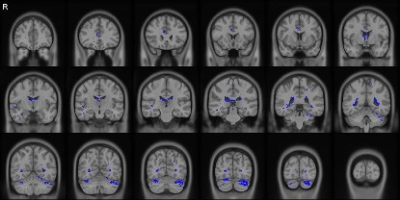 |
Combined 18F-FDG PET/DTI reveals concurrent more extensive brain
damage in mesial temporal lobe epilepsy
Chuan Huang1,2,
Tianyun Zhao2,
Siyu Yuan3,
Hui Huang3,
Miao Zhang4,
and Jie Luo3
1Radiology, Stony Brook Medicine, Stony Brook, NY, United States, 2Biomedical Engineering, Stony Brook University, Stony Brook, NY, United States, 3School of Biomedical Engineering, Shanghai Jiao Tong University, Shanghai, China, 4Department of Nuclear Medicine, Ruijin Hospital, Shanghai, China Keywords: Epilepsy, Epilepsy Mesial temporal lobe epilepsy is the most common type of drug-resistant epilepsy, with hippocampus sclerosis (HS) being its most common pathology. Anterior temporal lobectomy is the most common surgical strategy for these patients, many still suffer from seizures post-surgery, which may relate to their extensive brain network damage. 18F-FDG-PET is sensitive to metabolic changes in the epileptogenic zone. DTI can assess the integrity of white-matter tracts. In this study, we investigated the changes in glucose uptake, white-matter tracts, and their differences in brain networks of MR-HS versus MR-negative patients using simultaneous PET/MR, and studied if these changes coincide topographically. |
| 17:05 |
1004.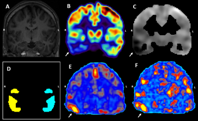 |
Can fMRI metrics lateralize epileptogenic hypometabolic FDG-PET
regions? A simultaneous PET/MR investigation in focal epilepsy
Daniel Uher1,2,3,
Gerhard S. Drenthen1,2,
Tineke van de Weijer2,
Jochem van der Pol2,
Rob Rouhl2,
Olaf E.M.G. Schijns1,3,4,
Albert J. Colon4,
Walter H. Backes1,2,
and Jacobus F.A. Jansen1,2,4,5
1School for Mental Health and Neuroscience, Maastricht University, Maastricht, Netherlands, 2Department of Radiology & Nuclear Medicine, Maastricht University Medical Centre, Maastricht, Netherlands, 3Department of Neurosurgery, Maastricht University Medical Centre, Maastricht, Netherlands, 4Academic Centre for Epileptology, Kempenhaeghe/Maastricht University Medical Centre, Maastricht, Netherlands, 5Department of Electrical Engineering, Eindhoven University of Technology, Eindhoven, Netherlands Keywords: Epilepsy, PET/MR Locally reduced glucose metabolism (i.e. hypometabolism) derived from the 18-FDG positron emission tomography (FDG-PET) is considered to be a valuable biomarker for epileptogenic zone localization. Spontaneous fluctuations in blood-oxygen-level-dependent fMRI (BOLD fMRI) can indirectly measure neuronal activity. Studies have suggested that the fMRI-derived metrics may be indicative of the epileptogenic zone localization, however the potential for fMRI to reflect and lateralize the hypometabolic FDG-PET regions remains underdetermined. Here, both static and dynamic fMRI-derived metrics were calculated and we assessed their potential for lateralizing the hypometabolic FDG-PET regions in patients with unilateral focal epilepsy. |
| 17:13 |
1005.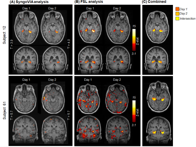 |
Clinical reliability of the “Home Town Walk” fMRI paradigm for
memory function localization in pre-surgical assessment of
patients with Epilepsy
Rosa Sanchez-Panchuelo1,
Nigel Paul Davies1,
Roya Jalali1,
Roman Wesolowski1,
Robert Flintham1,
and Vijay Sawlani2
1Medical Physics, University Hospitals Birmingham NHS Foundation Trust, Birmingham, United Kingdom, 2Radiology, University Hospitals Birmingham NHS Foundation Trust, Birmingham, United Kingdom Keywords: Epilepsy, fMRI (task based), Memory function; Pre-surgical assessment; Clinical implementation; Clinical reliability Memory-activated functional MRI (fMRI) is increasingly implemented in the clinic to assess memory function and inform pre-surgical decision making in refractory epilepsy. The Home Town Walking (HTW) fMRI paradigm has been shown to activate the parahippocampal gyri (PHG) and help determine memory lateralization in epilepsy patients. However, limited data are available on the reliability of this technique in clinical practice. This study aims to assess the robustness of the HTW paradigm for localising and lateralising memory function in a consecutive clinical series of 117 Temporal Lobe Epilepsy patients. Memory-related activation patterns were observed in 76% of cases with 94% reproducibility. |
| 17:21 |
1006.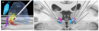 |
Direct visualization and automatic segmentation of the
centromedian nucleus for epilepsy deep brain stimulation
Manojkumar Saranathan1,
Chaitanya Ganne2,
and Sandipan Pati2 1Radiology, UMass Chan Medical School, Worcester, MA, United States, 2Neurology, University of Texas Health Science Center, Houston, TX, United States Keywords: Epilepsy, Segmentation, Deep brain stimulation targeting We present a patient-specific segmentation method for targeting the centromedian nucleus for epilepsy deep brain stimulation and compare it to an atlas-based indirect targeting method in 8 patients. |
| 17:29 |
1007.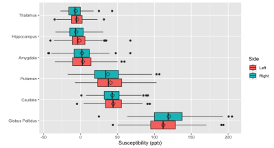 |
Quantitative Susceptibility of Subcortical Grey-Matter Regions
in Early versus Refractory Focal Epilepsy
David N Vaughan1,2,
Eric Y Pierre1,
Marty Bryant1,
David F Abbott1,
Heath R Pardoe1,
Warda T Syeda3,
Bahman Tahayori1,
Chris Tailby1,4,
and Graeme D Jackson1,2
1Florey Institute of Neuroscience and Mental Health, Melbourne, Australia, 2Department of Neurology, Austin Health, Melbourne, Australia, 3Division of Medicine, Dentistry and Health Sciences, University of Melbourne, Melbourne, Australia, 4Department of Neuropsychology, Austin Health, Melbourne, Australia Keywords: Epilepsy, Quantitative Susceptibility mapping Magnetic susceptibility of subcortical grey matter regions was assessed in participants of the Australian Epilepsy Project pilot study. Adults with medication-resistant focal epilepsy were compared to those with newly-diagnosed focal epilepsy, and to people with a single seizure but no epilepsy diagnosis. Basal ganglia, thalamus, hippocampi and amygdala showed susceptibility values consistent with published values, with a positive correlation to age seen at the basal ganglia. There was no significant difference in subcortical susceptibility between the groups. Although other brain network changes may progress with refractory seizures, mineralisation of subcortical regions appears generally stable over the course of focal epilepsy. |
| 17:37 |
1008.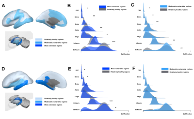 |
Transcriptomic Signatures of Brain Regions Vulnerable to
Anatomical and Metabolic Changes in Temporal Lobe Epilepsy
Jiwei Li1,
Hui Huang1,
Bingyang Cai1,
Siyu Yuan1,
and Jie Luo1
1School of Biomedical Engineering, Shanghai Jiao Tong University, Shanghai, China Keywords: Epilepsy, Tissue Characterization Imaging transcriptomics could bridge the gap between connectome and transcriptome. In this study, we selected brain regions that were vulnerable to hypometabolism, and those vulnerable to atrophy, then investigated transcriptional signatures and cell-type composition differences that may contribute to TLE-related brain structural and metabolic changes. We found hippocampus and entorhinal were found to be most vulnerable brain regions in both anatomical and metabolic changes in TLE patients. Enrichment analysis found that differential expression genes most significantly enriched in neuroactive ligand-receptor interaction pathway. Inhibitory neuron, microglia and oligodendrocyte precursor cells showed significant difference between vulnerable regions and relatively healthy regions. |
 Back to Meeting Home
 Back to the Program-at-a-Glance
The International Society for Magnetic Resonance in Medicine is accredited by the Accreditation Council for Continuing Medical Education to provide continuing medical education for physicians.
 Back to Meeting Home
 Back to the Program-at-a-Glance
 View Presentation Video