Oral Session
Parkinson’s Disease
ISMRM & ISMRT Annual Meeting & Exhibition • 03-08 June 2023 • Toronto, ON, Canada

16:00 |
1383.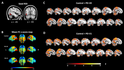 |
Progressive cortical cerebrovascular reactivity reduction occurs
in Parkinson's disease: a longitudinal study
Hongwei Li1,
Jian Wang2,3,
Jia Jia4,
Xiali Shao2,
He Wang1,5,6,
and Lirong Jin4
1Institute of Science and Technology for Brain-Inspired Intelligence, Fudan University, Shanghai, China, 2Department of Radiology, Zhongshan Hospital, Fudan University, Shanghai, China, 3Department of Radiology, Zhongshan Hospital, Fudan University (Xiamen Branch), Xiamen, China, 4Department of Neurology, Zhongshan Hospital, Fudan Universit, Shanghai, China, 5Human Phenome Institute, Fudan University, Shanghai, China, 6Key Laboratory of Computational Neuroscience and Brain-Inspired Intelligence (Fudan University), Ministry of Education, Shanghai, China Keywords: Parkinson's Disease, fMRI (resting state), cerebrovascular reactivity Increasing evidence showed subtle cerebrovascular reactivity (CVR) impairment in neurodegenerative disease. In the longitudinal study herein, we aimed to investigate regional CVR changes in the patients with Parkinson’s disease (PD) at baseline and 2 years follow up, and CVR was derived from resting state fmri scans. PD patients showed significantly reduced CVR in the left inferior occipital gyrus and right superior temporal cortex. In addition, the reduction of CVR may associate with executive function deficits. Our results also showed that there was a tendency for functional connectivity to be weakened from posterior to anterior with the progression of the disease. |
| 16:08 |
1384.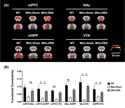 |
Therapy of Parkinson’s Disease Related Motor and Cognitive
Decline with Deep Brain Stimulation of Nucleus Accumbens
Ting-Chieh Chen1,
Ssu-Ju Li1,
Kai-Yun Chen2,
Yu-Chun Lo2,
Yi-Chen Lin1,
Ching-Wen Chang1,
Yao-Wen Liang1,
Tsai-Yu Cho1,
Mu-Hua Wang1,
Sheng-Huang Lin3,4,
and You-Yin Chen1,2
1Department of Biomedical Engineering, National Yang Ming Chiao Tung University, Taipei, Taiwan, 2PhD Program in Medical Neuroscience, College of Medical Science and Technology, Taipei Medical University, Taipei, Taiwan, 3Department of Neurology, Hualien Tzu Chi Hospital, Buddhist Tzu Chi Medical Foundation, Hualien, Taiwan, 4Department of Neurology, School of Medicine, Tzu Chi University, Hualien, Taiwan Keywords: Parkinson's Disease, Parkinson's Disease The progression of Parkinson′s disease (PD) is associated with mitochondrial dysfunction, and deep brain stimulation (DBS) of the nucleus accumbens (NAc) was found to enhance mitochondrial function. Therefore, the presented study aimed to explore the therapeutic effects of NAc-DBS on PD. In this study, the NAc-DBS treatment, behavioral tests, brain magnetic resonance imaging analysis, and mitochondrial respiratory assay were conducted on the MitoPark PD mouse model. After NAc-DBS, the PD mouse model showed improvements in motor and cognitive functions with increased functional connectivity and promoted aerobic metabolism in the dopaminergic (DA) pathways. |
| 16:16 |
1385.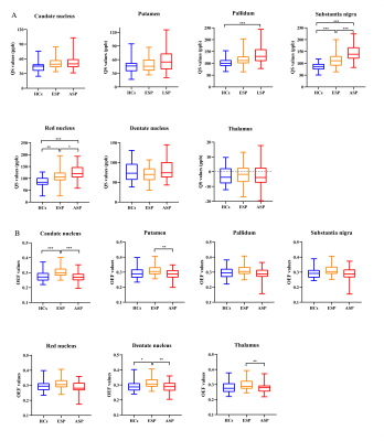 |
Spatiotemporal Patterns of Iron Accumulation and Oxygen
Metabolism in Patients with Parkinson's Disease
Su Yan1,
Jun Lu1,
Weiyin Vivian Liu2,
and Wenzhen Zhu1
1Department of Radiology, Tongji Hospital, Tongji Medical College, Huazhong University of Science and Technology, Wuhan, China, 2MR Research, GE Healthcare, Beijing, China Keywords: Parkinson's Disease, Quantitative Susceptibility mapping The purpose of this study was to employ QSM and OEF maps to quantitatively measure iron content and oxygen metabolism levels in deep gray matter of the Parkinson’ s disease brains in different stages and explore their relationship with clinical features. Iron accumulation in SN, RN and GP significantly increased with the progression of PD severity. In contrast, OEF in CAU, PT, and DN elevated in early PD and then gradually decreased with worsening symptoms, indicating a different spatiotemporal pattern. These findings may facilitate to understand the neurobiological mechanisms in PD progression and offered complementary indicators for diagnosis and prediction. |
| 16:24 |
1386.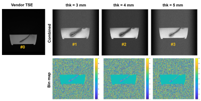 |
Signal recovery around DBS leads using 2D MSI
Gehua Tong1,
John Thomas Vaughan, Jr.1,2,
and Sairam Geethanath2,3
1Biomedical Engineering, Columbia University, New York, NY, United States, 2Columbia Magnetic Resonance Research Center, Columbia University, New York, NY, United States, 3Accessible MR Laboratory, BioMedical Engineering and Imaging Institute, Dept. of Diagnostic, Molecular and Interventional Radiology, Icahn School of Medicine at Mt. Sinai, New York, NY, United States Keywords: Parkinson's Disease, Pulse Sequence Design 2D MSI was tested for recovering the closest layer of off-resonant signals near a DBS lead. The effects of slice thickness and RF profile were measured in an ASTM gel phantom with an in-plane DBS lead. Increasing the slice thickness from 1.5x to 2.5x the lead diameter reduced the apparent lead width by 8.35% and increasing the time-bandwidth product of the RF pulse by four times improved SSIM with a reference TSE image by 88.5%. A trade off between bin definition and echo times limited signal recovery at a given bin bandwidth (800 Hz / 9 bins). |
| 16:32 |
1387.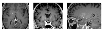 |
Enzyme Delivery to the Putamen in Parkinson’s Disease Patients
by MR-Guided Focused Ultrasound
Yuexi Huang1,
Ying Meng2,3,
Christopher B. Pople3,
Allison Bethune3,
Ryan M. Jones1,
Agessandro Abrahao3,4,
Clement Hamani2,3,
Suneil K. Kalia5,6,
Lorraine V. Kalia5,7,
Nir Lipsman2,3,
and Kullervo Hynynen1,8,9 1Physical Sciences, Sunnybrook Research Institute, Toronto, ON, Canada, 2Division of Neurosurgery, Sunnybrook Health Sciences Centre, Toronto, ON, Canada, 3Hurvitz Brain Sciences Research Program, Sunnybrook Research Institute, Toronto, ON, Canada, 4Department of Medicine, University of Toronto, Toronto, ON, Canada, 5Krembil Research Institute, University Health Network, Toronto, ON, Canada, 6Division of Neurosurgery, Toronto Western Hospital, Toronto, ON, Canada, 7Division of Neurology, Toronto Western Hospital, Toronto, ON, Canada, 8Department of Medical Biophysics, University of Toronto, Toronto, ON, Canada, 9Institute of Biomedical Engineering, University of Toronto, Toronto, ON, Canada Keywords: Parkinson's Disease, Focused Ultrasound The phase I clinical trial demonstrated the successful application of microbubble-assisted MR-guided Focused Ultrasound for blood-brain barrier (BBB) opening in the putamen to facilitate biweekly therapeutic drug delivery in patients with Parkinson's disease. BBB permeability within the targeted putamen was elevated successfully in all treatments, as revealed by Gd-enhanced T1-weighted MRI immediately post treatment. No contrast enhancement was observed in the treated putamen on MR imaging scans acquired one day following each treatment session, indicating closure of the BBB. FDG-PET revealed a reduction of glucose metabolism of the treated putamen relative to the contralateral putamen in all patients. |
|
16:40 |
1388.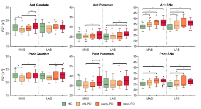 |
Nigrostriatal iron accumulation in Parkinson's disease
progression
Miguel López-Aguirre1,2,3,
Noelia Esteban-García1,4,
Tiziano Balzano1,
María Ciorraga1,
Javier Blesa1,3,
José A. Obeso1,3,5,
and José A. Pineda-Pardo1,3,5
1HM CINAC, Hospital Universitario HM Puerta del Sur, Móstoles, Spain, 2Universidad Complutense de Madrid, Madrid, Spain, 3Center for Networked Biomedical Research on Neurodegenerative Diseases (CIBERNED), Instituto de Salud Carlos III, Madrid, Spain, 4Universidad Autónoma de Madrid, Madrid, Spain, 5Universidad San Pablo-CEU, Madrid, Spain Keywords: Parkinson's Disease, Relaxometry, Iron Iron has an important role in Parkinson’s disease (PD) pathophysiology. However, it is still uncertain how iron accumulates within the nigrostriatal circuit along with PD progression. Here we assessed iron content from early to moderate PD stages using R2* relaxometry, and histologically using a macaque MPTP (1-metil-4-fenil6-tetrahidropiridina) model. Our analyses revealed that in both human PD and MPTP model, iron accumulates progressively within SNpc during early stages , reaching a plateau before moderate PD. Meanwhile, iron followed a V-shaped progression in the striatum. These results will contribute to improve our understanding of nigrostriatal vulnerability and the course of neurodegeneration in PD. |
16:48 |
1389.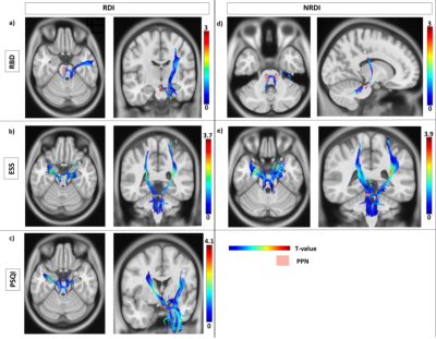 |
Non-motor Correlates of Pedunculopontine Nucleus Projection
Denervation in Parkinson’s Disease with Sleep Disturbances
Pohchoo Seow1,
Yao-Chia Shih2,
Septian Hartono3,
Pik Hsien Chai4,
Weiling Lee4,
Celeste Yan Teng Chen 3,
Eng King Tan3,
and Ling Ling Chan1
1Diagnostic Radiology, Singapore General Hospital, Singapore, Singapore, 2Graduate Institute of Medicine, Yuan-Ze University, Taoyuan City, Taiwan, 3Department of Neurology, National Neuroscience Institute, Singapore, Singapore, 4Radiography, Singapore General Hospital, Singapore, Singapore Keywords: Parkinson's Disease, Diffusion/other diffusion imaging techniques The mechanism by which involvement of the cholinergic pathways underlies the non-motor manifestations of Parkinsonian disorders remains unclear. We mapped and investigated the pedunculopontine nucleus (PPN) projections that exhibit significant correlations with sleep disturbances in 80 Parkinson’s disease (PD) patients and 110 healthy controls (HC) using correlational tractography. Intracellular and extracellular diffusivity (RDI and NRDI metrics respectively) demonstrated significant correlations (r=0.06-0.14, FDR<0.05) with sleep abnormalities in the PPN projections of PD compared to HC. Our findings established non-motor markers of PPN system denervation with sleep disturbances to further understand the pathophysiology of non-motor symptoms in PD. |
| 16:56 |
1390.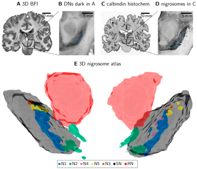 |
A 3D nigrosome atlas based on multi-modal histology and
multi-parametric quantitative MRI
Malte Brammerloh1,
Evgeniya Kirilina1,2,
Anneke Alkemade3,
Pierre-Louis Bazin1,3,
Caroline Jantzen1,
Sara Schaumberg1,
Carsten Jäger1,4,
Andreas Herrler5,
Kerrin J. Pine1,
Markus Morawski1,4,
Birte Forstmann3,
and Nikolaus Weiskopf1,6
1Department of Neurophysics, Max Planck Institute for Human Cognitive and Brain Sciences, Leipzig, Germany, 2Center for Cognitive Neuroscience Berlin, Freie Universität Berlin, Berlin, Germany, 3Integrative Model-based Cognitive Neuroscience Research Unit, University of Amsterdam, Amsterdam, Netherlands, 4Paul Flechsig Institute - Center of Neuropathology and Brain Research, Leipzig University, Leipzig, Germany, 5Department of Anatomy and Embryology, Maastricht University, Maastricht, Netherlands, 6Felix Bloch Institute for Solid State Physics, Leipzig University, Leipzig, Germany Keywords: Parkinson's Disease, Multimodal, Atlas, Subcortex MRI holds great promise to unravel the selective vulnerability and functional differentiation of the nigrosomes in the human substantia nigra pars compacta. Based on block-face imaging and calbindin-D28K immunohistochemistry, we constructed a 3D nigrosome atlas. Using this atlas, we demonstrate several nigrosomes to show increased R2* values in post mortem tissue. Our results further challenge the common identification of the nigrosomes with hyperintense structures in in vivo MRI, particularly nigrosome 1 and the swallow tail sign. |
| 17:04 |
1391.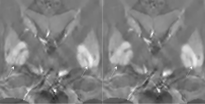 |
Improved Visualization of the Medial Medullary Lamina with Phase
Prior Reconstruction in Quantitative Susceptibility Mapping
Alexandra Grace Roberts1,2,
Ilhami Kovanlikaya2,
Brian Koppel3,
Pascal Spincemaille2,
Thanh Nguyen2,
and Yi Wang1,2
1Electrical Engineering, Cornell University, Ithaca, NY, United States, 2Radiology, Weill Cornell Medicine, New York, NY, United States, 3Radiology, Mount Sinai Hospital, New York, NY, United States Keywords: Parkinson's Disease, Brain Morphology Enabled Dipole Inversion (MEDI) is an iterative reconstruction algorithm for Quantitative Susceptibility Mapping (QSM) that is effective in suppressing artifacts by exploiting the magnitude image as a morphological prior. Use of a phase prior such as the local field to determine the $$$L_1$$$ regularization term results in improved visualization of the medial medullary lamina as measured by the contrast-to-noise ratio and was preferred by a radiologist over the magnitude prior in each case. |
| 17:12 |
1392.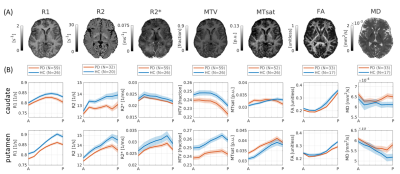 |
Multiparametric qMRI and dMRI gradients in the striatum are
associated with Parkinson’s disease motor dysfunction
Elior Drori1,
Lee Cohen1,
Einat Kohn2,
David Arkadir3,
Gilad Yahalom2,
and Aviv A Mezer1
1The Edmond and Lily Safra Center for Brain Sciences, The Hebrew University of Jerusalem, Jerusalem, Israel, 2The Department of Neurology, Shaare Zedek Medical Center, Jerusalem, Israel, 3The Department of Neurology, Hadassah Medical Center, The Hebrew University of Jerusalem, Jerusalem, Israel Keywords: Parkinson's Disease, Quantitative Imaging, striatum Parkinson’s disease (PD) is associated with degeneration in the striatum, a deep brain structure. It was recently shown with clinical MRI that in vivo gradients in the striatum of PD patients are related to the early-stage symptoms and pathology. However, the spatial variation in quantitative biophysical properties in PD is still unknown. Here, we collected multiparametric quantitative MRI of PD patients. We reveal distinct spatial profiles in the striatum that corroborate previous findings and show a relationship between putamen water fraction and motor behavior. Thus, our study provides new insights for the biological mechanisms associated with PD behavior. |
| 17:20 |
1393.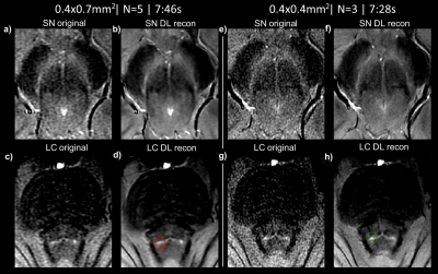 |
Neuromelanin MRI using 2D GRE and deep learning: considerations
for improving the visualization of substantia nigra and locus
coeruleus
Samy Abo Seada1,
Anke W. van der Eerden1,
Agnita J.W. Boon2,
and Juan Antonio Hernandez-Tamames1,3
1Radiology and nuclear medicine, Erasmus MC, Rotterdam, Netherlands, 2Department of Neurology, Erasmus MC, Rotterdam, Netherlands, 3Department of Imaging Physics, TU Delft, Delft, Netherlands Keywords: Parkinson's Disease, Normal development An optimized clinically feasible neuromelanin MRI imaging protocol for visualising the SN and LC simultaneously using deep learning reconstruction is presented. For a 2D sequence we set out to optimize the flip-angle for optimal combined SN and LC depiction. We also experimented with combinations of anisotropic and isotropic in-plane resolution, partial vs full echoes and the number of averages. Phantom and in-vivo experiments on three healthy volunteers illustrate that high-resolution imaging combined with deep-learning denoising shows good depiction of the SN and LC with a clinically feasible sequence of 7 minutes. |
| 17:28 |
1394.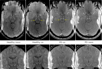 |
Visualizing Neuromelanin in Parkinson’s disease in the presence
of motion
Mikael Skorpil1,
Henric Rydén2,
Per Svenningsson2,
and Adam van Niekerk2 1Department of Molecular Medicine and Surgery, Karolinska Institutet, Stockholm, Sweden, 2Department of Clinical Neuroscience, Karolinska Institutet, Stockholm, Sweden Keywords: Parkinson's Disease, Motion Correction Neuromelanin-MRI can detect reductions in volume and intensity of the substantia nigra in Parkinson’s disease (PD). High-resolution magnetization transfer (MT) contrast T1-weighted sequences are used, and the MT pulse is time-consuming. We evaluated the degree of motion during a long (~ 12 min) scan in a PD patient and a healthy volunteer, and the effect of motion correction using a wireless RF-triggered acquisition device (WRAD) developed in our group. Despite only minor motion, for both participants, motion correction improved image quality. |
| 17:36 |
1395.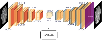 |
Robustness of Autoencoder-based Classifier for fMRI-based
Optimization of Single-sided Deep Brain Stimulation
Afis Ajala1,
Jianwei Qiu1,
John Karigiannis1,
Brendan Santyr2,
Jurgen Germann2,
Alexandre Boutet2,
Luca Marinelli1,
Chitresh Bhushan1,
Radhika Madhavan1,
Desmond Yeo1,
and Andres Lozano2
1GE Global Research, Niskayuna, NY, United States, 2University Health Network and University of Toronto, Toronto, ON, Canada Keywords: Parkinson's Disease, fMRI Successful treatment of Parkinson’s disease using deep brain stimulation (DBS) of the sub-thalamic nucleus (STN) requires an optimal set of DBS parameters that involves time-consuming programming sessions (~1 year) by the current standard-of-care optimization protocol. Functional magnetic resonance imaging (fMRI) and deep learning with autoencoder-based feature extraction from DBS-fMRI responses have provided a way to rapidly optimize the DBS parameters. In this work, we examine the robustness of the unsupervised autoencoder-based feature extraction method to changes in the activation patterns of the DBS-fMRI responses, which may be caused by patient motion, difference in stimulation side and disease condition. |
| 17:44 |
1396.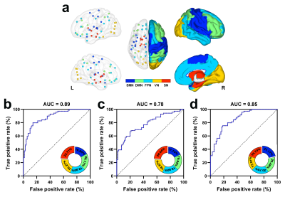 |
Optimization of structural connectomes and scaled patterns of
structural-functional decoupling in Parkinson’s disease
Song'an Shang1,
Weiqiang Dou2,
and Ye Jing1
1Clinical Medical College, Yangzhou University, Yangzhou, China, 2GE Healthcare, MR Research China, Beijing, China Keywords: Parkinson's Disease, Neurodegeneration Parkinson’s disease (PD) is manifested with disrupted topology of structural connection network (SCN) and functional connection network (FCN). However, SCN and its interactions with FCN remain to be further investigated. This multimodality study attempted to precisely characterize the SCN using diffusion kurtosis imaging (DKI) and further identify the neuropathological pattern of SCN-FCN decoupling. Our study verified the optimization of SCN by applying DKI metrics and its implications for the clinical diagnosis of PD. Moreover, SCN-FCN decoupling is consequent from mismatched disruptions of the SCN and FCN, identifying pathophysiological neuroimaging features for disturbed neural circuits in PD. |
| 17:52 |
1397.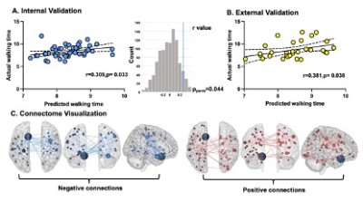 |
Functional connectome allows individual prediction of gait
function within Parkinson’s disease and is linked to molecular
architecture
Haoting Wu1 and
Minming Zhang1
1Radiology, Medical school of Zhejiang university, Hangzhou, China Keywords: Parkinson's Disease, fMRI (resting state) The predictive connectome of PD gait overlapped with the dopaminergic and cholinergic systems. |
 Back to Meeting Home
 Back to the Program-at-a-Glance
The International Society for Magnetic Resonance in Medicine is accredited by the Accreditation Council for Continuing Medical Education to provide continuing medical education for physicians.
 Back to Meeting Home
 Back to the Program-at-a-Glance
 View Presentation Video