Oral Session
Functional & Quantitative Imaging of the Spinal Cord
ISMRM & ISMRT Annual Meeting & Exhibition • 03-08 June 2023 • Toronto, ON, Canada

Oral Session
Functional & Quantitative Imaging of the Spinal Cord
| 13:30 |
0559.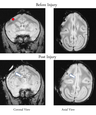 |
Alterations in intrinsic functional networks in squirrel monkey
spinal cord after focal damage to primary motor cortex using
resting state fMRI
Anirban Sengupta1,
Pai-Feng Yang1,
Jamie L Reed1,
Arabinda Mishra1,
Feng Wang1,
Li Min Chen1,
and John C Gore1 1Vanderbilt University Medical Center, Nashville, TN, United States Keywords: Spinal Cord, fMRI (resting state), Functional Connectivity The goal of this study was to detect and quantify how functional connectivity within networks in squirrel monkey spinal cord changes after a focal lesion of the primary motor cortex. We first used independent component analysis to identify 7 spatially independent functional hubs within each segment of the cervical spinal cord. The overall connectivity of these spinal cord networks increased after a lesion within M1, indicating plastic changes in spinal cord due to cortical injury. Distinct functional communities confined to neighboring spinal cord segments were identifiable one week after the injury, confirming spinal cord functional networks reorganized over that time. |
| 13:38 |
0560.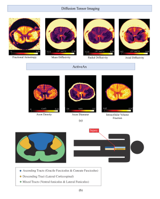 |
Diffusion Tensor Imaging and ActiveAx analysis of Post-Mortem
Human Cervical Spinal Cord Injury
Nikolai I Lesack1,2,3,
Sarah Rosemary Morris1,2,3,
Taylor Swift-LaPointe2,
Andrew Yung1,3,4,
Valentin Prevost1,3,4,
Shana George5,
Andrew Bauman4,
Piotr Kozlowski1,2,3,4,
Farah Samadi1,5,
Caron Fournier1,5,
Lisa Parker6,
Kevin Dong1,
Femke Streijger1,
Veronica Hirsch-Reinshagen1,5,6,
G. R. Wayne Moore1,5,6,
Brian Kwon1,7,
and Cornelia Laule1,2,3,5 1International Collaboration on Repair Discoveries, Vancouver, BC, Canada, 2Physics & Astronomy, University of British Columbia, Vancouver, BC, Canada, 3Radiology, University of British Columbia, Vancouver, BC, Canada, 4UBC MRI Research Centre, University of British Columbia, Vancouver, BC, Canada, 5Pathology & Laboratory Medicine, University of British Columbia, Vancouver, BC, Canada, 6Vancouver General Hospital, Vancouver, BC, Canada, 7Orthopaedics, University of British Columbia, Vancouver, BC, Canada Keywords: Spinal Cord, Diffusion/other diffusion imaging techniques, trauma, spinal cord injury, post-mortem, wallerian degeneration, axons, ActiveAx, DTI 7T Diffusion Tensor Imaging (DTI) and ActiveAx analysis techniques were used to probe the microstructural properties of post-mortem human spinal cord injury tissue. A decrease in DTI fractional anisotropy was observed caudal to (below) the injury epicenter for descending tracts and rostral to (above) the injury epicenter for ascending tracts. All cords displayed increased axon diameter in ascending tracts rostral to the injury site, which may be evidence of axonal swelling. A decrease in axon density for the two subjects with the longest injury-to-death interval may indicate the time scale of Wallerian degeneration-induced axonal damage observable using ActiveAx. |
| 13:46 |
0561.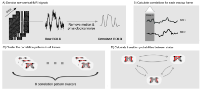 |
Sliding-Window Dynamic Functional Connectivity Of Resting-State
Spinal Cord fMRI Reveals Specific Interaction Patterns
Sukru Samet Dindar1,
Ilaria Ricchi2,3,
Nawal Kinany2,3,
and Dimitri Van De Ville2,3 1Electrical and Electronics Engineering, Bogazici University, Istanbul, Turkey, 2Neuro-X Institute, Ecole Polytechnique Fédérale de Lausanne (EPFL), Geneva, Switzerland, 3Department of Radiology and Medical Informatics, University of Geneva, Geneva, Switzerland Keywords: Spinal Cord, fMRI (resting state) In recent years, while the exploration of spontaneous brain activity has shifted from static methods (i.e., examining average connectivity patterns over the entire run) towards dynamic approaches (i.e., accounting for the non-stationarity of resting-state fluctuations), the non-stationary nature of intrinsic activity in the spinal cord has seldom been studied. Here, we propose to probe time-varying functional connectivity patterns in the spinal cord using a sliding-window correlation approach, as commonly employed in the brain. Our results suggest the potential of this approach to unravel ventral-ventral and dorsal-dorsal correlation patterns, while also emphasizing their time-varying nature. |
13:54 |
0562.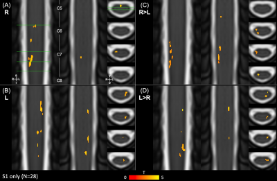 |
Spatial distribution of hand-grip motor task activity in spinal
cord fMRI
Kimberly J. Hemmerling1,
Mark A. Hoggarth1,
Milap S. Sandhu2,
Todd B. Parrish1,
and Molly G. Bright1 1Northwestern University, Chicago, IL, United States, 2Shirley Ryan AbilityLab, Chicago, IL, United States Keywords: Spinal Cord, fMRI A unilateral isometric hand-grip task was used to elicit motor neuron activation in spinal cord fMRI. As predicted, activation was lateralized to the ipsilateral hemicord. However, active voxels were also observed outside of the ventral horn and superior to C7. Reduced spatial precision and sensitivity in activation estimates, particularly along the cord axis, may be due to the combination of inter-individual differences in anatomy, imperfect registration to the standard spinal cord template, additional sensory activity associated with performing the task, and co-contraction of muscles not directly involved in hand-gripping. |
14:02 |
0563.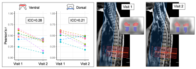 |
Intra- and inter-session reliability of lumbar spinal cord
resting-state fMRI
Anna JE Combes1,2,
Grace Sweeney1,
Delaney Houston1,
Logan Prock1,
Lipika Narisetti1,
Baxter P Rogers1,2,
Colin D McKnight2,
John C Gore1,2,3,
Seth A Smith1,2,3,
and Kristin P O'Grady1,2,3 1Vanderbilt University Institute of Imaging Science, Vanderbilt University Medical Center, Nashville, TN, United States, 2Radiology & Radiological Sciences, Vanderbilt University Medical Center, Nashville, TN, United States, 3Biomedical Engineering, Vanderbilt University Medical Center, Nashville, TN, United States Keywords: Spinal Cord, fMRI (resting state) We investigated the intra- and inter-session reliability of functional connectivity indices derived from resting-state fMRI in the lumbar spinal cord. Slicewise and average indices of connectivity for the ventral and dorsal networks were collected across three experiments in healthy participants at 3T. Intra-session reliability was high for back-to-back scans (ICC>0.9), and moderate-to-good when introducing repositioning, surveying and reshimming (COV=3-25%). Repeatability of indices across separate visits was low, although similar grey matter functional networks could be independently observed and showed high spatial overlap using ICA, a data-driven approach. This is the first evaluation of resting-state fMRI reliability in the lumbar cord. |
| 14:10 |
0564.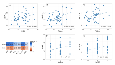 |
Neurodegeneration within upper spinal cord is associated with
brain gray matter volume atrophy in early stage of cervical
spondylotic myelopathy
Cuili Kuang1,
Yunfei Zha1,
and Weiyin Vivian Liu2
1Renmin Hospital of Wuhan University, Wuhan, China, 2GE Healthcare, MR Research, Beijing, China Keywords: Spinal Cord, Neurodegeneration, Cervical Spondylotic Myelopathy, Spinal Cord Toolbox Associations between cross-sectional area and white matter area within the rostral spinal cord and gray matter volume in left supplementary motor area and right middle cingulate gyrus suggest a concordant change pattern and adaptive mechanisms for neuronal plasticity underlying remote neurodegeneration in early cervical spondylotic myelopathy. The atrophy of cross-sectional area within the upper spinal cord and gray matter volume loss in right postcentral gyrus can serve as potential neuroimaging biomarkers of early structural changes within spinal cord and brain preceding marked clinical disabilities in patients with cervical spondylotic myelopathy. |
| 14:18 |
0565.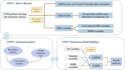 |
Predictive Value of Diffusion Magnetic Resonance Imaging for the
Postoperative Outcome of Cervical Spondylotic Myelopathy
Ming Ni1,
Xiaoyi Wen2,
Mengze Zhang1,
Chenyu Jiang1,
Yali Li1,
Xianchang Zhang3,
Ning Lang1,
Qiang Zhao1,
Yuqing Zhao1,
Wen Chen1,
Liang Jiang4,
and Huishu Yuan1 1Department of Radiology, Peking University Third Hospital, BeiJing, China, 2Institute of Statistics and Big Data, Renmin University of China, BeiJing, China, 3MR Collaboration, Siemens Healthineers Ltd., BeiJing, China, 4Department of Orthopedics, Peking University Third Hospital, BeiJing, China Keywords: Spinal Cord, Diffusion/other diffusion imaging techniques This study used multi-factorial linear quantile mixed-effects regression models to predict the outcome of cervical spondylotic myelopathy (CSM) patients one year after surgery based on MRI. Six models were constructed using the linear quantile mixed model and linear mixed-effects regression model based on the diffusion magnetic resonance imaging (dMRI) data, all the imaging data (dMRI & Conventional MRI), and all the registered data (dMRI & Conventional MRI & clinical data). We found that fractional anisotropy (FA) values quantified by preoperative dMRI could predict the surgical outcome of CSM and showed a significant positive correlation with the postoperative outcome. |
| 14:26 |
0566.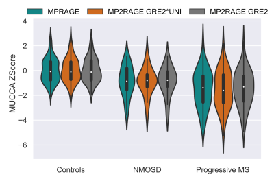 |
Automated cervical spinal cord atrophy detection from
conventional 3D T1w brain MRI
Jonathan A. Disselhorst1,2,3,
Michaela Andělová4,
Veronica Ravano1,2,3,
Gian Franco Piredda1,5,6,
Tobias Kober1,2,3,
Manuela Vaněčková7,
and Bénédicte Maréchal1,2,3 1Advanced Clinical Imaging Technology, Siemens Healthineers International AG, Lausanne, Switzerland, 2Department of Radiology, Lausanne University Hospital and University of Lausanne, Lausanne, Switzerland, 3LTS5, École Polytechnique Fédérale de Lausanne (EPFL), Lausanne, Switzerland, 4Department of Neurology and Center of Clinical Neuroscience, First Faculty of Medicine, Charles University and General University Hospital, Prague, Czech Republic, 5Human Neuroscience Platform, Fondation Campus Biotech Geneva, Geneva, Switzerland, 6CIBM-AIT, École Polytechnique Fédérale de Lausanne (EPFL), Lausanne, Switzerland, 7Department of Radiology, First Faculty of Medicine, Charles University and General University Hospital, Prague, Czech Republic Keywords: Spinal Cord, Segmentation, atrophy Detecting atrophy in the spinal cord (SC) is highly relevant in multiple sclerosis (MS) and other neuroinflammatory and neurodegenerative diseases. The most used method is measuring mean upper cervical cord area (MUCCA) from SC images. MRI of SC is time consuming and not always available. We recently developed a method to measure MUCCA directly from conventional T1w brain images. This work compares MUCCA estimates derived from MPRAGE and MP2RAGE sequences, determine reference ranges from 98 healthy subjects and show their value to detect atrophy in patients with progressive MS and neuromyelitis optica. We observe very high agreement between methods. |
| 14:34 |
0567.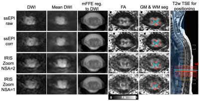 |
Application of multi-shot EPI to mitigate image distortion in
diffusion tensor imaging of the human lumbar spinal cord
Kristin P. O'Grady1,2,3,
Caroline Seehorn1,
Grace Sweeney1,
Logan Prock1,
Delaney Houston1,
Anna J.E. Combes1,
Kurt G. Schilling1,2,
Colin D. McKnight2,
Ryan K. Robison1,2,4,
and Seth A. Smith1,2,3
1Vanderbilt University Institute of Imaging Science, Vanderbilt University Medical Center, Nashville, TN, United States, 2Department of Radiology and Radiological Sciences, Vanderbilt University Medical Center, Nashville, TN, United States, 3Department of Biomedical Engineering, Vanderbilt University, Nashville, TN, United States, 4Philips, Nashville, TN, United States Keywords: Spinal Cord, Diffusion Tensor Imaging The lumbar spinal cord is significantly understudied with quantitative MRI methods such as diffusion tensor imaging (DTI), in part due to challenges of spatial and temporally-varying field inhomogeneities that cause distortion in commonly used single-shot EPI sequences. In this pilot study, we implemented IRIS Zoom, a multi-shot, reduced field-of-view method that corrects for shot-to-shot variations, phase errors (2D navigation), and T2* dephasing. Multi-shot EPI was successfully acquired in healthy volunteers at 3T, led to reduced geometric distortions, and provided quantitative DTI values comparable to those derived from single-shot EPI. Multi-shot EPI is feasible for high-resolution DTI of the lumbar cord. |
| 14:42 |
0568.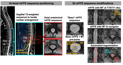 |
Effects of MT-weighting, respiratory navigation, and biological
variables on multi-echo gradient echo signal and contrast in the
lumbar cord
Kristin P. O'Grady1,2,3,
Kritin Vasamreddy1,
Grace Sweeney1,
Logan Prock1,
Delaney Houston1,
Anna J.E. Combes1,
Trey McGonigle4,
Simon Vandekar4,
Margareta Clarke1,
Atlee A. Witt1,
Colin D. McKnight2,
Erik Landman2,
Ryan K. Robison1,2,5,
Francesca Bagnato6,
and Seth A. Smith1,2,3
1Vanderbilt University Institute of Imaging Science, Vanderbilt University Medical Center, Nashville, TN, United States, 2Department of Radiology and Radiological Sciences, Vanderbilt University Medical Center, Nashville, TN, United States, 3Department of Biomedical Engineering, Vanderbilt University, Nashville, TN, United States, 4Department of Biostatistics, Vanderbilt University Medical Center, Nashville, TN, United States, 5Philips, Nashville, TN, United States, 6Department of Neurology, Vanderbilt University Medical Center, Nashville, TN, United States Keywords: Spinal Cord, Multiple Sclerosis, lumbar spinal cord Improvements are needed in advanced anatomical imaging of the lumbar spinal cord. We implemented a T2*-weighted multi-echo gradient echo (mFFE) sequence that enables visualization of CSF, gray and white matter with an in-plane resolution of 0.65x0.65mm2. Use of an MT-weighted pre-pulse and a respiratory navigator decreased SNR, but did not affect CNR, and qualitatively improved image quality and possibly multiple sclerosis lesion conspicuity. CNR/SNR were robust to biological factors known to affect signal quality (e.g. age, weight). The proposed sequence holds potential for improved visualization of anatomy, pathology, and morphometric measurements in the lumbar cord. |
| 14:50 |
0569.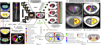 |
AMU7T: a 3D qT1 and T2*w high-resolution in vivo template with
refined white and gray matter parcellation dedicated to 7T
spinal cord MR analyses
Arnaud Le Troter1,2,
Nilser J. Laines Medina 1,2,3,
Samira Mchinda1,2,3,
Julien Cohen-Adad4,
and Virginie Callot1,2,3 1Aix Marseille Univ, CNRS, CRMBM, Marseille, France, 2APHM, CHU Timone, Pôle d’Imagerie Médicale, CEMEREM, Marseille, France, 3iLab-Spine, International Associated Laboratory, Marseille-Montreal, France, 4NeuroPoly Lab, Institute of Biomedical Engineering, Polytechnique Montréal, Montréal, QC, Canada Keywords: Spinal Cord, Software Tools Ultra-High-Field MRI has opened new perspectives for spinal cord exploration due to improved spatial resolution and contrast. The present work proposes a dedicated 7T multimodal 3D qT1 and T2*w template and a parcellation including eight substructures within gray matter, thirty WM tracts and three inter-hemispheric ROIs, for an accurate atlas-based segmentation in the subject space. This atlas was interpolated in the 3D PAM50 space to benefit from the advanced functions for registration implemented in the SCT. A preliminary segmentation result in healthy subject gives promising perspectives for group studies. |
| 14:58 |
0570.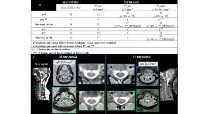 |
Spinal cord MS lesion detection at 7T - Added value of MP2RAGE
imaging and requirement for automatic lesion segmentation
Nilser LAINES MEDINA1,2,3,
Benoit TESTUD1,2,4,
Sarah DEMORTIERE5,
Samira MCHINDA1,2,3,
Arnaud LE TROTER1,2,
and Virginie CALLOT1,2,3 1Aix Marseille Univ, CNRS, CRMBM, Marseille, France, 2APHM, Hôpital Universitaire Timone, CEMEREM, Marseille, France, 3iLab-Spine, International Associated Laboratory, Montreal, Canada, Marseille, France, France, 4APHM, Hôpital Universitaire Timone, Department of Neuroradiology, Marseille, France, 5APHM, Hôpital Universitaire Timone, Department of Neurology, Marseille, France Keywords: Spinal Cord, Multiple Sclerosis In this exploratory study focusing on MP2RAGE and MS lesions, we demonstrated the added value of 7T imaging for lesion detection, with +20-to-30% of additional lesions seen compared to 3T. We also demonstrated that both currently available automatic segmentation model in SCT and an exploratory model build and trained based on MP2RAGE 3T images were not optimal for detecting lesions in 7T images (although a small contribution with slightly higher PPV was obtained with our model). As 7T opens great perspectives, further study should now focus on specific training and analysis tools to deal with 7T resolution and contrasts. |
| 15:06 |
0571.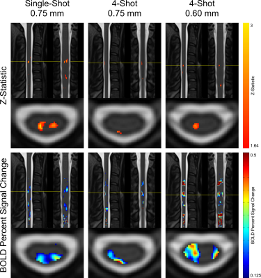 |
7T fMRI in the Cervical Spinal Cord Under Noxious Thermal
Stimulation
Alan C Seifert1,2,3 and
S Johanna Vannesjo4 1Biomedical Engineering and Imaging Institute, Icahn School of Medicine at Mount Sinai, New York, NY, United States, 2Department of Radiology, Icahn School of Medicine at Mount Sinai, New York, NY, United States, 3Graduate School of Biomedical Sciences, Icahn School of Medicine at Mount Sinai, New York, NY, United States, 4Department of Physics, Norwegian University of Science and Technology (NTNU), Trondheim, Norway Keywords: Spinal Cord, Spinal Cord FMRI of the spinal cord is challenging due to its small size, necessitating high resolution. Increasing B0 to 7T enables higher spatial resolution, and also enhances BOLD signal. However, challenges related to B0 homogeneity complicate spinal cord fMRI at 7T. We present group-level stimulus task fMRI results in the spinal cord at 7T, and compare the performance of single-shot and multi-shot 2D EPI protocols. Single-shot at 0.75mm in-plane resolution was most sensitive to activation, while multi-shot at 0.60mm provided the best-localized clusters. The best choice of protocol depends on the importance of sensitivity versus spatial localization for a given experiment. |
| 15:14 |
0572.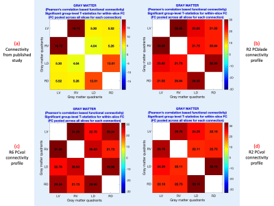 |
Realizing sub-second and sub-millimeter spinal cord fMRI at 7
Tesla
D Rangaprakash1 and
Robert L Barry1
1Athinoula A. Martinos Center for Biomedical Imaging, Massachusetts General Hospital, Harvard Medical School, Charlestown, MA, United States Keywords: Spinal Cord, fMRI (resting state) Spinal cord fMRI is an emerging field with clinical potential. Sub-millimeter in-plane fMRI acquisitions are desirable and achievable, but published studies have had modest temporal resolutions (>2s). Using a custom-built 7T spine coil, we demonstrate sub-second and sub-millimeter cervical cord fMRI for the first time. Employing a 3D multi-shot sequence with appropriate phase corrections and NORDIC denoising, our data demonstrated temporal signal-to-noise ratios similar to those of standard supra-second protocols, and we replicated functional connectivity patterns previously published in the cord. This opens new avenues of discovery similar to those realized through high spatiotemporal resolution brain fMRI. |
| 15:22 |
0573.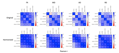 |
Harmonization of Spinal Cord Diffusion Tensor Imaging Data from
a Multi-Site, Multi-Scanner Study using Longitudinal ComBat
Devon M Middleton1,
Yutong Li2,
Andrew Chen3,
Russell T Shinohara3,
John H Woo3,
Adam E Flanders1,
Scott H Faro1,
Laura Krisa2,
and Feroze B Mohamed1
1Radiology, Thomas Jefferson University, Philadelphia, PA, United States, 2Thomas Jefferson University, Philadelphia, PA, United States, 3University of Pennsylvania, Philadelphia, PA, United States Keywords: Spinal Cord, Diffusion Tensor Imaging This study uses longitudinal ComBat data harmonization on a multi-site/multi-scanner spinal cord DTI study with a traveling cohort imaged on four scanners. The results show considerable improvement in correlation between scanners as well as decerased coefficient of variation. This approach has potential benefits for large, multi-site spinal cord studies. |
 Back to Meeting Home
 Back to the Program-at-a-Glance
The International Society for Magnetic Resonance in Medicine is accredited by the Accreditation Council for Continuing Medical Education to provide continuing medical education for physicians.
 Back to Meeting Home
 Back to the Program-at-a-Glance
 View Presentation Video