Oral Session
Brain Connectivity in Humans & Animals
ISMRM & ISMRT Annual Meeting & Exhibition • 03-08 June 2023 • Toronto, ON, Canada

Oral Session
Brain Connectivity in Humans & Animals
15:45 |
1024.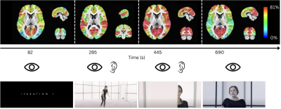 |
A Multi-Subject Deconvolution Algorithm for the Analysis of
Naturalistic fMRI data
Eneko Uruñuela1,
Clara Sava-Segal2,
Megan Leung2,
Emily S Finn2,
and César Caballero-Gaudes1
1Basque Center on Cognition, Brain and Language, Donostia - San Sebastián, Spain, 2Department of Psychological and Brain Sciences, Dartmouth College, Hanover, NH, United States Keywords: Brain Connectivity, fMRI, naturalistic paradigms Collecting fMRI data during naturalistic paradigms has drawn considerable attention in human neuroscience as a way to investigate brain function in ecologically valid conditions. We introduce a novel method (multi-subject paradigm free mapping) to decipher BOLD events in a temporally agnostic manner, and explore concordant group activations and individual idiosyncrasies. Besides, it can operate at the fastest temporal and spatial resolutions of the data. We validate it on simulated and real naturalistic fMRI data, revealing events that track expected features of the stimulus. Overall, this technique substantially increases sensitivity in linking moment-to-moment brain activity to its underlying cause(s). |
|
15:53 |
1025.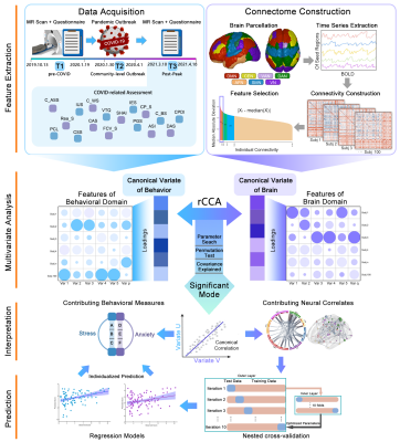 |
Multivariate Patterns of Brain Functional Connectome Underlying
COVID-related Negative Affect Symptoms
Nanfang Pan1,2,
Kun Qin1,2,
Song Wang1,
and Qiyong Gong1
1Department of Radiology, West China Hospital of Sichuan University, Chengdu, China, 2Department of Psychiatry and Behavioral Neuroscience, University of Cincinnati, CINCINNATI, OH, United States Keywords: Brain Connectivity, COVID-19 In the investigation of mental health issues following the unprecedented COVID-19 pandemic, we used the pre-COVID neuroimaging data to predict the severity of negative affect during the pandemic in a general population. Notably, covariation patterns of mode stress and mode anxiety were identified, and the brain DAN network plays a critical role in both modes. Based on individualized patterns, we may identify individuals who confer high vulnerability to pandemic-induced stress or anxiety symptoms. Our findings may facilitate the understanding of neural correlates underlying pandemic-induced negative affect, and the susceptibility neuromarkers may serve as targets for early prevention and psychological intervention. |
|
16:01 |
1026.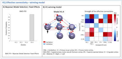 |
The cerebellum modulates non-linear behavior of motor planning
areas in variable grip-force visuomotor task
Roberta Maria Lorenzi1,
Gokce Korkmaz1,
Adnan Alahmadi2,
Letizia Casiraghi3,
Anita Monteverdi4,
Egidio D'Angelo1,4,
Fulvia Palesi1,
and Claudia A.M. Gandini Wheeler Kingshott1,4,5
1Department of Brain and Behavioral sciences, Università di Pavia, Pavia, Italy, 2Department of Diagnostic Radiology, College of Applied medical sciences, King Abdulaziz University, Jeddah, Saudi Arabia, 3Department of Mental Health and Dependence, ASST, Pavia, Italy, 4Brain Connectivity Center, IRCCS Mondino Foundation, Pavia, Italy, 5Department of Neuroinflammation, UCL Queen Square Institute of Neurology, NMR Research Unit, Queen Square Multiple Sclerosis Centre, London, United Kingdom Keywords: Brain Connectivity, fMRI (task based), Cerebellum The hemodynamic response to neuronal stimuli can be both linear and nonlinear with the applied grip-force of a “squeeze-ball” task, controlled by a visual cue. Dynamic Causal Modelling was applied here to understand the causal effective between-region connectivity shaping the hemodynamic response recorded by functional-MRI. Effective connectivity resulted non-symmetric, with strong excitation from visual to motor areas. Results indicated primary visual cortex linear modulation of the cerebellar response, which exerted nonlinear influence on cortical motor planning, suggesting activation hierarchy in the motor circuit with prominent cerebellar role. This result may have important implications for understanding pathological changes affecting neural mechanisms. |
| 16:09 |
1027.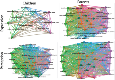 |
A Brain-Behavior Interaction Analysis for Dyadic Brain Responses
during Eye Contact between Parents and Children
Ray Lee1,
Joshua Friedman2,
Maria O'Brien3,
Zhihua Ren1,
Linbi Hong4,
Paul Sajda4,
and Nim Tottenham5 1Zuckerman Mind Brain Behavior Institute, Columbia University, New York, NY, United States, 2Teacher's College, Columbia University, New York, NY, United States, 3Teacher's College, Columbia Unviersity, New York, NY, United States, 4Biomedical Engineering, Columbia University, New York, NY, United States, 5Psychology, Columbia University, New York, NY, United States Keywords: Brain Connectivity, Neuroscience By combining hyperscan fMRI (hfMRI) and the facial action coding system (FACS), we introduce a new brain-behavior interaction analysis that can identify the underlying brain networks for perception and expression for different emotions during eye contact. It consists of three steps. 1) The affective facial muscle movements are encoded by facial action units (FAU) using FACS. 2) The specific emotional FAUs subserve the regressors for the univariant analysis on hfMRI data. 3) The brains’ activation maps are transformed into brain networks by multivariant analysis. It provides an approach to quantify affective interaction and emotional states. |
| 16:17 |
1028.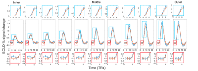 |
Examining the temporal evolution of top-down task dependent
modulations across cortical depths.
Luca Vizioli1,
Logan T Dowdle1,
Steen Moeller2,
Kamil Ugurbil1,
and Essa Yacoub1 1CMRR, University of Minnesota, Minneapolis, MN, United States, 2CMRR, University of Minnesota, Mineapolis, MN, United States Keywords: Brain Connectivity, Brain We demonstrate the feasibility of simultaneously acquiring submillimeter and subsecond functional images to examine the temporal evolution of task-dependent modulations across cortical depths elicited by socially relevant stimuli such as faces at the single subject level. We achieved this using NORDIC denoising. Our results highlight the complexity of laminar fMRI results in humans which can be better resolved by high spatiotemporal recordings. We suggest that, when studied with sufficient, sub-second temporal resolution, depth-dependent fMRI has the potential to address laminar communication within and across areas, which is crucial to understanding the complexity of human hierarchal organization at the mesoscale level. |
|
16:25 |
1029.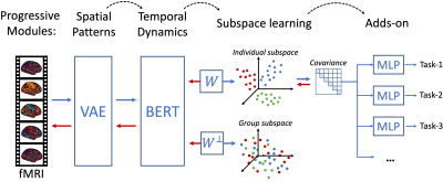 |
Individualized representation learning of resting-state fMRI
Kuan Han1,
Minkyu Choi1,
Xiaokai Wang1,
Amaya Murguia1,
and Zhongming Liu1
1University of Michigan, Ann Arbor, MI, United States Keywords: Brain Connectivity, fMRI (resting state) We describe a generalizable, modular and explainable model for individualized representation learning of resting-state fMRI. The model consists of a “deep” base which learns representations that are unique to each individual brain through self-supervised learning, and “shallow” adds-on which are trained with supervised learning for different tasks of behavior prediction. The model is scalable to allow some add-on modules to be trainable without affecting others, and is explainable to identify brain structures responsible for individualized behavioral prediction. |
| 16:33 |
1030.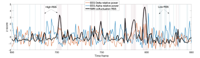 |
Electrophysiological correlates of BOLD events with high
cofluctuation amplitude in the resting human brain
Patricia Figueiredo1,
Inês Esteves1,
Ana Fouto1,
Amparo Ruiz-Tagle1,
Gina Caetano1,
and César Caballero-Gaudes2
1Institute for Systems and Robotics - Lisboa and Department of Bioengineering, Instituto Superior Técnico, University of Lisbon, Lisboa, Portugal, 2Basque Center on Cognition, Brain and Language, Donostia - San Sebastián, Spain Keywords: Brain Connectivity, fMRI (resting state) fMRI studies have shown that the large-scale organization of resting-state functional brain networks can be largely explained by a small fraction of events exhibiting high cofluctuation amplitude. However, their neurobiological relevance remains unclear. We investigated the electrophysiological origins of high cofluctuation amplitude BOLD events using concurrent EEG-fMRI data acquired from humans. We found that high amplitude cofluctuations were associated with higher delta power and lower alpha power. This association was specifically observed when considering delays ~6s between EEG and BOLD signals, supporting its neurovascular origin and suggesting that high cofluctuation BOLD events have a neurophysiological origin. |
| 16:41 |
1031.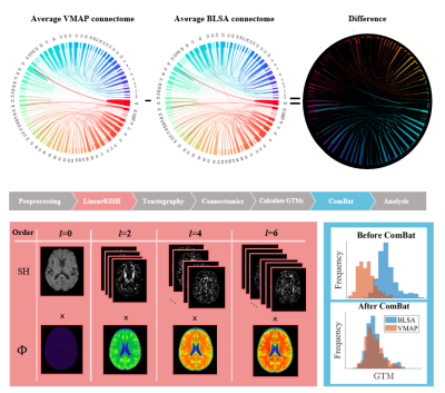 |
Comparing Techniques for Multi-Site Harmonization of Structural
Connectivity
Nancy Rose Newlin1,
Leon Cai2,
Derek Archer3,4,5,
Kimberly R Pechman3,
Kurt G Schilling6,
Angela Jefferson3,5,7,
Susan M Resnick8,
Timothy J Hohman3,4,5,
Andrea Shafer9,
and Bennett Landman2,5,6,10 1Computer Science, Vanderbilt University, Nashville, TN, United States, 2Biomedical Engineering, Vanderbilt University, Nashville, TN, United States, 3Vanderbilt Memory and Alzheimer's Center, Vanderbilt University Medical Center, Nashville, TN, United States, 4Vanderbilt Genetics Institute, Vanderbilt University School of Medicine, Nashville, TN, United States, 5Department of Neurology, Vanderbilt University Medical Center, Nashville, TN, United States, 6Department of Radiology and Radiological Sciences, Vanderbilt University Medical Center, Nashville, TN, United States, 7Department of Medicine, Vanderbilt University Medical Center, Nashville, TN, United States, 8Laboratory of Behavioral Neuroscience, National Institute on Aging, National Institutes of Health, Baltimore, MD, United States, 9Laboratory of Behavioral Neuroscience, National Institute on Aging, Baltimore, MD, United States, 10Vanderbilt University Institute of Imaging Science, Vanderbilt University, Nashville, TN, United States Keywords: Brain Connectivity, Diffusion/other diffusion imaging techniques Tractography is a method to reconstruct white matter microstructure from DWI information and connectomics maps this reconstruction to a graph representation. We compute modularity, assortativity, global efficiency, and average betweenness centrality on this graph. We model changes in these measures with age and sex using DWI for healthy patients from two sites. Data from different sites requires harmonization to remove site-effects. We compare performances of ComBat and LinearRISH harmonization techniques at reducing CoV and removing confounding site-effects from associated linear models. We find that ComBat is effective at both and using LinearRISH in addition acts synergistically at harmonizing site differences. |
|
16:49 |
1032.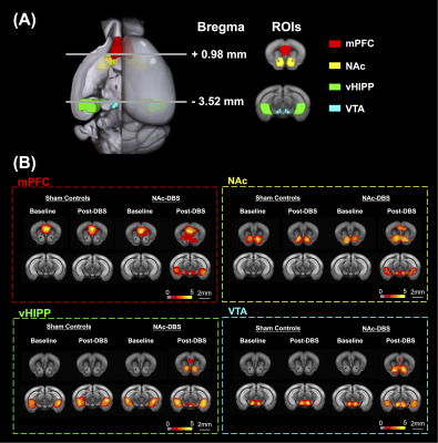 |
Deep Brain Stimulation of Nucleus Accumbens Alters Brain
Functional Connectivity and Metabolism to Enhance Memory-Related
Cognitive Function
Ssu-Ju Li1,
Ting-Chieh Chen1,
Yu-Chun Lo2,
Yi-Chen Lin1,
Ching-Wen Chang1,
Mu-Hua Wang1,
Tsai-Yu Cho1,
Sheng-Huang Lin3,4,
and You-Yin Chen1,2 1Department of Biomedical Engineering, National Yang Ming Chiao Tung University, Taipei City, Taiwan, 2PhD Program in Medical Neuroscience, Taipei Medical University, Taipei City, Taiwan, 3Department of Neurology, Buddhist Tzu Chi Medical Foundation, Hualien County, Taiwan, 4Department of Neurology, Tzu Chi University, Hualien County, Taiwan Keywords: Brain Connectivity, fMRI (resting state), deep brain stimulation, memory, cognition Deep brain stimulation (DBS) has been a well-established treatment for cognitive dysfunction. However, cognitive dysfunctions were demonstrated to be associated with the metabolic syndrome. Nucleus accumbens (NAc) in the dopaminergic pathway is associated with glucose metabolism, considered to be a promising DBS targeted region to be investigated. Resting-state functional MRI, behavioral test, bioenergetic analysis and electron microscopy were applied in this study. We found increased functional connectivity, enhancement in cognitive behavior, increased energy metabolism and mitochondrial biomass after NAc-DBS. |
| 16:57 |
1033.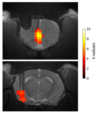 |
Zero echo time MB-SWIFT functional MRI of orientation selective
deep brain stimulation of the rat infralimbic cortex using
tetrahedral electrode
Hanne Laakso1,
Ekaterina Paasonen1,
Irina Gureviciene1,
Omar Narvaez1,
Lauri J Lehto1,
Jaakko Paasonen1,
Raimo Salo1,
Kestutis Gurevicius1,
Silvia Mangia2,
Shalom Michaeli2,
Heikki Tanila1,
Alejandra Sierra1,
and Olli Gröhn1 1A.I. Virtanen Institute, Kuopio, Finland, 2Center for Magnetic Resonance Research, Minneapolis, MN, United States Keywords: Brain Connectivity, fMRI In this study, the response of the amygdala to 3D orientation selective deep brain stimulation of the infralimbic cortex (IL) was followed using MB-SWIFT fMRI. The aims were to investigate how the fMRI response of the amygdala changes whilst changing the stimulation angle in the IL, and to detect the effects in resting-state functional connectivity when sustained 130 Hz stimulation of the IL at the most effective stimulation angle is used. The fMRI response of the amygdala showed dependence on the stimulation angle and inter-animal variation, which can be exploited for optimizing the stimulation effects. |
17:05 |
1034.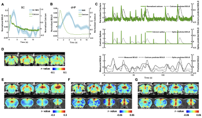 |
Gaining insight into the neural basis of resting-state fMRI
signal
Zilu Ma1,2,
Qingqing Zhang1,
Wenyu Tu3,
and Nanyin Zhang1,3
1Department of Biomedical Engineering, The Pennsylvania State University, University Park, PA, United States, 2National Institute on Drug Abuse, National Institutes of Health, Baltimore, MD, United States, 3The Huck Institutes of the Life Sciences, The Pennsylvania State University, University Park, PA, United States Keywords: Multimodal, fMRI (resting state), Awake, Calcium fiber photometry, BOLD Understanding the relationships between neuronal, vascular and Blood oxygenation level-dependent (BOLD) signals is essential for the appropriate interpretation of functional magnetic resonance imaging (fMRI) results in relation to the corresponding neuronal activity. In this study, we utilized multimodal imaging technique that concurrently measures BOLD fMRI and calcium-based fiber photometry signal to examine the relationship between BOLD and neural spiking activity in awake rats. Our results demonstrated significant correspondence between the BOLD and calcium signals at both evoked and resting state, suggesting critical role of spiking activity in the neural mechanism underlying BOLD signal. |
|
17:13 |
1035. |
Interrogation of resting-state effective connectivity by
whole-brain fMRI with optogenetic silencing
Hyun Seok Moon1,2,3,
Thanh Tan Vo1,2,3,
and Seong-Gi Kim1,2,3
1Center for Neuroscience Imaging Research, Institute for Basic Science (IBS), Suwon, Korea, Republic of, 2Department of Biomedical Engineering, Sungkyunkwan University, Suwon, Korea, Republic of, 3Department of Intelligent Precision Healthcare Convergence, Sungkyunkwan University, Suwon, Korea, Republic of Keywords: Brain Connectivity, fMRI Mapping resting-state effective connectivity by cortical optogenetic activation of inhibitory neurons in mice |
| 17:21 |
1036.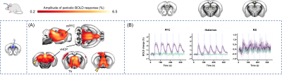 |
Optogenetic modulation of the mouse default mode network with a
single tapered fiber
Elizabeth de Guzman1,
Barbara Spagnolo2,
Filippo Pisano2,
Marco Pisanello2,
Alberto Galbusera1,
Luigi Balasco3,
Yuri Bozzi3,
Massimo De Vittorio2,4,
Tommaso Fellin5,
Ferruccio Pisanello2,
and Alessandro Gozzi1 1Functional Neuroimaging Lab, Istituto Italiano di Tecnologia, Rovereto, Italy, 2Center for Biomolecular Nanotechnologies, Istituto Italiano di Tecnologia, Arnesano (Lecce), Italy, 3Center for Mind/Brain Sciences (CiMEC), University of Trento, Rovereto, Italy, 4Dipartimento di Ingegneria dell’Innovazione, Università del Salento, Lecce, Italy, 5Optical Approaches to Brain Function Laboratory, Istituto Italiano di Tecnologia, Genova, Italy Keywords: Brain Connectivity, fMRI, preclinical, functional connectivity, neural oscillations, DMN The default mode network (DMN) is a distributed functional system of the human brain widely studied with fMRI due to its involvement in advanced cognitive processes and its dysregulation in a variety of brain disorders. Causal perturbations of this network in physiologically accessible species are critically required to probe its circuit organization and the underpinnings of its (dys)function. Here we show that tapered fiber optogenetic technology enables the reliable stimulation of key DMN nodes in a frequency dependent fashion, overcoming the limitations of traditional optogenetic approaches. |
| 17:29 |
1037.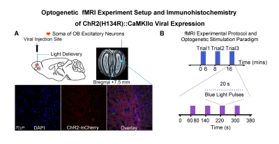 |
Optogenetic fMRI reveals neural adaptation properties beyond
local olfactory circuits
Teng Ma1,2,3,
Xunda Wang1,2,
Linshan Xie1,2,
Junjian Wen1,2,
Pit Shan Chong4,
Peng Cao3,
Lee Wei Lim4,
Ed X. Wu1,2,4,
and Alex T. L. Leong1,2
1Laboratory of Biomedical Imaging and Signal Processing, The University of Hong Kong, Hong Kong SAR, China, 2Department of Electrical and Electronic Engineering, The University of Hong Kong, Hong Kong SAR, China, 3Department of Diagnostic Radiology, Li Ka Shing Faculty of Medicine, The University of Hong Kong, Hong Kong SAR, China, 4School of Biomedical Sciences, Li Ka Shing Faculty of Medicine, The University of Hong Kong, Hong Kong SAR, China Keywords: Brain Connectivity, Neuroscience Olfactory adaptation due to repeated odor cues has been studied extensively by fMRI or electrophysiology studies in several primary olfactory regions (i.e., anterior olfactory nucleus, AON, and piriform cortex, Pir). However, the modulatory role of other primary olfactory regions (e.g., amygdala and entorhinal cortex) and their integrations with high-order olfactory regions during olfactory adaptation is likely underestimated due to the documented weak and unstable responses at regions beyond AON and Pir with conventional presentation of odor stimuli. Here, we deployed an optogenetic fMRI approach to improve sensitivity in detecting olfactory responses and examine their adaptation at the systems level. |
| 17:37 |
1038.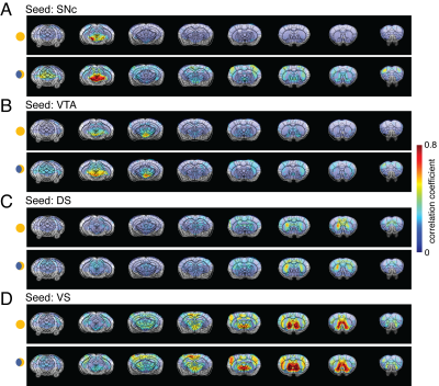 |
Circadian influence on brain connectivity revealed by
resting-state fMRI in awake mice
Muditha Bandara Rathnayaka1,
Rui Yang1,
Yeison Rodriguez1,
Janaka Wansapura1,
and Nan Li1 Circadian influence on brain connectivity revealed by resting-state fMRI in awake mice
Muditha Bandara Rathnayaka1,
Rui Yang1,
Yeison Rodriguez1,
Janaka Wansapura1,
and Nan Li1
1Advanced Imaging Research Center, University of Texas Southwestern Medical Center, Dallas, TX, United States Keywords: Brain Connectivity, f |
 Back to Meeting Home
 Back to the Program-at-a-Glance
 View Presentation Video