Oral Session
Imaging Inflammation Across Neurological Diseases
ISMRM & ISMRT Annual Meeting & Exhibition • 03-08 June 2023 • Toronto, ON, Canada

Oral Session
Imaging Inflammation Across Neurological Diseases
| 08:15 |
0024.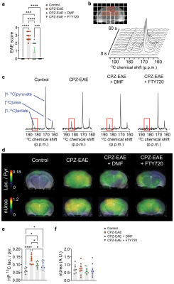 |
Hyperpolarized 13C MRSI detects immunomodulatory responses to
dimethyl fumarate and fingolimod therapies in a model of
multiple sclerosis
Caroline Guglielmetti1,2,
Christian Cordano3,
Chloe Najac4,
Ari Green3,
and Myriam Chaumeil1,2
1Department of Physical Therapy and Rehabilitation Science, University of California San Francisco, San Francisco, CA, United States, 2Department of Radiology and Biomedical Imaging, University of California San Francisco, San Francisco, CA, United States, 3Department of Neurology, University of California San Francisco, San Francisco, CA, United States, 4Department of Radiology, C.J. Gorter Center for High Field MRI, Leiden University Medical Center, Leiden, Netherlands Keywords: Multiple Sclerosis, Hyperpolarized MR (Non-Gas) We used hyperpolarized 13C MR spectroscopy imaging (MRSI) in a multiple sclerosis model and showed that we could monitor immune cell activation by measuring hyperpolarized [1-13C]pyruvate conversion to lactate. We further demonstrated that this approach detected response to two existing treatments, fingolimod and dimethyl fumarate. We observed a reduction of pyruvate-to-lactate flux after treatment, that can be explained by increased pyruvate dehydrogenase activity and decrease of immune cells. In addition, we evaluated brain perfusion using hyperpolarized [13C]urea, but saw no therapy effect. Altogether, we demonstrated that hyperpolarized 13C MRSI has potential to monitor immunomodulatory therapies within the central nervous system. |
| 08:23 |
0025.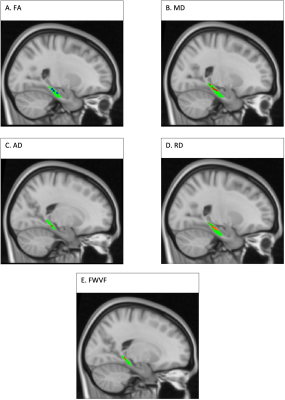 |
Free water diffusion volume fraction from NODDI suggests
inflammation may drive decreased memory performance in
Subjective Cognitive Decline
Ryn Flaherty1,
Yu Sui1,
and Mariana Lazar1
1Radiology, New York University Grossman School of Medicine, New York, NY, United States Keywords: Alzheimer's Disease, Neuroinflammation Evidence suggests subjective cognitive decline (SCD) is an early risk factor for Alzheimer’s disease. We analyzed the associations between memory and diffusion microstructure in the lower cingulum white matter bundle in SCD with DTI and NODDI. Better memory performance was associated with decreased free water volume fraction (FWVF) in the SCD group but not the control group. To the best of our knowledge, this is the first study to find differences in the associations between NODDI FWVF and memory in SCD in the lower cingulum. This finding supports prior findings of increased neuroinflammation in the earliest stages of Alzheimer’s Disease. |
| 08:31 |
0026. |
Multiple Sclerosis (MS): High Contrast Visualisation of
Abnormalities in Normal Appearing White Matter Using MASDIR
Sequences
Paul Condron1,2,
Haribalan Kumar 1,3,4,
Samantha Holdsworth1,2,
Daniel Cornfeld1,2,
and Graeme Bydder1,5
1Mātai Medical Research Institute, Gisborne, New Zealand, 2Univeristy of Auckland, Auckland, New Zealand, 3Auckland Bioengineering Institute, Auckland, New Zealand, 4GE Healthcare, Victoria, Australia, 5University of California San Diego, San Diego, CA, United States Keywords: Neuroinflammation, Multiple Sclerosis, neurodegeneration, MASDIR, Inversion Recovery (IR) sequences, tissue property filters Keywords; Neuroinflammation, Multiple Sclerosis, Neuro, Drugs. MASDIR sequences can show up to 15 times the contrast of T1-weighted MP-RAGE sequences. This is a consequence of using changes in T1 synergistically 3-4 times in the same sequence to detect subtle changes due to neuroinflammation and lesions. Extensive high contrast focal and diffuse abnormalities in white matter were demonstrated in areas that showed no abnormality with state-of-the-art T2-weighted spin echo and T2-FLAIR sequences in patients with multiple sclerosis. This demonstrated good correlation with advanced sequences such as T1 and myelin maps. |
| 08:39 |
0027. |
Neuroinflammation and amyloid deposition in the progression of
mixed Alzheimer and vascular dementia
Chunwei Ying1,
Peter Kang2,
Michael M. Binkley2,
Andria L. Ford1,2,
Yasheng Chen2,
Jason Hassenstab2,3,
Qing Wang1,3,
Jeremy Strain2,
John C. Morris3,
Jin-Moo Lee1,2,
Tammie L. S. Benzinger1,3,4,
and Hongyu An1,2
1Mallinckrodt Institute of Radiology, Washington University School of Medicine, St Louis, MO, United States, 2Department of Neurology, Washington University School of Medicine, St Louis, MO, United States, 3Knight Alzheimer Disease Research Center, Washington University School of Medicine, St Louis, MO, United States, 4Department of Neurosurgery, Washington University School of Medicine, St Louis, MO, United States Keywords: Dementia, Dementia Alzheimer's disease (AD) and vascular contributions to cognitive impairment and dementia (VCID) pathologies commonly coexist in community-dwelling elderly. It is not discernible whether neuroinflammation and amyloid beta (Aβ) deposition are distinct or entangled pathophysiological mechanisms in patients with mixed AD and VCID pathologies. In this study, we found that neuroinflammation (measured by 11C-PK11195 uptake) but not Aβ deposition (measured by 11C-PiB binding), contributes to white matter hyperintensities baseline volume and progression. Both neuroinflammation and Aβ deposition independently contribute to cognitive impairment progression. Neuroinflammation and Aβ deposition represent two distinct pathophysiological pathways in elderly participants with mixed AD and VCID pathologies. |
| 08:47 |
0028.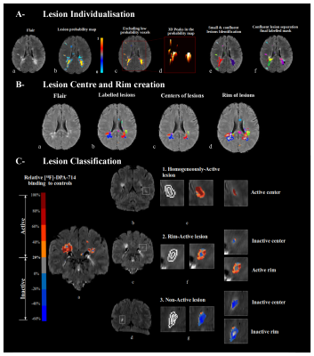 |
Positron emission tomography with [ 18F]-DPA-714 unveils a
smoldering component in most multiple sclerosis lesions driving
disease progression
Mariem Hamzaoui1,
Jeanne Garcia1,2,
Giacomo Boffa1,3,
Andrea Lazzarotto1,2,4,
Vito A G Ricigliano2,
Arya Yazdan Panah1,
Théodore Soulier1,
Celine Louapre1,
Benedetta Bodini1,2,
and Bruno Stankoff1,2
1Sorbonne Université, Paris Brain Institute, ICM, CNRS, Inserm, Paris, France, paris, France, 2Department of Neurology, Saint- Antoine Hospital, APHP, Paris, France, paris, France, 3Department of Neurology, Rehabilitation, Ophthalmology, Genetics, Maternal and Child Health, University of Genoa, Genoa, Italy, genoa, Italy, 4Padova Neuroscience Center, University of Padua, Padua, Italy, padua, Italy Keywords: Multiple Sclerosis, Neuroinflammation, [¹⁸F]-DPA-714, TSPO, Lesion individualization and phenotyping, disease progression Positron emission tomography with18kDa-translocator (TSPO) tracers opens the perspective to image innate immune cells underlying the smoldering component of multiple sclerosis (MS), that currently mostly escape from MRI evaluation. Using [18F]-DPA-714-PET, we developed a novel lesion TSPO based classification of MS lesions and showed that an unexpectedly high proportion have a persistent neuroinflammatory content. A longitudinal follow up of subjects unraveled that this lesional smoldering component predicted atrophy and clinical progression. Following the acute phase, most lesions may therefore develop a chronic inflammatory component which can persist for several years, subsequently promoting neurodegeneration and clinical progression in MS. |
| 08:55 |
0029.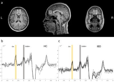 |
Disruption to normal excitatory and inhibitory function within
the medial prefrontal cortex in patients with IBD
Jun Wang1,2,
Guangyao Liu2,3,
Kai Ai4,
Pengfei Zhang1,
Wenjing Huang1,
and Jing Zhang2,3
1Second Clinical School, Lanzhou University, Lanzhou, China, 2Department of Magnetic Resonance, Lanzhou University Second Hospital, Lanzhou, China, 3Gansu Province Clinical Research Center for Functional and Molecular Imaging, Lanzhou University Second Hospital, Lanzhou, China, 4Department of clinical science, Philips Healthcare, Xi’an, China Keywords: Neuroinflammation, fMRI The etiology of IBD complicated with anxiety, depression and other psychological comorbidities is still unclear. The purpose of this study was to quantify neurotransmitter (GABA and Glx) in the medial prefrontal cortex (mPFC) of IBD patients with MEGA-PRESS spectroscopy and explore its relationship with clinical scores. We found that mPFC GABA+ and Glx concentration were significantly decreased in IBD patients and were associated with abdominal pain and depressive symptoms separately, suggesting psychological comorbidities in IBD patients were related to mPFC neurotransmitter dysregulation. It provides a new explanation for the neuropsychological mechanism of IBD in the development of the disease. |
| 09:03 |
0030. |
Quantifying Cervical Spinal Cord Pathology of Multiple Sclerosis
Patients Using Advanced MRI
Osman Hatipoglu1,2,3,
Tsagkas Charidimos 1,2,3,4,
Mario Ocampo-Pineda1,2,3,
Lester Melie-Garcia1,2,3,
Matthias Weigel1,2,3,5,
Po-Jui Lu1,2,3,
Muhamed Barakovic1,2,3,
Julien Cohen-Adad6,7,8,
Ludwig Kappos2,3,
Jens Kuhle2,3,
and Cristina Granziera1,2,3
1Translational Imaging in Neurology (ThINk) Basel, Department of Biomedical Engineering, University Hospital Basel and University of Basel, Basel, Switzerland, 2Research Center for Clinical Neuroimmunology and Neuroscience Basel (RC2NB), University Hospital Basel and University of Basel, Basel, Switzerland, 3Neurologic Clinic and Policlinic, Departments of Head, Spine and Neuromedicine, University Hospital Basel, Basel, Switzerland, 4Translational Neuroradiology Section, National Institute of Neurological Disorders and Stroke, National Institutes of Health (NIH), Bethesda, MD, United States, 5Division of Radiological Physics,Department of Radiology, University Hospital Basel, Basel, Switzerland, 6Functional Neuroimaging Unit, CRIUGM, Université de Montréal, Montreal, QC, Canada, 7NeuroPoly Lab, Institute of Biomedical Engineering, Polytechnique Montreal, Montreal, QC, Canada, 8Mila, Quebec AI Institute, Montreal, QC, Canada Keywords: Multiple Sclerosis, Spinal Cord The utility of advanced quantitative MRI for assessment of spinal cord tissue damage in multiple sclerosis has not yet been established. In this work, we used T1-mapping as well as quantitative magnetization transfer saturation and echo-planar imaging to quantify the extent of pathologic changes in the cervical cord of multiple sclerosis patients. Our results point to extensive demyelination and axonal loss both in the normal-appearing and lesional cervical cord, as well as to and chronic inflammation of cSCWM lesions in secondary progressive multiple sclerosis. Hence, quantitative spinal cord MRI may provide valuable information about the pathologic substrate of this disease. |
| 09:11 |
0031.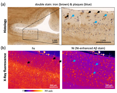 |
Iron accumulation and MRI iron contrast are not driven by
amyloid plaques in posterior cortical atrophy
Evgeniya Kirilina1,
Luke Edwards1,
Carsten Jäger1,2,
Tilo Reinert1,
Anna Jauch1,
Malte Brammerloh1,3,
Karl-Heinz Herrmann4,
Patrick Scheibe1,
Felix Büttner1,3,
Dennis Brückner5,
Gerald Falkenberg5,
Jürgen R. Reichenbach4,
and Nikolaus Weiskopf1
1Neurophysics, Max Planck Institute for Human Cognitive and Brain Sciences, Leipzig, Germany, 2Paul Flechsig Institute - Center of Neuropathology and Brain Research, Leipzig, Germany, 3Faculty of Physics and Earth Sciences, Leipzig University, Leipzig, Germany, 4Medical Physics Group, Institute of Diagnostic and Interventional Radiology, Jena University Hospital, Jena, Germany, 5PETRA III, Deutsches Elektronen-Synchrotron DESY, Hamburg, Germany Keywords: Alzheimer's Disease, Contrast Mechanisms, iron, plaques, posterior cortical atrophy, cortex We combined ultra-high resolution quantitative MRI, X-ray fluorescence, and biophysical modeling to study iron-induced MRI contrast in the visual cortex of patients with Alzheimer’s disease (AD) and its variant posterior cortical atrophy (PCA). The iron content of amyloid plaques in patients with PCA and AD exceeded the iron concentration in the surrounding tissue by less than 15%, constituting only a minor contribution to the intracortical T2* contrast. The elevated levels of brain iron detected by iron-sensitive MRI contrasts are not indicative of plaques but may reflect other processes, such as inflammation or leakage in the blood-brain barrier. |
| 09:19 |
0032.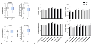 |
Regional Changes in Brain Viscoelasticity in Multiple Sclerosis
Assessed with Three-Dimensional MR Elastography
Ling Fang1,
Matthew C. Murphy2,
Sha Tan3,
Qiuxia Luo1,
Jingbiao Chen1,
Bingjun He1,
Jun Chen2,
Jonathan M. Scott2,
Kevin J. Glaser2,
Richard L. Murphy2,
Wei Qiu3,
and Jin Wang1
1Department of Radiology, the Third Affiliated Hospital, Sun Yat-sen University, Guangzhou, China, 2Department of Radiology, Mayo Clinic, Rochester, MN, United States, 3Department of Neurology, the Third Affiliated Hospital, Sun Yat-sen University, Guangzhou, China Keywords: Multiple Sclerosis, Elastography Multiple sclerosis (MS) is a chronic inflammatory demyelinating disease of the central nervous system. MR Elastography (MRE) can quantitatively assess biomechanical tissue properties in vivo noninvasively. We investigated the potential value of MRE to reveal changes in tissue viscoelasticity in regions and the whole brain in MS patients and to analyze the relevance to clinical manifestations. Our results suggest that the damping ratio and loss modulus are promising quantitative biomarkers for evaluating tissue damage in MS. |
| 09:27 |
0033. |
Elevated free-water volume in the brains of HIV+ alcohol
drinkers compared to HIV- drinkers as an indication of
neuro-inflammation
Teddy Salan1,
Varan Govind1,
Joseph Gullett2,
Eric Porges2,
Zhigang Li2,
Ronald Cohen2,
and Robert Cook2 1University of Miami, Miami, FL, United States, 2University of Florida, Gainesville, FL, United States Keywords: Neuroinflammation, Infectious disease, Alcohol Use Chronic alcohol use and HIV infection can have synergistic effects that negatively impact the brain leading to abnormalities such as neuro-inflammation. Free-water eliminated DTI (FWE-DTI) has been suggested as a biomarker of neuro-inflammation. This study compare DTI and FWE-DTI derived metrics in the brains of HIV+/HIV- chronic drinkers to determine whether HIV infection or drinking behavior is a more significant factor contributing to brain microstructural abnormalities and neuro-inflammation. Our results suggest that HIV is a more significant factor than drinking level contributing to elevated free water in the brain as a sign of neuro-inflammation. |
| 09:35 |
0034.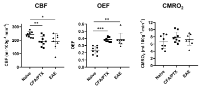 |
Multimodal 9.4T MRI and Near-Infrared Spectroscopy shows reduced
perfusion and abnormal oxidative metabolism in the EAE mouse
model of MS
Mada Hashem1,2,3,
A. Max Hamilton1,2,3,
and Jeff F. Dunn1,2,3
1Department of Radiology, University of Calgary, Calgary, AB, Canada, 2Hotchkiss Brain Institute, University of Calgary, Calgary, AB, Canada, 3Experimental Imaging Centre, University of Calgary, Calgary, AB, Canada Keywords: Multiple Sclerosis, Animals, arterial spin labeling, high field MRI While multiple sclerosis (MS) is typically considered a white matter disease, there is prominent evidence that gray matter pathology, such as hypoxia, disruptions in metabolic rate and mitochondrial dysfunction are involved as well. We are applying a novel multimodal technique combining near-infrared spectroscopy and high-field MRI to study the mitochondrial status as well as oxygen delivery and consumption in the cortex of the EAE mouse model of MS. Reduced perfusion, and consequent hypoxia, with abnormal mitochondrial regulation and no change in consumption rate were found in EAE mice. Hypoxia and mitochondrial damage are likely to exacerbate the pathology in MS. |
| 09:43 |
0035.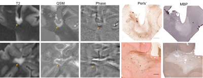 |
Pathological Validation of Multiple Sclerosis Lesion Rims on
Phase and Quantitative Susceptibility Mapping (QSM) images
Kelly M Gillen1,
Thanh D Nguyen1,
Alexey Dimov1,
Emily Demmon2,
Ilhami Kovanlikaya1,
Francesca Bagnato3,4,
Dominick Romano1,
David Pitt5,
Susan Gauthier2,
and Yi Wang1
1Department of Radiology, Weill Cornell Medicine, New York, NY, United States, 2Department of Neurology, Weill Cornell Medicine, New York, NY, United States, 3Department of Neurology, TN Valley HealthCare VA Medical System, Nashville, TN, United States, 44Department of Neurology, Vanderbilt University Medical Center, Nashville, TN, United States, 5Department of Neurology, Yale School of Medicine, New Haven, CT, United States Keywords: Multiple Sclerosis, Quantitative Susceptibility mapping, Phase, iron Multiple Sclerosis (MS) is an autoimmune disorder characterized by focal inflammatory demyelination. Chronic MS lesions can contain chronically activated, iron-laden microglia and macrophages. By comparing rim status on quantitative susceptibility mapping (QSM) and phase imaging with histopathology that identifies iron, we demonstrate that QSM is a more reliable indicator of iron status than phase. QSM is a valuable clinical tool to identify iron positive smoldering lesions not visible using conventional MRI techniques. |
| 09:51 |
0036.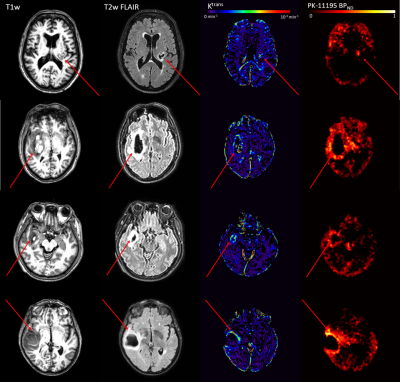 |
Association between PET-MR imaging measures of blood-brain
barrier leakage and immune cell activation in acute
intracerebral hemorrhage
Olivia Jones1,2,
Saffwan Mohamed3,
Rainer Hinz2,4,
Ben Dickie2,4,
Laura Parkes1,2,
and Adrian Parry-Jones2,3,5
1Division of Psychology, Communication and Human Neuroscience, University of Manchester, Manchester, United Kingdom, 2Geoffrey Jefferson Brain Research Centre, Manchester, United Kingdom, 3Division of Cardiovascular Sciences, University of Manchester, Manchester, United Kingdom, 4Division of Informatics, Imaging & Data Sciences, University of Manchester, Manchester, United Kingdom, 5Manchester Centre for Clinical Neurosciences, Northern Care Alliance NHS Foundation Trust, Manchester, United Kingdom Keywords: Neuroinflammation, PET/MR Intracerebral hemorrhage is a severe form of stroke. Secondary injury involves a cascade of pathophysiological changes thought to begin with microglial activation and result in blood-brain barrier(BBB) breakdown and edema formation. In this study, patients underwent dynamic contrast-enhanced MRI and [11C](R)-PK11195 PET, which measure BBB leakage and binding to the translocator protein 18kDa (TSPO) expressed on activated microglia, respectively. BBB leakage and TSPO binding were elevated in the edema. Mean co-localisation of BBB leakage and TSPO binding was 5.6%; drugs targeting inflammation may cross the BBB into 0-28% of target tissue. Modified Rankin Scale scores negatively correlated with BBB leakage. |
09:59 |
0037. |
Brain Metabolite Concentrations of White Matter and Grey Matter
in Perinatal HIV Infected Adults Using a Whole-Brain MR
Spectroscopic Imaging
Teddy Salan1,
Elizabeth Willen2,
Anai Cuadra1,
Sulaiman Sheriff1,
Andrew Maudsley1,
and Varan Govind1 1Univerity of Miami, Miami, FL, United States, 2University of Missouri-Kansas City School of Medicine, Kansas City, MO, United States Keywords: Infectious disease, Spectroscopy With the advent of antiretroviral therapy (ART), perinatally HIV-infected children have been able to transition into adulthood. However, the long term impact of HIV infection and the potential toxic effects of long term use of antiretroviral therapeutics on brain metabolites are not completely characterized. In this study, a whole-brain MRSI method is used to quantitate changes in brain metabolites, N-acetyl aspartate (NAA), total-creatine (Cr) and total-choline (Cho), in PHIV+ adults compared to matched controls. Results show significant alterations of proton MRS-observed metabolites in the whole brain of PHIV+ as they reach adulthood despite their adherence to ART. |
| 10:07 |
0038. |
Brain diffusion alterations in patients with COVID-19 pathology
and neurological manifestations
Anna Caroli1,
Serena Capelli1,
Angela Napolitano2,
Giulia Cabrini3,
Alberto Arrigoni1,
Giulio Pezzetti2,
Mattia Previtali1,
Luca Giovanni Longhi4,
Rosalia Zangari5,
Ferdinando Luca Lorini6,
Maria Sessa7,
Andrea Remuzzi3,
and Simonetta Gerevini2
1Bioengineering Department, Istituto di Ricerche Farmacologiche Mario Negri IRCCS, Ranica (BG), Italy, 2Department of Neuroradiology, ASST Papa Giovanni XXIII, Bergamo, Italy, 3University of Bergamo, Bergamo, Italy, 4Neurosurgical Intensive Care Unit, Department of Anesthesia and Critical Care Medicine, ASST Papa Giovanni XXIII, Bergamo, Italy, 5FROM Research Foundation, ASST Papa Giovanni XXIII, Bergamo, Italy, 6Department of Emergency and Critical Care Area, ASST Papa Giovanni XXIII, Bergamo, Italy, 7Department of Neurology, ASST Papa Giovanni XXIII, Bergamo, Italy Keywords: Infectious disease, COVID-19 This study aimed at quantifying brain diffusion alterations on DWI scans in 215 COVID-19 patients with neurological manifestations as compared with 36 normal controls. The Apparent Diffusion Coefficient (ADC) was quantified in brain tissues and regions, using an in-house MRI processing procedure. In COVID-19 patients, a widespread significant increase in ADC was found in white matter. ADC values were significantly correlated with MRI time from disease onset. ADC alteration was highest in hospitalized patients. Patients with neurological disorders showed significantly higher ADC than those with olfactory loss only. DWI shows potential as a non-invasive marker of neuroinflammation in COVID-19. |
 Back to Meeting Home
 Back to the Program-at-a-Glance
The International Society for Magnetic Resonance in Medicine is accredited by the Accreditation Council for Continuing Medical Education to provide continuing medical education for physicians.
 Back to Meeting Home
 Back to the Program-at-a-Glance
 View Presentation Video