Oral Session
Blood Vessels in the Brain
ISMRM & ISMRT Annual Meeting & Exhibition • 03-08 June 2023 • Toronto, ON, Canada

| 08:15 |
0774.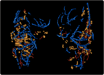 |
Enlarged Perivascular Spaces in the Basal Ganglia Surround
Arteries, not Veins
Jan Oltmer1,
Julia Beck2,
Hendrik Mattern3,4,
Renat Yakupov3,5,
Corinne Auger6,
Emrah Düzel3,5,7,8,
Susanne van Veluw6,9,10,
Stefanie Schreiber2,3,5,7,
and Valentina Perosa3,9 1Athinoula A. Martinos Center, Massachusetts General Hospital/Harvard medical school, Charlestown, MA, United States, 2Department of Neurology, Otto-von-Guericke University, Magdeburg, Germany, 3German center for neurodegenerative diseases (DZNE), Magdeburg, Germany, 4Department of Biomedical Magnetic Resonance (BMMR), Institute for Physics, Otto-von-Guericke-University, Magdeburg, Germany, 5Institute of Cognitive Neurology and Dementia Research (IKND), Otto-von-Guericke University, Magdeburg, Germany, 6MassGeneral Institute for Neurodegenerative Disease, Massachusetts General Hospital/Harvard Medical School, Charlestown, MA, United States, 7Center for Behavioral Brain Sciences (CBBS), Magdeburg, Germany, 8Institute of Cognitive Neuroscience, University College London, London, United Kingdom, 9J. Philip Kistler Stroke Research Center, Department of Neurology, Massachusetts General Hospital/Harvard Medical School, Boston, MA, United States, 10Department of Radiology, Leiden University Medical Center, Leiden, Netherlands Keywords: Blood vessels, High-Field MRI, Histopathology Fluid-filled perivascular spaces (PVS) surround brain vessels. Their enlargement is a common hallmark of cerebral small vessel disease (CSVD) and has been related to impaired clearance of toxic proteins from the brain. It is unclear whether PVS enlarge around arteries, veins, or both. Combining ultra-high resolution 7T MRI angiography, venography and histology, we show that in healthy controls and patients with CSVD, PVS enlarge around arteries more than veins within the basal ganglia. A better understanding of the anatomy and distribution of enlarged PVS can contribute to the understanding of perivascular clearance and disease mechanisms. |
08:23 |
0775.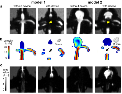 |
BB MRI for monitoring aneurysm treatment – a novel biomarker?
Mariya S. Pravdivtseva1,
Hivnu Toraman2,
Jana Korte3,
Franziska Gaidzik3,
Philipp Berg3,
Lana Bautz1,
Fritz Wodarg2,
Jan-Bernd Hövener1,
Olav Jansen2,
and Naomi Larsen2
1Department of Radiology and Neuroradiology, Section Biomedical Imaging, Molecular Imaging North Competence Center (MOIN CC), University Medical Center Schleswig-Holstein (UKSH), Kiel University, Kiel, Germany, Kiel, Germany, 2Department of Radiology and Neuroradiology, University Medical Center Schleswig-Holstein (UKSH), Kiel University, Kiel, Germany, Kiel, Germany, 3Research Campus STIMULATE, University of Magdeburg, Magdeburg, Germany, Magdeburg, Germany Keywords: Blood vessels, Phantoms, blood vessels, aneurysms, diagnostics, treatment evaluation Treating life-threatening intracranial aneurysms with flow modulating devices (FMDs) is mostly successfull, but may lead to complications and even delayed aneurysm rupture. A biomarker indicating treatment success is direly needed. Flow MRI can detect flow reduction, but it is impaired by metal artifacts from FMDs. Black-blood (BB) MRI is less sensitive to metal artifacts and often results in poor slow-flow suppression. We hypothesize that BB contrast changes after aneurysm treatment. Here, after placing FMDs in 3D-printed aneurysm models, an enhanced signal was found on BB MRI, associated with the reduced aneurysmal flow after flow modulation and thus possible treatment success. |
|
08:31 |
0776.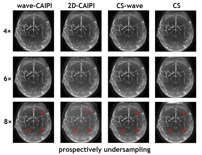 |
Accelerated intracranial time-of-flight magnetic resonance
angiography using wave-encoding
Yang Ji1,
Wenchuan Wu1,
Matthijs H. S. de Buck 1,
Thomas Okell 1,
and Peter Jezzard1
1Wellcome Centre for Integrative Neuroimaging, FMRIB Division, Nuffield Department of Clinical Neurosciences, University of Oxford, Oxford, United Kingdom Keywords: Blood vessels, Parallel Imaging 3D time-of-flight (TOF) can be used to acquire a volume with high spatial resolution, making it a preferable choice for depicting smaller vascular structures. However, 3D TOF requires long acquisition times when acquiring multiple slabs and covering a large field of view, especially for high spatial resolution imaging. To accelerate acquisition and to improve image quality of TOF MRA, we developed an accelerated 3D intracranial TOF MRA sequence with wave-encoding (referred to as 3D wave-TOF) and evaluated two variants – wave-CAIPI and compressed-sensing wave (CS-wave). |
| 08:39 |
0777.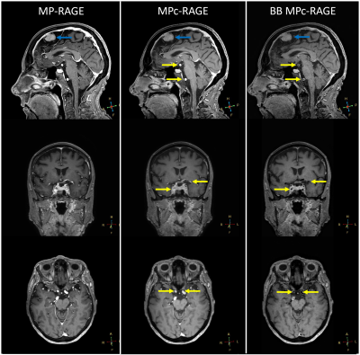 |
MPc-RAGE: Simultaneous MP-RAGE and Phase Contrast Angiography
Acquisition
Brian Johnson1,2,
Sandeep Ganji1,3,
and Nandor Pinter4,5
1Philips, Cleveland, OH, United States, 2Department of Radiology, University of Texas Southwestern Medical Center, Dallas, TX, United States, 3Department of Radiology, Mayo Clinic College of Medicine, Rochester, MN, United States, 4Dent Neurologic Institute, Amherst, NY, United States, 5Department of Neurosurgery, University at Buffalo, Buffalo, NY, United States Keywords: Blood vessels, Blood vessels The 3D magnetization-prepared rapid gradient-echo (MP-RAGE) and 3D phase contrast angiography (PCA) are widely used sequences in both clinical and research settings, due to high spatial resolution, excellent contrast, and clinically feasible scan time. Recent image acceleration techniques, such as compressed sensing and artificial intelligence reconstruction can drastically reduce scan times, allowing to create multi-domain imaging approaches. Here we introduce MPc-RAGE, a simultaneous MP-RAGE and PCA acquisition that combines the benefits of these two sequences in a single scan. Furthermore, we explore the possibility of the MPc-RAGE as a potential black blood technique for use in vessel wall imaging. |
| 08:47 |
0778.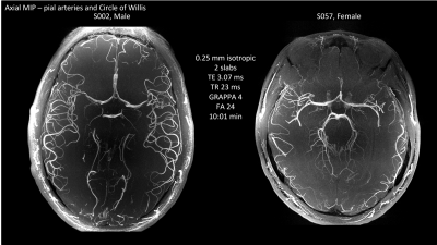 |
Utilizing High Performance Gradients to Image Human Brain
Arterial Vasculature with High Resolution 7T Time-of-flight MR
Angiography
Samantha J Ma1,
Alexander JS Beckett2,3,
Gerhard Laub4,
and David A Feinberg2,3
1Siemens Medical Solutions USA, Inc., Berkeley, CA, United States, 2Helen Wills Neuroscience Institute, University of California, Berkeley, Berkeley, CA, United States, 3Advanced MRI Technologies, Sebastopol, CA, United States, 4Dr. Laub Consulting, LLC, San Mateo, CA, United States Keywords: Blood vessels, High-Field MRI Time of flight (TOF) 3D MR Angiography (MRA) has been shown to effectively image small (50-300um diameter) pial vessels. However, such high resolution requires strong flow compensation (flow-comp) gradient pulses and short echo times which are limited by peripheral nerve stimulation and hardware capability in most scanners. We demonstrate high resolution TOF MRA acquisition using an advanced head gradient system (200mT/m, 900 T/m/s) at ultrahigh field 7T. |
|
08:55 |
0779.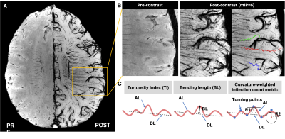 |
Detection of Age-related Cerebral Microvessel Tortuosity and Its
Association with Dilated Perivascular Space Using USPIO-enhanced
7T MRI
Zhe Sun1,2,
Chenyang Li1,2,
E. Mark Haacke3,
and Yulin Ge1
1Department of Radiology, New York University Grossman School of Medicine, New York, NY, United States, 2Vilcek Institute of Graduate Biomedical Sciences, New York University Grossman School of Medicine, New York, NY, United States, 3Department of Radiology, Wayne State University, Detroit, MI, United States Keywords: Blood vessels, Aging Microvessel tortuosity, which is lack detection on clinical imaging, may have direct detrimental effects on capillary flow and nutrient supply which is critical in maintaining neural functions. A high-resolution USPIO-enhanced 7T MRI demonstrated corkscrew appearing medullary arterioles (about 50 µm) in white matter with some enclosed in dilated perivascular space (PVS). The number of tortuous arterioles increased with aging and more tortuous vessels with dilated PVS were found in elderly people (P<0.05). The dynamic interactions between vulnerable tortuous arteries and PVS dilation may be the basis of age-related hypoperfusion, white matter pathology, and waste clearance dysfunction in the elderly. |
| 09:03 |
0780.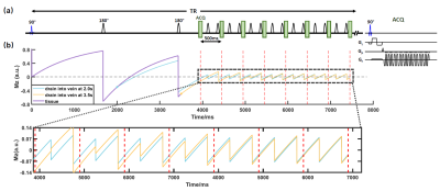 |
VICTR: Venous transit time (VTT) Imaging by Changes in T1
Relaxation
Wen Shi1,
Dengrong Jiang2,
Abhay Moghekar3,
Zhiyi Hu1,
and Hanzhang Lu1,2
1Department of Biomedical Engineering, Johns Hopkins University School of Medicine, Baltimore, MD, United States, 2Department of Radiology & Radiological Science, Johns Hopkins University School of Medicine, Baltimore, MD, United States, 3Department of Neurology, Johns Hopkins University School of Medicine, Baltimore, MD, United States Keywords: Blood vessels, Blood Venous hemodynamics is not well-studied despite it is a crucial part of human vascular system and involves in many brain vascular diseases. Here, we developed a novel technique dubbed Venous transit time Imaging by Changes in T1 Relaxation (VICTR) to measure the time for blood to travel from capillary to veins, i.e., VTT. We verified VICTR on different locations of superior sagittal sinus and VTT showed regional dependency. The caffeine challenge study also validated VTT with the vasoconstriction effect. With excellent test-retest reproducibility, VTT may be a potential physiological biomarker to evaluate vessel tortuosity and detect venous pathologies. |
| 09:11 |
0781.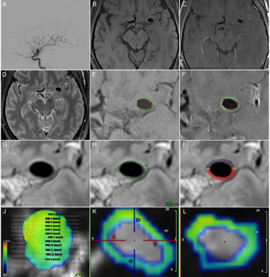 |
4D-Flow and DCE-MRI Study on the Wall Enhancement of Unruptured
Intracranial Aneurysms
Qichang Fu1,
Yong Zhang1,
Chengcheng Zhu2,
Sheng Guan1,
and Jingliang Cheng1
1The First Affiliated Hospital of Zhengzhou University, Zhengzhou, China, 2University of Washington, Seattle, WA, United States Keywords: Blood vessels, Stroke, unruptured intracranial aneurysms; aneurysmal wall enhancement; wall enhancement index; Ktrans; wall shear stress This study used 4D-flow-MRI, dynamic contrast-enhanced MRI, and vessel wall MRI to explore the relationship between wall shear stress (WSS) and Ktrans and aneurysm wall enhancement (AWE) to understand the functional changes in the unruptured intracranial aneurysms (UIAs). Seventy-eight patients were enrolled, including 96 UIAs. All patients completed examinations based on a 3.0T Siemens Prisma magnetic resonance. WSS with the AWE group was lower, and Ktrans with the AWE group were higher than those without AWE. The wall enhancement index (WEI) was a quantitative index of AWE. WEI had a negative correlation with WSS and a positive correlation with Ktrans. |
| 09:19 |
0782.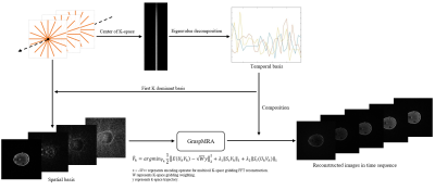 |
An advanced reconstruction framework in stack-of-stars
golden-angle radial non-contrast enhanced 4D MRA with ultra-high
temporal resolution
Tianrui Zhao1,2,
Li Feng3,
and Lirong Yan2
1Department of Biomedical Engineering, Northwestern University, Chicago, IL, United States, 2Department of Radiology, Northwestern University, Chicago, IL, United States, 3BioMedical Engineering and Imaging Institute (BMEII), Department of Radiology, Icahn School of Medicine at Mount Sinai, New York, NY, United States Keywords: Blood vessels, Image Reconstruction In the present study, we developed an advanced reconstruction framework for ASL-based 4-dimensional (4D) MRA dubbed GraspMRA, which combines stack-of-stars golden-angle radial sampling with low-rank subspace-based image reconstruction to achieve ultra-high temporal resolution. The performance of GraspMRA was evaluated by comparison with three other reconstruction methods at different acceleration rates. Our results have demonstrated that GraspMRA has superior performance to the other methods, and it provides real flow dynamics at an ultra-high temporal resolution of up to 25ms per 3D volume while preserving good image quality. |
| 09:27 |
0783.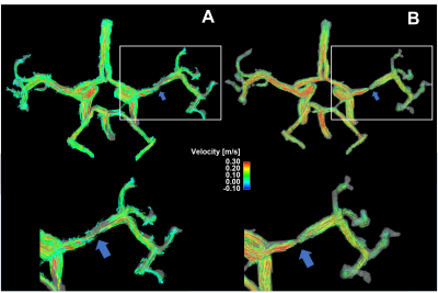 |
Super-resolution assessment of relative pressure in intracranial
atherosclerosis using ML-enhanced 4D Flow MRI
Patrick Winter1,2,
David Marlevi3,4,
Maria Aristova2,
Edward Ferdian5,
Jonas Schollenberger6,
Alireza Vali2,
Jackson Moore2,
Michael Markl2,
Ramez Abdallah2,
Sameer Ansari2,
C Alberto Figuerora6,
David Nordsletten6,
Alistair Young6,
and Susanne Schnell1,2
1Department of Medical Physics, University of Greifswald, Greifswald, Germany, 2Department of Radiology, Northwestern University Feinberg School of Medicine, Chicago, IL, United States, 3Dept Molecular Medicine and Surgery, Karolinska Institutet, Solna, Sweden, 4Institute for Medical Engineering and Science, Massachusetts Institute of Technology, Cambridge, MA, United States, 55Department of Anatomy and Medical Imaging, University of Auckland, Auckland, New Zealand, 6Department of Anatomy and Medical Imaging, University of Michigan, Ann Arbor, MI, United States Keywords: Stroke, Atherosclerosis, intracranial atherosclerotic disease, stenosis, pressure differences In vivo measurements of intracranial pressure differences using 4D flow are useful to assess health risks associated with intracranial atherosclerotic disease (ICAD). In the clinical routine, approximations of the Navier-Stokes equation are used to derive relative pressure values, which can be inaccurate in intracranial vessels. Recently we presented an algorithm using a virtual work-energy formulation(vWERP). While this technique yields more accurate pressure estimations in intracranial settings, still systematic errors due to insufficient spatial resolution were observed. Here, we apply artificial intelligence-based super resolution for more accurate assessments of pressure values near the stenosis in a cohort of ICAD patients. |
|
09:35 |
0784.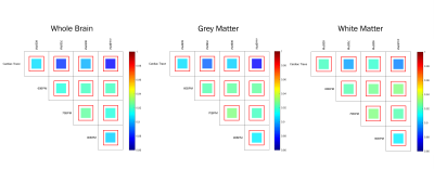 |
A novel method of calculating cardiac pulsatility from resting
state fMRI data 1CUBRIC, School of Physics and Astronomy, Cardiff University, Cardiff, United Kingdom, Cardiff, United Kingdom, 2CUBRIC, School of Psychology, Cardiff University, Cardiff, United Kingdom, Cardiff, United Kingdom, 3Department of Bioengineering, McGill University, Montreal, QC, Canada, Montreal, QC, Canada, 4Graduate Program in Biological and Biomedical Engineering, McGill University, Montreal, QC, Canada, Montreal, QC, Canada Keywords: Blood vessels, fMRI (resting state) Measures of cardiac pulsatility were generated from resting-state fMRI data using ICA information and processed HRV traces. This was achieved by performing a correlation between the ICA component and HRV time series to isolate cardiac-related components from non-cardiac-related components. Classified datasets were used to train the FIX noise correction algorithm. FIX was used to classify cardiac components in 4123 rfMRI datasets. Measures of voxelwise cardiac pulsatility were generated be determining the R2 variance explained in the voxel time series by the classified cardiac components. Comparisons with gold standard pulsatility measures derived from the HRV traces showed high correlation values. |
| 09:43 |
0785.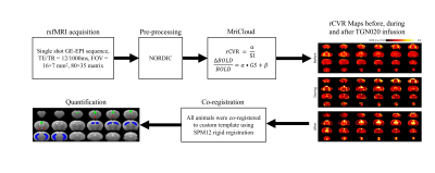 |
Effects of Aquaporin-4 Inhibition on Relative Cerebrovascular
Reactivity using Resting-state Functional MRI 1Ophthalmology, New York University Grossman School of Medicine, New York, NY, United States, 2Radiology, New York University Grossman School of Medicine, New York, NY, United States Keywords: Blood vessels, fMRI (resting state), Cerebrovscular reactivity Aquaporin-4 (AQP4) is most abundant water channel responsible for cerebrospinal fluid influx in the brain, and has important bearings on cerebrovascular diseases. However, it remains unclear how aquaporin-4 functions in the cerebrovasculature. We used relative cerebrovascular reactivity (rCVR) mapping derived from task-free, resting-state blood-oxygenation-level-dependent functional MRI to determine the effects of AQP4 suppression on the healthy mouse brains using the AQP4 inhibitor TGN020. We observed different patterns of rCVR responses across cortical and subcortical brain regions, indicating the heterogeneity of AQP4 functions in the cerebrovasculature, which may explain the varying vulnerability of different brain regions to cerebrovascular diseases. |
| 09:51 |
0786.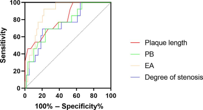 |
Can HR-VWI pre-operatively predict Postoperative Restenosis of
Intracranial Atherosclerotic Disease Treated by Drug-Coated
Balloon?
Shu Jiang1,
Weiqiang Dou2,
Xinyi Wang1,
and Chao Zhang1
1The First Affiliated Hospital of Shandong First Medical University, Jinan, China, 2GE Healthcare, Nanjing, China Keywords: Stroke, Stroke This study mainly investigated if high-resolution vessel wall MRI (HR-VWI) can predict postoperative restenosis before drug-coated balloon (DCB) treatment. We included 76 patients who underwent HR-VWI examination before DCB treatment. DSA measurement was assessed 6 months after operation to determine vessel restenosis, classifying patients into three groups of no stenosis, mild stenosis (<50%), and restenosis (>50%). Significant differences among three groups were observed in plaque length, lumen area of MLN, degree of stenosis, enhancement amplitude and plaque burden. Plaque length and EA were independent prognostic factors of postoperative restenosis. Therefore, HR-VWI has potential to predict postoperative restenosis before DCB treatment. |
| 09:59 |
0787.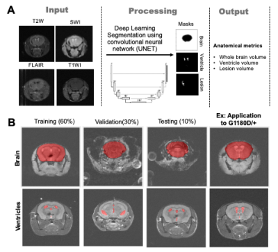 |
Multimodal neuroimaging of Col4A1-mutant mouse models of Gould
Syndrome 1UCSF, San Francisco, CA, United States Keywords: Blood vessels, Rare disease Gould syndrome is a multisystem disorder whose manifestations are highly variable. Animal studies suggest that allelic heterogeneity and genetic context contribute to the clinical variability. Clinical manifestations associated with Gould syndrome include blood–brain barrier (BBB) leakage, microbleeds, and white matter lesions. Here, we use multimodal MRI at 14.1Tesla to characterize radiological findings observed in mouse models of Gould syndrome caused by mutations in Collagen type IV alpha 1 (Col4a1). We show that multimodal MRI can successfully differentiate between disease subtypes based on anatomical changes as well as prevalence, number, volume and type of brain lesions. |
| 10:07 |
0788.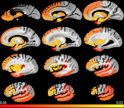 |
Cerebral Amyloid Angiopathy (CAA) is associated with gray matter
R2 relaxation rate: An ex-vivo MRI and pathology study
Md Tahmid Yasar1,
Arnold M. Evia2,
David A. Bennett2,
Julie A. Schneider2,
and Konstantinos Arfanakis1,2
1Biomedical Engineering, Illinois Institute of Technology, Chicago, IL, United States, 2Rush Alzheimer's Disease Center, Rush University Medical Center, Chicago, IL, United States Keywords: Blood vessels, Aging, Alzheimer’s disease, Dementia, Ex-vivo applications Cerebral amyloid angiopathy (CAA) is characterized by accumulation of amyloid-β protein in the walls of cortical and leptomeningeal small vessels. CAA is common in older adults and is associated with intracerebral hemorrhage, microbleeds, cognitive decline and dementia. The present study in a large number of community-based older adults (N=802) combined ex-vivo MRI and detailed neuropathology and showed for the first time that CAA is associated with higher transverse relaxation rate, R2, in gray matter, independent of other neuropathologies and demographics. The regions that showed this association included cortical regions in the temporal and frontal lobes as well as subcortical structures. |
 Back to Meeting Home
 Back to the Program-at-a-Glance
The International Society for Magnetic Resonance in Medicine is accredited by the Accreditation Council for Continuing Medical Education to provide continuing medical education for physicians.
 Back to Meeting Home
 Back to the Program-at-a-Glance
 View Presentation Video