Oral Session
Advanced Cerebrovascular MRI: Pipes, Perfusion, Parametric Imaging & Predictive Models
ISMRM & ISMRT Annual Meeting & Exhibition • 03-08 June 2023 • Toronto, ON, Canada

Oral Session
Advanced Cerebrovascular MRI: Pipes, Perfusion, Parametric Imaging & Predictive Models
| 13:30 |
0874.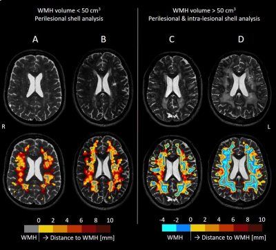 |
Microstructural and microvascular alterations in small vessel
diseases using multi-shell DTI inside and outside white matter
hyperintensities
Paulien Voorter1,
Maud van Dinther2,
Michael Stringer3,
Danielle Kerkhofs2,
Anna Dewenter4,
Gordon Blair3,
Daniela Jaime Garcia3,
Francesca Chappell3,
Anna Kopczak4,
Julie Staals2,
Michael Ingrisch4,
Marco Duering4,
Michael Thrippleton3,
Fergus Doubal3,
Martin Dichgans4,
Joanna Wardlaw3,
Robert van Oostenbrugge2,
Jacobus Jansen1,
and Walter Backes1
1Department of Radiology and Nuclear Medicine, School for Mental Health and Neuroscience, Maastricht University Medical Center, Maastricht, Netherlands, 2Department of Neurology, CARIM School for Cardiovascular Diseases, Maastricht University Medical Center, Maastricht, Netherlands, 3Brain Research Imaging Centre, Centre for Clinical Brain Sciences, UK Dementia Institute Centre at the University of Edinburgh, Edinburgh, United Kingdom, 4Institute for Stroke and Dementia Research (ISD), University Hospital, LMU Munich, Munich, Germany Keywords: Stroke, Stroke, small vessel disease The pathophysiology underlying white matter hyperintensities (WMHs) and changes in perilesional white matter are not fully understood in cerebral small vessel disease (SVD). Using multi-shell diffusion tensor imaging (DTI), we studied the mean (parenchymal) diffusivity (MD) and microvascular perfusion (f) inside and outside WMHs in sporadic and monogenetic (CADASIL) SVDs. The microstructure (expressed by MD) was most damaged inside WMHs and extended beyond the WMHs. Furthermore, f was highest at the WMH border, decreased towards the WMH center, and decreased outside WMHs, possibly reflecting multiple SVD-related alterations in the microvasculature, such as dilated microvessels, capillary rarefaction, and reduced microvascular integrity. |
| 13:38 |
0875.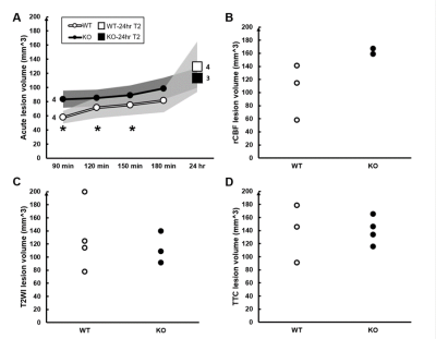 |
The role of FK506 binding protein 5 in the early development of
ischemic lesion using multi-parametric MRI
Zih-Rong Lai1,
Yu-Ping Kang2,
Chia-Feng Lu1,
Bao-Yu Hsieh3,4,
Yi-Hsuan Lee2,5,
and Yu-Chieh Jill Kao1
1Department of Biomedical Imaging and Radiological Sciences, National Yang Ming Chiao Tung University, Taipei, Taiwan, 2Department and Institute of Physiology, College of Medicine, National Yang Ming Chiao Tung University, Taipei, Taiwan, 3Department of Medical Imaging and Radiological Sciences, College of Medicine, Chang Gung University, Taoyuan, Taiwan, 4partment of Medical Imaging and Intervention, Chang Gung Memorial Hospital at Linkou, Taoyuan, Taiwan, 5Brain Research Center, National Yang Ming Chiao Tung University, Taipei, Taiwan Keywords: Stroke, Preclinical, Transgenic mice To explore how the FK506 binding protein 5 (FKBP5) affect the ischemic stroke outcome, we employed Fkbp5-knock out (KO) mice and evaluated the ADC- and CBF-deficit volume, as well as angiography in the acute phase and the final lesion volume at 24 hours after the occlusion of middle cerebral artery (MCAO). The discent lesion volume and loss of collaterals after MCAO in the Fkbp5-KO animals suggested the early determination of the stroke outcome in the Fkbp5-KO animals after ischemic insult. |
| 13:46 |
0876.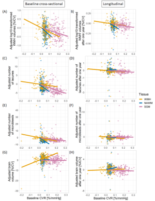 |
Cerebrovascular reactivity is a predictor of small vessel
disease severity after one year in patients with mild stroke
Emilie Sleight1,2,
Michael S Stringer1,2,
Una Clancy1,2,
Carmen Arteaga1,2,
Daniela Jaime Garcia1,2,
Will Hewins1,2,
Angela CC Jochems1,2,
Rachel Penman1,2,
Yajun Cheng1,2,3,
Dillys Liu1,2,4,
Junfang Zhang1,2,5,
Iona Hamilton6,
Charlotte Jardine6,
Rosalind Brown1,2,
Eleni Sakka1,2,
Agniete Kampaite1,2,
Stewart Wiseman1,2,
Maria Valdes-Hernandez1,2,
Francesca Chappell1,2,
Fergus Doubal1,2,
Ian Marshall1,2,
Michael J Thrippleton1,2,6,
and Joanna M Wardlaw1,2,6
1Centre for Clinical Brain Sciences, University of Edinburgh, Edinburgh, United Kingdom, 2UK Dementia Research Institute, University of Edinburgh, Edinburgh, United Kingdom, 3Department of Neurology, West China Hospital of Sichuan University, Chengdu, China, 4Department of Medicine, University of Hong Kong, Hong Kong, Hong Kong, 5Department of Neurology, Shanghai Jiao Tong University, Shanghai, China, 6Edinburgh Imaging Facility RIE, University of Edinburgh, Edinburgh, United Kingdom Keywords: Stroke, Blood vessels, Cerebrovacular reactivity Small vessel disease (SVD) causes stroke and dementia, but its pathogenesis remains poorly understood. We measured cerebrovascular reactivity (CVR) in SVD patients with minor stroke using 3T BOLD MRI and investigated its relation to baseline and progression of SVD features after one year adjusting for age and vascular risk factors. Patients with lower CVR had higher baseline white matter hyperintensity (WMH) volume, more lacunes and microbleeds and lower brain volume. After one year, patients with lower baseline CVR had increased WMH burden. In conclusion, CVR impairment in SVD patients is associated with higher SVD burden and predicts worsening of SVD. |
| 13:54 |
0877.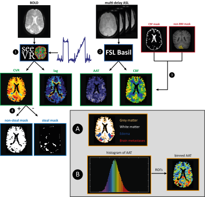 |
Hemodynamic imaging parameters in brain metastases patients –
Agreement between multi-delay ASL and hypercapnic BOLD
Eva Elisabeth van Grinsven1,
Marielle Philippens2,
Jeroen Siero3,4,
and Alex Bhogal3
1Department of Neurology & Neurosurgery, UMC Utrecht, Utrecht, Netherlands, 2Department of Radiation Oncology, UMC Utrecht, Utrecht, Netherlands, 3Department of Radiology, UMC Utrecht, Utrecht, Netherlands, 4Spinoza Center for Neuroimaging, Amsterdam, Netherlands Keywords: Blood vessels, Neuroscience This study compared baseline ASL with BOLD cerebrovascular reactivity (BOLD-CVR) parameters in the brain under different hemodynamic circumstances in patients with brain metastases. There was a strong relationship between baseline cerebral blood flow and BOLD-CVR measurements and the temporal metrics of ASL and BOLD-CVR. However, the relationship between baseline ASL and BOLD-CVR does not hold in tissue with exhausted cerebral autoregulation (i.e. vascular steal regions). Thereby, BOLD-CVR may be able to flag at-risk areas with depleted vascular reserve capacity before they become visible on ASL MRI. |
14:02 |
0878.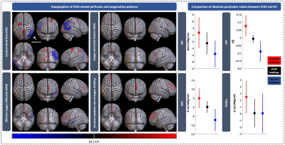 |
Multivariate analysis of perfusion and oxygenation patterns in
asymptomatic unilateral carotid artery stenosis
Jan Kufer1,
Jens Göttler1,2,
Gabriel Hoffmann1,
Lena Schmitzer1,
Claus Zimmer1,
Fahmeed Hyder2,
Christine Preibisch1,3,
and Stephan Kaczmarz1,2,4
1Department of Diagnostic and Interventional Neuroradiology, School of Medicine, Technical University of Munich (TUM), Munich, Germany, 2MRRC, Department of Radiology & Biomedical Imaging, Yale University, New Haven, CT, United States, 3Department of Neurology, School of Medicine, Technical University of Munich (TUM), Munich, Germany, 4Philips GmbH Market DACH, Hamburg, Germany Keywords: Blood vessels, Oxygenation Asymptomatic internal carotid artery stenosis (ICAS) is linked with increased stroke risk and cognitive decline. Physiological MRI of cerebral hemodynamic changes in ICAS patients is promising to inform treatment decisions. Here, we investigated the complex pathophysiology of ICAS using principal component analysis of cerebral perfusion and oxygenation changes to establish disease-specific patterns of CBF, OEF, CMRO2 and the effective oxygen diffusivity of the capillary bed. Regression models revealed the association of pattern expression with sonography-based degree of stenosis and flow velocity in the carotid artery, as well as systemic blood pressure. |
| 14:10 |
0879.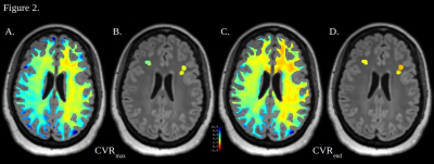 |
White Matter Reactivity to Hemodynamic Stimuli: Insights from
Dynamic and Maximal Cerebrovascular Reserve after Acetazolamide
Provocation
James Michael Gee1,
Xiuyuan Wang2,
Siddhant Dogra1,
Jelle Veraart1,
Koto Ishida3,
Deqiang Qiu4,
and Seena Dehkharghani1,3 1Radiology, New York University, New York, NY, United States, 2Radiology, Weill Cornell Medical College, New York, NY, United States, 3Neurology, New York University, New York, NY, United States, 4Radiology and Imaging Sciences, Emory University, Atlanta, GA, United States Keywords: White Matter, fMRI (resting state), Cerebrovascular reactivity We explored cerebrovascular reactivity (CVR) of white matter in patients with evidence for micro- and macrovascular disease using our previously described Blood Oxygen Level Dependent – acetazolamide paradigm. Subjects consistently demonstrated lower CVR in voxels with microangiopathic white matter when present in hemispheres with coexistent proximal, large-vessel steno-occlusive disease (SOD). The relationship of CVR values in normal appearing white matter and contralateral white matter lesions in patients with unilateral SOD is more complex and warrants further investigation. |
| 14:18 |
0880.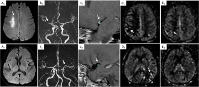 |
Arterial transit artifacts may be easier to identify transient
ischemic attack and stroke in intracranial stenosis than
High-resolution MRI
Ling Li1,
Xiaoling Zhang1,
Min Tang1,
Xiaoyan Lei1,
Jing Zhang1,
Xuejiao Yan1,
Yu Wen1,
and Kai Ai2
1Department of MRI, Shaanxi Provincial People's Hospital, Xi’an, China, 2Philips Healthcare, Xi’an, China Keywords: Stroke, Arterial spin labelling, Arterial transit artifacts, transient ischemic attack, stroke High-resolution magnetic resonance imaging (HRMRI) can be used to characterize the differences in plaque characteristics between transient ischemic attack (TIA) and stroke populations, but it is a time consuming and complicated method.. In this study, we compared the diagnostic performance of HRMRI and arterial spin labeling (ASL) with two post labeling delay times (PLD) combined with clinical risk factors to distinguish TIA and stroke in intracranial stenosis. The results showed that there was no statistical difference between the two methods. In addition, combined model has a good diagnostic performance in predicting stroke occurrence in TIA patients. |
| 14:26 |
0881.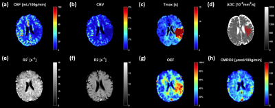 |
Quantification of oxygen metabolism in acute ischemic stroke
with multi-parametric quantitative BOLD MRI
Hongwei Li1,
Ying-Hua Chu2,
Yu Luo3,
and He Wang1,4,5
1Institute of Science and Technology for Brain-Inspired Intelligence, Fudan University, Shanghai, China, 2MR Collaboration, Siemens Healthineers Ltd., Shanghai, China, 3Department of Radiology, Shanghai Fourth People's Hospital Affiliated to TongjiUniversity School of Medicine, Shanghai, China, 4Human Phenome Institute, Fudan University, Shanghai, China, 5Key Laboratory of Computational Neuroscience and Brain-Inspired Intelligence (Fudan University), Ministry of Education, Shanghai, China Keywords: Stroke, Oxygenation Multi-parametric quantitative BOLD (Mq-BOLD), a biophysical model-based technology, has shown promising results for measuring brain oxygenation in healthy subjects. Our purpose is to explore Mq-BOLD in evaluating the changes of oxygen metabolism in early stage of acute ischemic stroke. There is compensatory elevated oxygen extraction fraction (OEF) in the hypoperfused tissue, but decreased cerebral metabolic rate of oxygen (CMRO2) present in the central part of the infarct core. These alternative biomarkers may provide intuitive diagnostic information of ischemic penumbra in stroke. Moreover, Mq-BOLD only depends on standard clinical imaging protocols, which has a great potential for future routine application. |
| 14:34 |
0882.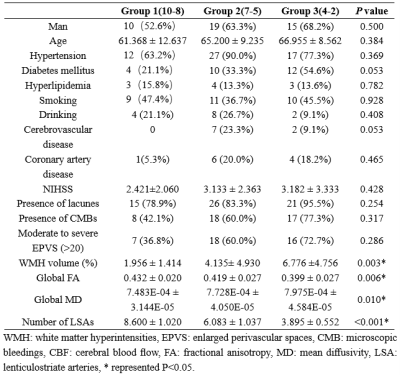 |
Relationship between Lenticulostriate Arteries Numbers and White
Matter Changes in Cerebral Small Vessel Disease Patients
Yukun Zhang1,
Peipei Chang1,
Na Liu1,
Yiming Wang2,
Liangjie Lin3,
Qingwei Song1,
and Yanwei Miao1
1the First Affiliated Hospital of Dalian Medical University, Dalian, China, 2Clinical and Technical Support, Philips Healthcare, shanghai, China, 3Clinical and Technical Support, Philips Healthcare, beijing, China Keywords: Stroke, White Matter, Cerebral Small Vessel Disease In this study, according to the number of lenticulostriate arteries (LSAs), cerebral small blood vessel disease (CSVD) was divided into three groups. Differences of typical CSVD markers and whole brain analysis based on diffusion tensor imaging (DTI) were explored among three groups. It was found that with the decrease of the number of LSAs, the volume of WMH and the scope and extent of fiber tract damage increased. Results show that the change of LSAs have the potential to represents extensive subcortical microvascular damage of brain tissues in CSVD. |
| 14:42 |
0883. |
Microvascular response of an oxygen carrier in the penumbral
tissue of fast and slow progressors in a canine large vessel
occlusion model
Mohammed Salman Shazeeb1,
Robert King1,
Zeynep Vardar1,
Josephine Kolstad1,
Anna Kuhn1,
Vania Anagnostakou1,
Christopher Raskett1,
Jonathan Winger2,
Ana Krtolica3,
Nils Henninger1,
and Matthew Gounis1
1University of Massachusetts Chan Medical School, Worcester, MA, United States, 2Omniox, Inc., Palo Alto, CA, United States, 3Oryn Therapeutics, Redwood City, CA, United States Keywords: Stroke, Diffusion/other diffusion imaging techniques In acute ischemic stroke due to large vessel occlusion (LVO), information about the penumbral tissue can be vital in making decisions on how to treat ischemic stroke patients in the clinic. This study investigated the use of intravoxel incoherent motion (IVIM) MRI in a canine LVO model to quantify the perfusion information in penumbral tissue of fast and slow progressors that received an oxygen carrier drug. The IVIM parameters were assessed to predict the onset of penumbral tissue death. The IVIM parameters showed a better utility in predicting penumbral tissue death in fast progressors compared to slow progressors. |
| 14:50 |
0884. |
Quantitative evaluation of mannitol induced effects on ischemic
rat brains using free water elimination and mapping with
explicit T2 attenuation
Chia-Wen Chiang1,
Ezequiel Farrher2,
Kuan-Hung Cho1,
Sheng-Min Huang1,
N. Jon Shah2,3,4,5,6,
Chang-Hoon Choi2,
and Li-Wei Kuo1,7
1Institute of Biomedical Engineering and Nanomedicine, National Health Research Institutes, Miaoli, Taiwan, 2Institute of Neuroscience and Medicine – 4, Medical Imaging Physics, Forschungszentrum Jülich, Jülich, Germany, 3Department of Neurology, Faculty of Medicine, RWTH Aachen University, Aachen, Germany, 4JARA – BRAIN – Translational Medicine, RWTH Aachen University, Aachen, Germany, 5Institute of Neuroscience and Medicine – 11, Forschungszentrum Jülich, Jülich, Germany, 6Monash University, Melbourne, Australia, 7National Taiwan University College of Medicine, Taipei, Taiwan Keywords: Stroke, Treatment Cerebral edema occurs after stroke. Mannitol is commonly used dehydrating agent for effectively improving cerebral edema. However, the edema state after mannitol administration and its effectiveness at the post-acute stages remain unclear. Recently, we have demonstrated free-water elimination and mapping (FWET2) can be used to characterize vasogenic edema in stroke animal models. The aim of this study was to quantitatively evaluate mannitol-induced effects on ischemic rat brains at different time points by using FWET2. We attempted to examine if free water fraction change could reflect mannitol-induced effects on infraction and correlate free water fraction change with total infarct volume. |
| 14:58 |
0885.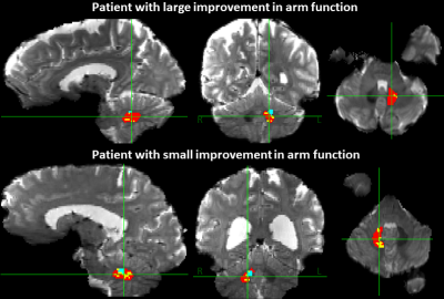 |
Pre-surgical structural and functional MRI to optimize
cerebellar stimulation electrode placement for chronic stroke
therapy
Jacqueline Chen1,
Ajay Nemani1,
Julio Cesar de Almeida2,3,
Xuemei Huang1,
Leonardo Favi Bocca4,
Kenneth B Baker1,
Mark J Lowe1,
Stephen E Jones1,
and Andre G Machado1
1Cleveland Clinic, Cleveland, OH, United States, 2Santa Casa de Belo Horizonte Hospital, Belo Horizonte, Brazil, 3Universidade Federal de Minas Gerais, Belo Horizonte, Brazil, 4Universidade Federal de Sao Paulo, Sao Paulo, Brazil Keywords: Stroke, fMRI (resting state), deep brain stimulation To improve reproducibility of positive outcomes from cerebellar dentate stimulation for chronic stroke, we hypothesized that the volume of tissue activated (VTA) (dentateVTA) proximity to the volume functionally connected to ipsilesional motor-associated cortices (dentateFC-ipsi-motor) would be associated with improved arm impairment/function metrics. DentateVTA was estimated using computed tomography and T1-weighted MRI. DentateFC-ipsi-motor was estimated from fMRI (resting state). Significant correlations with arm metric improvement were not found for dentateVTA proximity to dentateFC-ipsi-motor, but were found for dentateVTA size, distance from center, and percentage VTA in the inferior-lateral-posterior region. These findings suggest stimulating a small target at the inferior-posterior-lateral dentate edge. |
| 15:06 |
0886.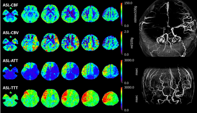 |
The validation of ASL-aCBV measured by Hadamard encoded ASL
imaging evaluating moyamoya disease correlative study with
15O-H2O PET-aCBV.
Hirohiko Kimura1,2,
Makoto Isozaki3,
Shota Ishida4,
Naoyuki Takei5,
Yasuhiro Fujiwara6,
Yuki Matta7,
HIdehikko Okazawa8,
and Tetsuya Tsujikawa9
1Faculty of Medical Sciences, University of Fukui, Eiheiji, Japan, 2Radiology, National Health Insurance Echizen-cho Ota Hospital, Fukui, Japan, 3Neurosurgery, University of Fukui, Eiheiji, Japan, 4Department of Radiological Technolog, Kyoto College of Medical Science, Kyoto, Japan, 5GE Healthcare, Hino, Japan, 6Kumamoto University, Kumamoto, Japan, 7University of Fukui Hospital, Eiheiji, Japan, 8Biomedical Imaging Research Center, University of Fukui, Eiheiji, Japan, 9Department of Radiology, University of Fukui, Eiheiji, Japan Keywords: Stroke, Perfusion, Arterial spin labeling, CBF, CBV Positron emission computed tomography (PET) has been used for evaluating cerebral blood flow (CBF) in moyamoya disease to diagnose and assess revascularization result for the patient. Using Hadamard encoded method and DANTE vascular suppression, arterial transit time (ATT) and delay corrected both CBF and aCBV could be obtained in clinical setting. This study aimed to clarify whether ASL- aCBV hemodynamically related to PET-V0 obtained from PET data. There was a significant correlation between PET and CBF both on CBF and aCBV. This may suggest that aCBV could become an additional hemodynamic parameter related to aCBV in completely non-invasive way. |
| 15:14 |
0887.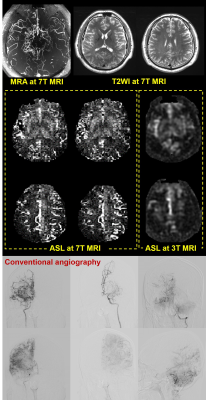 |
Pseudo-continuous arterial spin labeling evaluation of
collateral circulation at 7T and 3T MRI in Moyamoya disease
Jinhao Lyu1,
Qi Duan2,
Caohui Duan2,
Xiangbing Bian2,
Danny JJ Wang3,
Chenyang Zhao3,
Jianxun Qu4,
and Lou Xin2
1Radiology, Chinese PLA General Hospital, Beijing, China, 2Chinese PLA General Hospital, Beijing, China, 3Mark & Mary Stevens Neuroimaging and Informatics Institute Keck School of Medicine University of Southern California (USC), Los Angeles, CA, United States, 4MR Collaboration, Siemens Healthineers Ltd., Beijing, China Keywords: Stroke, Perfusion We compared the performance of pseudo-continuous arterial spin labeling (ASL) between ultrahigh field 7T MRI and 3T MRI in evaluating collateral circulation based on arterial transit artifact (ATA) in Moyamoya disease (MMD). 7T ASL shows more subtle hypoperfusion and more prominent ATA as compared with 3T ASL. The performance of collateral circulation assessment is more favorable by 7T ASL. These findings render 7T ASL a competing none-invasive approach in the management of MMD. |
| 15:22 |
0888.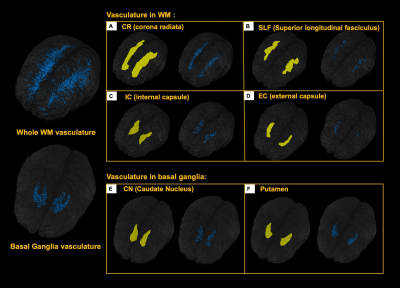 |
Mapping the vascular reserve in white matter and basal ganglia
using Ferumoxytol-enhanced MRI at 7T
Chenyang Li1,2,
Li Jiang1,
Zhe Sun1,2,
Marco Muccio1,
Yongsheng Chen3,
Sagar Buch3,
E.Mark Haacke4,
Jiangyang Zhang1,
and Yulin Ge1
1Department of Radiology, NYU Grossman School of Medicine, New York, NY, United States, 2Vilcek Institute of Graduate Biomedical Sciences, NYU Grossman School of Medicine, New York, NY, United States, 3Department of Neurology, Wayne State University School of Medicine, Detroit, MI, United States, 4Department of Radiology, Wayne State University School of Medicine, Detroit, MI, United States Keywords: Blood vessels, Susceptibility, Cerebral blood volume In this work, we use high resolution susceptibility weighted imaging (SWI) and ΔR2* to investigate the cerebral blood volume (CBV) in white matter tracts and basal ganglia after the administration of blood-pool contrast agent Ferumoxytol. Utilizing the merit of ultrahigh field and strong T1 and T2* shortening contrast agent, we further probe the effect of microvascular density on capillary CBV in white matter tracts and basal ganglia, which provides additional insights to the vascular reserve at capillary level in white matter and basal ganglia. |
 Back to Meeting Home
 Back to the Program-at-a-Glance
The International Society for Magnetic Resonance in Medicine is accredited by the Accreditation Council for Continuing Medical Education to provide continuing medical education for physicians.
 Back to Meeting Home
 Back to the Program-at-a-Glance
 View Presentation Video