Oral Session
White Matter
ISMRM & ISMRT Annual Meeting & Exhibition • 03-08 June 2023 • Toronto, ON, Canada

| 08:15 |
1124.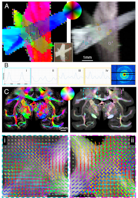 |
Detecting crossing fibers in animal and human brain using small
angle X-ray scattering and comparison to diffusion MRI
Marios Georgiadis1,2,
Miriam Menzel3,4,
Jan Andre Reuter3,
Aileen Schroeter2,
Zirui Gao2,
Sophie Kovacevich1,
Dario Alvarez1,
Manuel Guizar-Sicairos5,
Markus Rudin2,
Donald Born1,
Thomas M Weiss6,
Ivan Rajkovic6,
Markus Axer3,
and Michael M Zeineh1 1Stanford University School of Medicine, Stanford, CA, United States, 2ETH Zurich, Zurich, Switzerland, 3Forschungszentrum Jülich, Jülich, Germany, 4Delft University of Technology, Delft, Netherlands, 5Paul Scherrer Institute, Villigen, Switzerland, 6SLAC National Accelerator Laboratory, Menlo Park, CA, United States Keywords: White Matter, Tractography & Fibre Modelling Mapping neuronal trajectories requires accurate determination of fiber crossings. Diffusion MRI detects fiber orientations but is affected by multiple brain structures and requires constant validation. Small-angle X-ray scattering (SAXS) can specifically image myelinated axons exploiting myelin’s periodic nanostructure. However, its capability to detect crossing fibers is still unexplored. We show that SAXS detects multiple crossing fibers using human corpus callosum strips, and in white and gray matter of mouse, vervet monkey, and human brain. We compare results to polarized light and tracer experiments and show that SAXS more sensitively detects fiber crossings compared to diffusion MRI on the same samples. |
| 08:23 |
1125.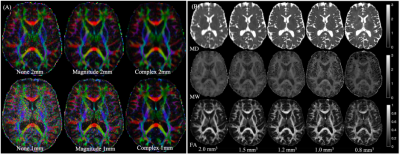 |
Direct visualization of small anisotropic brain structures using
high resolution, multi-shell diffusion MRI
Benjamin Ades-Aron1,
Valentin Stepanov1,
Santiago Coelho1,
Alon Mogilner2,
Dmitry S. Novikov1,
Timothy M. Shepherd1,
and Els Fieremans1
1Radiology, Center for biomedical imaging, NYU Grossman School of Medicine, New York, NY, United States, 2Neurosurgery, NYU Grossman School of Medicine, New York, NY, United States Keywords: White Matter, Data Analysis, high-resolution, visualization In vivo visualization and quantitative assessment of small anisotropic brain structures is challenging with diffusion MRI because of signal-to-noise limitations. We show that direct visualization of small structures, like the fornix or stria medullaris, dramatically improves using denoised 1-mm isotropic resolution complex-valued dMRI data. This approach also reduces the bias and variance of diffusion parameter estimations for small structures. Increased resolution with denoising decreases parameter variance due to thermal noise and partial volume effects – these data also can be used to better estimate the true biological variance in a structure by extrapolating the data to the infinite SNR limit. |
| 08:31 |
1126.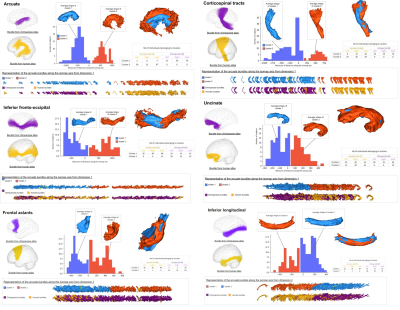 |
Morphological comparison of deep white matter bundles between
the human and chimpanzee brain using a geometrical approach
Maelig Chauvel1,
Ivy Uszynski1,
Marco Pascucci1,
Bastien Herlin1,
Yann Leprince2,
Jean-François Mangin1,
William Hopkins3,
and Cyril Poupon1
1Université Paris-Saclay, CEA, CNRS, UMR 9027, BAOBAB, NeuroSpin , Gif-sur-Yvette, France, Saclay, France, 2UNIACT, NeuroSpin, Université Paris-Saclay, CNRS, CEA, Gif-sur-Yvette, France, Saclay, France, 3Michele E Keeling Center for Comparative Medicine and Research, The University of Texas MD Anderson Cancer Center, Bastrop, Texas, TX, United States Keywords: White Matter, Brain Connectivity, chimpanzee, white matter atlas Humans and chimpanzees are related by a common ancestor that lived around 6 to 7 millions years ago. From then, a cascade of acquired brain features have occurred and the scientific community has tried for years to capture them. Providing measures of the hominin brain divergences or conserved characters can be challenging considering the multitude of variables involved. We propose here a study relying on an intuitive and yet innovative morphological analysis of the deep white matter (DWM) bundles of the human and chimpanzee brain using isomap algorithm. |
| 08:39 |
1127.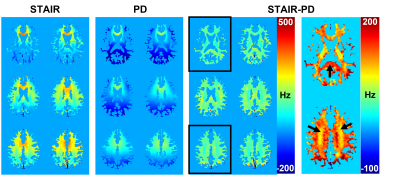 |
3D volumetric myelin water phase mapping via short-TR adiabatic
inversion recovery (STAIR) MRI
Soo Hyun Shin1,
Dina Moazamian1,
James Lo1,2,
Hyungseok Jang1,
Michael Carl3,
Eric Y. Chang1,4,
Jiang Du1,2,4,
and Yajun Ma1
1Department of Radiology, University of California, San Diego, La Jolla, CA, United States, 2Department of Bioengineering, University of California, San Diego, La Jolla, CA, United States, 3GE Healthcare, San Diego, CA, United States, 4Radiology Service, Veterans Affairs San Diego Healthcare System, La Jolla, CA, United States Keywords: White Matter, Contrast Mechanisms Demyelination is a common hallmark of neurodegenerative diseases such as multiple sclerosis. Thus, selective imaging of myelin will greatly enhance the diagnosis of such diseases. Here, we demonstrate the feasibility of acquiring whole-brain 3D myelin water phase maps via short-TR adiabatic inversion recovery (STAIR) MRI. The phase of proton density-weighted (PD) images was subtracted from the phase of STAIR images to remove the background field. The overall positive frequency shift, with a larger shift in the splenium of the lower slice, matches the white matter fiber orientation. |
| 08:47 |
1128.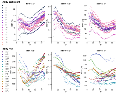 |
The apparent orientation dependence of MTR, ihMTR and MWF is
affected by local microstructural properties in white matter
Sarah Rosemary Morris1,2,3,
Irene M. Vavasour3,4,
Anastasia Smolina5,6,
Erin MacMillan4,7,
Guillaume Gilbert7,
Michelle Lam1,4,
Piotr Kozlowski1,2,3,4,
Carl A. Michal1,
Alan Manning1,
Alex L. MacKay1,3,4,
and Cornelia Laule1,2,3,4,8
1Physics & Astronomy, University of British Columbia, Vancouver, BC, Canada, 2International Collaboration on Repair Discoveries, Vancouver, BC, Canada, 3Radiology, University of British Columbia, Vancouver, BC, Canada, 4UBC MRI Research Centre, Vancouver, BC, Canada, 5Medical Biophysics, University of Toronto, Toronto, ON, Canada, 6The Hospital for Sick Children, Toronto, ON, Canada, 7MR Clinical Science, Philips Healthcare Canada, Markham, ON, Canada, 8Pathology & Laboratory Medicine, University of British Columbia, Vancouver, BC, Canada Keywords: White Matter, CEST & MT, inhomogeneous Magnetization Transfer, ihMT, myelin water fraction, MWF, magnetization transfer, MT, fibre direction, orientation dependence, diffusion tensor imaging (DTI), myelin We measured MTR, ihMTR, MWF and fibre orientation from DTI in 17 white matter regions in 17 healthy adults at 3 T. All three metrics showed an apparent orientation dependence: MWF and ihMTR were lower in fibres perpendicular to B0 by 6% and 1% respectively compared to those parallel, while MTR was lower by 0.5% at ~40°, with the highest values in fibres perpendicular to B0. However, separating the apparent orientation dependence by region revealed large variation in the trends, suggesting that real differences in myelination and other microstructural properties are confounding the apparent orientation dependence measured using this method. |
| 08:55 |
1129. |
7T MR DTI and Tractography of a 1-Week Postmortem Fixed Human
Brainstem-Cerebellum: Novel Imaging Methodology Producing
Extraordinary Images
Sahin Hanalioglu1,
Giancarlo Mignucci-Jiménez2,
Siyar Bahadir1,
Alberto Fuentes3,
Ethan Mathew3,
Gregory Turner3,
and Mark C Preul2 1Department of Neurosurgery, Hacettepe University, Ankara, Turkey, 2Department of Neurosurgery, Barrow Neurological Institute, Phoenix, AZ, United States, 3Barrow Neurological Institute, Phoenix, AZ, United States Keywords: White Matter, Diffusion Tensor Imaging, 7T MRI, Tractography, Connectivity, Postmortem Diffusion-tensor imaging (DTI) is routinely used to estimate the pattern, orientation, and directionality of white matter tracts in human living subjects. However, diffusion imaging of ex-vivo brain specimens is complex due to the diffusivity properties of fixed brain tissue. We developed a novel methodology for tractography of a 1-week postmortem fixed human brainstem and cerebellum specimen utilizing a deterministic fiber tracking algorithm on a 7T MR DTI diffusion scheme registered to an MNI space through landmark-based affine registration. The results showed highly detailed tractography images and agreement between left and right crossing and non-crossing fibers. |
| 09:03 |
1130.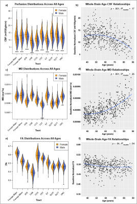 |
Tractwise perfusion-microstructure relationships in the aging
white matter based on the Lifespan Human Connectome Project -
Aging
Tyler D. Robinson1,
Yutong L. Sun1,
Paul T. H. Chang1,
and J. Jean Chen1,2,3
1Rotman Research Institute, Baycrest, Toronto, ON, Canada, 2Medical Biophysics, University of Toronto, Toronto, ON, Canada, 3Biomedical Engineering, University of Toronto, Toronto, ON, Canada Keywords: White Matter, Perfusion This study examined baseline and age-related differences in white matter perfusion and microstructural integrity across ten tracts of interest using 535 adult subjects of the Human Connectome Project in Aging. Whole-brain and tractwise relationships were identified between age, cerebral blood flow, mean diffusivity, and fractional anisotropy. Additionally, regional differences in age-related perfusion and microstructural trajectories were identified, representing one of the first direct examinations of regional variation in white matter perfusion in aging. |
| 09:11 |
1131.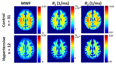 |
Adults with hypertension exhibit lower cerebral myelin content.
John Laporte1,
Mary Faulkner1,
Zhaoyuan Gong1,
Maryam Alsameen1,
Mohammad A.B.S Akhonda1,
Curtis Triebswetter1,
Matthew Kiely1,
and Mustapha Bouhrara1
1Laboratory of Clinical Investigations, National Institute on Aging, Baltimore, MD, United States Keywords: White Matter, Relaxometry Hypertension is a major risk factor for a myriad of neurodegenerative diseases. However, the effect of hypertension on cerebral microstructure, especially myelination, remains poorly understood. We employed multicomponent MR relaxometry and DTI to investigate the association between hypertension and cerebral microstructural integrity, with a focus on myelin content, in a cohort of cognitively unimpaired adults. Our results indicate that adults with hypertension exhibit significantly lower cerebral microstructural integrity and axonal myelination as compared to controls across, several white matter structures. |
09:19 |
1132.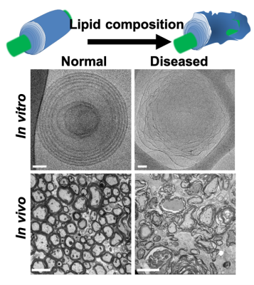 |
In-vivo water gap mapping as a new myelin packing marker
Rona Shaharabani1 and
Aviv Mezer1
1Edmond & Lily Safra Center for Brain Science, The Hebrew University, Jerusalem, Israel Keywords: White Matter, Multiple Sclerosis, Myelin water imaging, water content We established a new in-vivo measurement of the myelin water gap that can serve as a biomarker for myelination packing. In postmortem studies, it was shown that the water layer gap between healthy myelin membranes is compact. However, it changes during the demyelination and remyelination processes in MS patients. We developed a biophysical model based on the water fraction and multi-compartment T2 to estimate the water gap. Next, we designed a lipid phantom system with varying water gaps and validated it using Cryo-TEM. Our model successfully estimates the water gap in-vitro and shows a reliable estimation for in-vivo healthy volunteers. |
| 09:27 |
1133.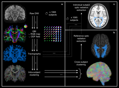 |
Cross-subject variability of the optic radiation anatomy: a
large-scale analysis
Bastien Herlin1,2,3,
Ivy Uszynski1,
Maëlig Chauvel1,
Cyril Poupon1,
and Sophie Dupont2,3
1BAOBAB, NeuroSpin, Université Paris-Saclay, CNRS, CEA, Gif-sur-Yvette, France, 2AP-HP, Epilepsy Unit, GH Pitié-Salpêtrière-Charles Foix, Paris, France, 3Sorbonne Université, Paris, France Keywords: White Matter, Brain Optic radiations are tracts of importance for neurosurgery, especially their path within the temporal lobe. Using an advanced analysis pipeline relying on probabilistic tractography and fiber clustering, we processed the diffusion MRI data of the 1065 subjects of the HCP cohort and reconstructed a reference optic radiation bundle. From it, we subsequently extracted the optic radiations of each subject to study the variability of their morphometry. We identified a higher variability of their rostral extent, with a significant left-right difference (median distance to the temporal pole +/- standard deviation: left: 28.8 +/- 2.3 mm, right: 29.2 +/- 2.1 mm, p=1.10-8). |
| 09:35 |
1134.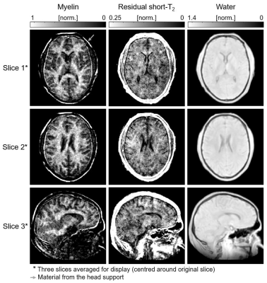 |
Mapping the myelin bilayer with short-T2 MRI: Translation to in
vivo application
Emily Louise Baadsvik1,
Markus Weiger1,
Romain Froidevaux1,
Christoph Michael Schildknecht1,
Benjamin Victor Ineichen2,
and Klaas Paul Pruessmann1
1Institute for Biomedical Engineering, ETH Zurich and University of Zurich, Zurich, Switzerland, 2Department of Neuroradiology, Clinical Neuroscience Center, University Hospital Zurich, University of Zurich, Zurich, Switzerland Keywords: White Matter, Multiple Sclerosis, Myelin imaging Direct access to myelin content through the detection of signals from the myelin lipid-protein bilayer can be achieved using advanced short-T2 techniques. Here, we translate an existing procedure for mapping the myelin bilayer ex vivo to in vivo human application. Myelin maps are generated by fitting a three-component complex model to multi-TE (20–800µs) data acquired using the HYFI variant of the zero-TE sequence. The presented myelin maps exhibit expected white/grey matter contrast and are of reasonable quality. The step to in vivo represents an important advancement for myelin bilayer mapping as a promising emerging technique. |
| 09:43 | 1135. |
Measuring water exchange in myelinated white matter using
Magnetization Transfer (MT)-weighted constant gradient diffusion
MRI (MT-cgdMRI)
Chenyang Li1,2,
Els Fieremans1,
Dmitry S. Novikov3,
and Jiangyang Zhang1
1Department of Radiology, NYU Grossman School of Medicine, New York, NY, United States, 2Vilcek Institute of Graduate Biomedical Sciences, NYU Grossman School of Medicine, New York, NY, United States, 3NYU Grossman School of Medicine, New York, NY, United States Keywords: White Matter, Contrast Mechanisms, Water exchange In white matter, myelin forms a barrier for water exchange between the intra-axonal and extra-axonal compartments. Although myelin is not directly visible in conventional diffusion MRI (dMRI), it may affect dMRI measurements via exchange. In this study, we compared two ways to combine magnetization transfer (MT) preparation with constant gradient dMRI (cg-dMRI) to study the effects of myelin on water exchange rate. Our results demonstrate that MT preparation can modulate the sensitivity of cg-dMRI to exchange by suppressing signals from a portion of exchanging spins and that placing MT preparation is more effective before diffusion encoding than during diffusion encoding. |
| 09:51 |
1136.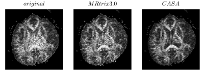 |
Component Analysis based on Standard-deviation Attenuation
(CASA): a new algorithm for the denoising of Diffusion MRI data
Mauro Zucchelli1,
Christos Papageorgakis1,
and Stefano Casagranda1
1Department of R&D Advanced Applications, Olea Medical, La Ciotat, France Keywords: White Matter, Diffusion Tensor Imaging, Denoising, CASA Noise is a crucial problem that affects even the most advanced MRI techniques based on model fitting. It is therefore important to act on the raw data to remove as much noise as possible, while preserving the anatomical structures. Many techniques based on Principal Component Analysis (PCA) take advantage of the redundancy of information contained in multiphase data, to perform robust denoising. In this work, we introduce a new denoising method based on PC images. We show the added value of our technique on both the raw data and the derived fractional anisotropy map. |
| 09:59 |
1137.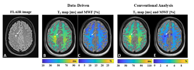 |
A data-driven paradigm for myelin water imaging – validation on
multicompartment phantom and multiple sclerosis patient
Sharon Zlotzover1,
Noam Omer1,
Neta Stern1,
Dvir Radunsky1,
Tamar Blumenfeld-Katzir1,
and Noam Ben-Eliezer1,2,3
1The Department of Biomedical Engineering, Tel-Aviv University, Tel-Aviv, Israel, 2Sagol School of Neuroscience, Tel-Aviv University, Tel Aviv, Israel, 3Center for Advanced Imaging Innovation and Research (CAI2R), New-York University Langone Medical Center, New York, NY, United States Keywords: White Matter, Multiple Sclerosis, Myelin Multicomponent (MC) T2 analysis is a common technique for probing sub-voxel compartmentation in myelinated tissues. However, the task of resolving the T2 spectra from a single voxel is ill-posed and highly sensitive to noise. Applying a spatially-global MC analysis of the tissue prior to the voxel-wise analysis promotes more stable solutions. Specifically, this approach identifies a specific set of MC T2 features which are then used for fitting the signal in each voxel. Preliminary results suggest that the new data-driven approach can accurately estimate myelin content and correctly estimate myelin content in healthy tissue and in multiple sclerosis lesions. |
| 10:07 |
1138.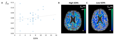 |
Assessing microstructural and microvascular abnormalities in
hospitalized COVID-19 patients using intravoxel incoherent
motion imaging
Noa van der Knaap1,2,3,
Simona Klinkhammer1,4,
Alida A. Postma1,3,
Arjen J.C. Slooter5,6,
Janneke Horn7,8,
Caroline M. van Heugten4,9,
Paulien H.M. Voorter1,3,
Merel M. van der Thiel1,3,
Gerhard S. Drenthen1,3,
Walter H. Backes1,3,
David E.J. Linden1,
Marcel J.H. Ariës1,2,
and Jacobus F.A. Jansen1,3,10
1School for Mental Health & Neuroscience, Maastricht University, Maastricht, Netherlands, 2Department of Intensive Care, Maastricht University Medical Center, Maastricht, Netherlands, 3Department of Radiology and Nuclear Medicine, Maastricht University Medical Center, Maastricht, Netherlands, 4Limburg Brain Injury Center, Maastricht University, Maastricht, Netherlands, 5Department of Intensive Care, University Medical Center Utrecht, Utrecht, Netherlands, 6UMC Utrecht Brain Center, University Medical Center Utrecht, Utrecht, Netherlands, 7Department of Intensive Care, University Medical Center Amsterdam, Amsterdam, Netherlands, 8Amsterdam Neuroscience, University Medical Center Amsterdam, Amsterdam, Netherlands, 9Department of Neuropsychology and Psychopharmacology, Maastricht University, Maastricht, Netherlands, 10Department of Electrical Engineering, Eindhoven University of Technology, Eindhoven, Netherlands Keywords: Infectious disease, COVID-19 Cerebral abnormalities are common in (severely affected) COVID-19 patients, although most reports only cover macrostructural abnormalities. Zooming in on microstructural abnormalities may better explain persisting COVID-19-related symptoms in COVID-19 survivors. Using intravoxel incoherent motion (IVIM) imaging, this study explored potential differences in microstructural and microvascular diffusivity between COVID-19 patients. No differences were found between patients admitted to the intensive care unit (ICU) (n=40) and general ward (n=38). However, increased disease severity in COVID-19 ICU patients was found to be associated with increased interstitial fluid content in the normal appearing white matter. |
 Back to Meeting Home
 Back to the Program-at-a-Glance
The International Society for Magnetic Resonance in Medicine is accredited by the Accreditation Council for Continuing Medical Education to provide continuing medical education for physicians.
 Back to Meeting Home
 Back to the Program-at-a-Glance
 View Presentation Video