Oral Session
Pediatric Neuroimaging
ISMRM & ISMRT Annual Meeting & Exhibition • 03-08 June 2023 • Toronto, ON, Canada

08:15 |
0039.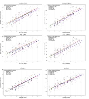 |
Total and regional brain volumes in fetuses with congenital
heart disease
Daniel Cromb1,2,
Alena Uus1,2,
Milou Van Poppel3,
Johannes Steinweg3,
Alexandra Bonthrone1,2,
Alessandra Maggioni1,
Paul Cawley1,4,
Vanessa Kyriakopoulou1,
Jacqueline Matthew1,
Anthony Price1,2,
A David Edwards1,2,
Maria Deprez1,2,
Joseph V Hajnal1,2,
David F Lloyd1,3,5,
Kuberan Pushparajah1,3,5,
John Simpson1,3,5,
Mary Rutherford1,4,
and Serena J Counsell1,2
1Centre for the Developing Brain, School of Biomedical Engineering and Imaging Sciences, King's College London, London, United Kingdom, 2Biomedical Engineering Department, School of Biomedical Engineering and Imaging Sciences, King's College London, London, United Kingdom, 3Department of Cardiovascular Imaging, School of Biomedical Engineering & Imaging Science, King's College London, London, United Kingdom, 4MRC Centre for Neurodevelopmental Disorders, King's College London, London, United Kingdom, 5Paediatric Cardiology, Evelina London Children's Hospital, London, United Kingdom Keywords: Fetal, Brain, Brain Volumes, Congenital Heart Disease Total and regional brain volumes, derived from automatically segmented, motion-corrected, 3D fetal brain MR images were obtained in 45 healthy fetuses and 305 fetuses with isolated congenital heart disease (CHD) in the third trimester. Total brain tissue, cortical and deep grey matter, and white matter volumes are significantly lower in fetuses with CHD where cerebral oxygenation and substrate delivery are likely to be reduced. Brain volumes appear normal in fetuses with CHD but an otherwise expected normal cerebral oxygenation. |
| 08:23 |
0040.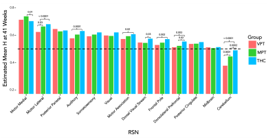 |
Altered Brain Signaling Complexity in Preterm-born and Term-born
Infants at Term Age: a Functional MRI Study
Allison Eve Mella1 and
Alexander Weber2
1Neuroscience, University of British Columbia, Vancouver, BC, Canada, 2Pediatrics, University of British Columbia, Vancouver, BC, Canada Keywords: Neonatal, fMRI (resting state), Neuroscience We compared Hurst exponent (H) values in preterm and term healthy controls to quantify brain signaling criticality using rsfMRI scans. Neonatal data from the Developing Human Connectome Project was analyzed. We found that H significantly increased in the preterm groups longitudinally in all resting state networks. Motor and sensory networks were found to have the greatest increase in H. At term age, very preterm, moderately preterm, and health controls displayed different H values in 8 of the 13 networks examined. |
| 08:31 |
0041.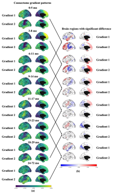 |
Development of Infant Brain Functional Connectome Gradients
during Age 0-6 Years
Xinyi Cai1,
Lianghu Guo1,
Mianxin Liu1,
Feihong Liu1,
Jiawei Huang1,
Jiameng Liu1,
Qing Yang1,
Lang Mei1,
Tianli Tao1,
Zhuoyang Gu1,
Xiaozhao Liu1,
Yuxiao Liu1,
Xiangnan Tian1,
Qian Chen1,
Ruoming Wang1,
Yizhou Shi1,
Qian Wang1,
Han Zhang1,
and Dinggang Shen1
1School of Biomedical Engineering, ShanghaiTech University, Shanghai, China Keywords: Normal development, Brain, Infant Studies have revealed a particular functional connectome gradient (FC-grad) pattern in the human brain, reflecting delicate organization of brain connectome. Interestingly, the pattern can be observed along childhood-adolescent, even in neonates. However, FC-grad changes from infancy to childhood remain unraveled, with significant disparities between neonates and school-aged children not explained. We explored the early development trajectories of the FC-grads between age 0-5 years. We found that the neonatal “prototypic” connectome gradients undergoes rapid changes in this period, especially the first 12 months, reflecting maturing functional integration. Our study filled the gap of FC-grad development in early infancy. |
| 08:39 |
0042.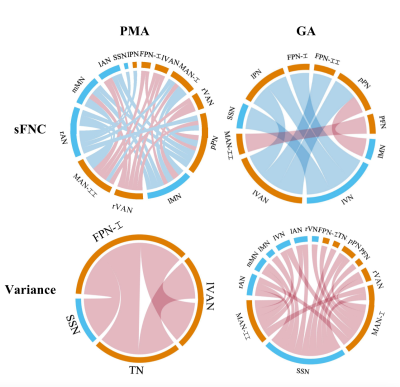 |
Dynamic functional network connectivity in neonatal brain
Ruolin Li1,
Yihan Wu1,
Yifan Shuai1,
Zhiyong Zhao1,
and Dan Wu1
1Key Laboratory for Biomedical Engineering of Ministry of Education, Department of Biomedical Engineering, College of Biomedical Engineering & Instrument Science, Zhejiang University, Hangzhou, China Keywords: Neonatal, Normal development, brain network The study aimed to explore the changes of dynamic functional network connectivity (FNC) with age in neonatal brain. We used independent component analysis to extract 17 resting-state networks, and calculated their static and dynamic FNC (sFNC/dFNC). We found that the variance of dFNC significantly correlated with GA, and the sFNC significantly correlated with PMA controlling GA as a covariate. Moreover, the preterm- than term-born neonates showed decreased variances of dFNC between almost all networks, and altered sFNC between several networks. These findings suggest that the time-varying FNCs are modulated more by maternal environment than postnatal experience during early brain development. |
| 08:47 |
0043.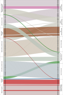 |
Structural reorganisation in higher-order brain networks of the
common marmoset from infancy to adulthood revealed by DTI
tractography
Stephen Sawiak1,
Michal Graczyk1,
Gemma J Cockcroft1,
Lauren McIver1,
Edward Bullmore2,
and Angela Roberts1
1Translational Neuroimaging Laboratory; Physiology, Development and Neuroscience, University of Cambridge, Cambridge, United Kingdom, 2University of Cambridge, Cambridge, United Kingdom Keywords: Brain Connectivity, Brain Connectivity Adolescence is a critical period in development where neuropsychiatric symptoms emerge. The most striking morphological changes in the brain throughout this time are seen in white matter. Here we present a large longitudinal non-human primate study mapping detailed changes in the structural DTI connectome from infancy to adulthood, showing changes in connectivity across this period in higher order association areas, particularly in prefrontal cortex. We demonstrate integration of subcortical structures across hemispheres in the adult brain alongside differentiation of right and left prefrontal areas in their community structure. |
| 08:55 |
0044.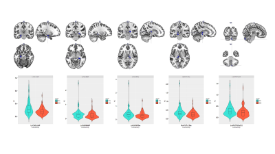 |
Causal evidence for cerebello-limbic-striatal circuit dynamics
supporting depression
Ruiping Zheng1,
Jingliang Cheng1,
and Yong Zhang1
1MRI, the first affiliated hospital of zhengzhou university, zhengzhou, henan, China Keywords: Adolescents, Neuroscience, Resting-state functional connectivity The striatum is known to be impaired in MDD patients. Abnormal structure or function of the striatum may disturb the top-down regulation of negative emotions among persons more vulnerable to developing depressive state, especially adolescent. Here, we investigated (1) voxel-wise FC between the striatal nucleus and the whole brain; (2) region of interest wise effective connectivity using DCM analysis, according to the between-group differences in FC of the striatal nucleus. We found decreased FC in cerebello-limbic-striatal circuit, and further DCM analysis showed the dysregulation of vrPUT nucleus disturb the top-down regulation of cerebello-limbic-striatal circuit during reward processing in adolescent MDD. |
| 09:03 |
0045.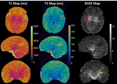 |
Motion robust MR Fingerprinting scans for non-sedated infant
imaging
Chaitra Badve1,
Jessie EP Sun2,
Ameya Nayate1,
Michael Wien1,
Douglas Martin1,
Jared Durieux1,
Chris Flask2,
Deanne Wilson Costello3,
and Dan Ma4
1Radiology, University Hospitals Cleveland Medical Center, Cleveland, OH, United States, 2Radiology, Case Western Reserve University, Cleveland, OH, United States, 3Neonatology, University Hospitals Cleveland Medical Center, Cleveland, OH, United States, 4Biomedical Engineering, Case Western Reserve University, Cleveland, OH, United States Keywords: Neonatal, MR Fingerprinting The 5 min high resolution MRF scans coupled with low-rank iterative reconstruction successfully generated perfectly co-registered T1, T2 maps, synthetic MR contrast images, R1R2 maps, and myelin water fraction maps. Image Quality Assessment analysis with three pediatric neuroradiologists found that MRF based synthetic T1w and T2w images quality were superior in quality to MRI T1w and T2w (p<.0001) with lower image artifacts in the MRF synthetic images as compared to standard of care MRI. MRF T1w images demonstrated better myelin visualization compared to clinical T1w, and MRF T2w demonstrated improved tissue structure visualization as compared to clinical T2w images. |
|
09:11 |
0046.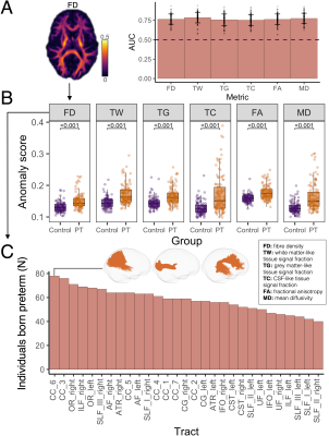 |
Detecting individual variation in white matter microstructure in
a high-risk infant population using deep normative modelling
Claire E Kelly1,2,3,
Peter J Anderson1,2,
Sila Genc3,4,
Thijs Dhollander3,
Deanne K Thompson2,3,5,
Alice C Burnett2,5,6,7,
Lex W Doyle2,5,6,8,
and Jeanie LY Cheong2,6,8
1Turner Institute for Brain and Mental Health, School of Psychological Sciences, Monash University, Melbourne, Australia, 2Victorian Infant Brain Studies (VIBeS), Murdoch Children's Research Institute, Melbourne, Australia, 3Developmental Imaging, Murdoch Children's Research Institute, Melbourne, Australia, 4Neurosurgery Advanced Clinical Imaging Service (NACIS), Department of Neurosurgery, The Royal Children's Hospital, Melbourne, Australia, 5Department of Paediatrics, The University of Melbourne, Melbourne, Australia, 6The Royal Women’s Hospital, Melbourne, Australia, 7Neonatal Medicine, The Royal Children's Hospital, Melbourne, Australia, 8Department of Obstetrics and Gynaecology, The University of Melbourne, Melbourne, Australia Keywords: Neonatal, Neuro There is large inter-individual variation in cognitive outcomes of adults born preterm, which may relate to under-recognised heterogeneity in white matter tract microstructural disturbances. This study created a normative model of white matter tract profiles using deep learning in term-born adults (n=95). The model was then applied to detect anomalies in tract profiles of preterm adults (n=111). The location and extent of anomalies varied across preterm adults. Further, tract anomalies were correlated with neonatal brain injury and IQ. Thus, this study demonstrates inter-individual variation in white matter abnormalities in preterm adults, helping to explain variation in cognitive outcomes. |
| 09:19 |
0047.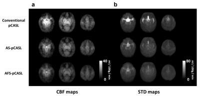 |
Macrovasculature-suppressed ASL MRI in neonates: quantification
of cerebral blood flow and arterial transit time
Zhiyi Hu1,
Dengrong Jiang2,
Jennifer Shepard3,
Yuto Uchida2,
Kenichi Oishi2,
Peiying Liu2,4,
Doris Lin2,
Vivek Yedavalli2,
Aylin Tekes2,
W. Christopher Golden3,
and Hanzhang Lu1,2,5
1Department of Biomedical Engineering, Johns Hopkins University School of Medicine, Baltimore, MD, United States, 2Department of Radiology, Johns Hopkins University School of Medicine, Baltimore, MD, United States, 3Department of Pediatrics, Johns Hopkins University School of Medicine, Baltimore, MD, United States, 4Department of Diagnostic Radiology and Nuclear Medicine, University of Maryland School of Medicine, Baltimore, MD, United States, 5F.M. Kirby Research Center for Functional Brain Imaging, Kennedy Krieger Research Institute, Baltimore, MD, United States Keywords: Neonatal, Perfusion A prominent feature in neonatal cerebral blood flow (CBF) measurement is the hyperperfusion in the deep brain region. Given the rich presence of large arteries in this region, it is plausible that macrovascular artifacts may play a major role in the hyperintense signals observed. This study presented a new MRI technique, pCASL with arterial suppression and flow suppression (AFS-pCASL) to minimize the macrovascular artifacts in neonates. We demonstrated that macrovascular artifacts in neonatal pCASL can be substantially suppressed, from which quantitative CBF and arterial transit time can be measured when applying the sequence in a multi-delay setting. |
| 09:27 |
0048.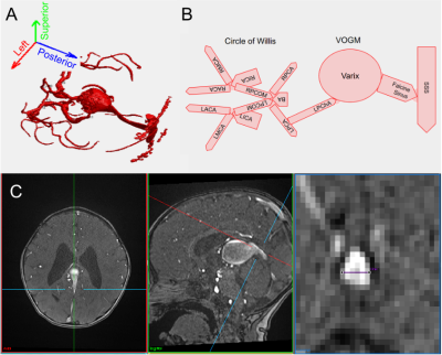 |
Comparing vascular morphology and hemodynamics in patients with
Vein of Galen Malformations using intracranial 4D flow MRI
Jeffrey N Stout1,
Julie Swanson1,
Alfred P See2,
Shivani Rangwala1,
and Darren B Orbach1
1Cerebrovascular Surgery and Interventions Center, Boston Children's Hospital, Boston, MA, United States, 2Department of Neurosurgery, CSIC, Boston Children's Hospital, Boston, MA, United States Keywords: Blood vessels, Blood vessels, 4D Flow, Vein of Galen Malformation, blood flow Vein of Galen Malformation is the most common congenital cerebrovascular malformation and patients have variable mortality risk and cognitive outcomes. Tools are needed to guide treatment decisions, so we investigated the hemodynamic underpinnings of a morphological metric used for clinical prognostication. We used 4D flow MRI to quantify flow through the venous sinus that drains the malformation, and compared this to the maximum mediolateral diameter of the same sinus. A significant positive correlation between flow and diameter was observed when controlling for prior embolization; this finding motivates continued efforts to develop hemodynamic metrics of disease severity and treatment progress. |
| 09:35 |
0049.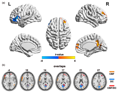 |
Altered neurovascular coupling in children with idiopathic
generalized epilepsy
HaiFeng Ran1,
Jie Hu1,2,
GuiQin Chen1,
YuLun He1,
Heng Liu1,
and TiJiang Zhang1
1Department of Radiology, The Affiliated Hospital of Zunyi Medical University, ZunYi, China, 2Department of Radiology and Nuclear Medicine, Xuanwu Hospital, Capital Medical University, BeiJing, China Keywords: Neuro, Epilepsy, Neurovascular coupling This study investigated the neurovascular coupling of children with idiopathic epilepsy using resting-state functional Magnetic Resonance Imaging and arterial spin labeling. Children with IGE present altered global neurovascular coupling, and higher regional neurovascular coupling in in the right medial frontal gyrus associated with lower performance intelligence quotient scores. The study shed a new insight into the pathophysiology of epilepsy and provided the potential imaging biomarkers of cognitive performances in children with IGE. |
| 09:43 |
0050.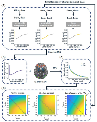 |
Optimization of the T2-weighted MRI contrast in 0-6-month-old
infant brain based on extended phase graph theory
Jiani Wu1,
Hongxi Zhang2,
Haotian Li1,
Siyifei Wang1,
Xingwang Yong1,
and Dan Wu1
1Key Laboratory for Biomedical Engineering of Ministry of Education, Department of Biomedical Engineering, College of Biomedical Engineering & Instrument Science, Zhejiang University, Hangzhou, China, 2Department of Radiology, Children's Hospital, Zhejiang University School of Medicine, Hangzhou, China Keywords: Pulse Sequence Design, Contrast Mechanisms 3D T2-weighted MRI using fast spin-echo (FSE) with variable flip angles has been widely for anatomical imaging. However, 3D FSE of infant brains exhibits poor contrast due to the inherently close and rapidly changing T2 relaxation times between white matter and grey matter. Here we proposed an extended-phase-graph-based method to optimize the flip angles in FSE sequence for maximizing the white/grey matter contrast in 0 to 6 month-old infant brains at 3T, based on T2 values acquired from 37 infant brains. Results demonstrated improved relative contrasts in infant brains by 1.6-2 folds at different ages in different regions of interest. |
| 09:51 |
0051.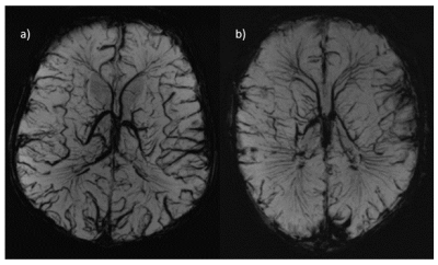 |
Development of safe operating procedures and early experience of
scanning Newborn Infants at 7T
Philippa Bridgen1,2,
Tomoki Arichi 2,3,4,
Megan Quirke2,3,5,
Jennifer Almabis2,3,5,
Daniel Cromb3,
Paul Cawley3,
Raphael Tomi-Tricot1,5,6,
Enrico De Vita7,8,
Anthony N Price2,3,
Alena Uus3,7,
Maira Deprez3,7,
Lucilio Cordero-Grande3,7,9,
Sharon Giles1,2,7,
Serena Counsell3,
Tom Finck3,10,
Mary A Rutherford3,
A David Edwards3,4,
Joseph V Hajnal1,3,7,
and Shaihan J Malik1,3,7
1London Collaborative Ultra high field System (LoCUS), King's College London, London, United Kingdom, 2Guys and St Thomas’ NHS Foundation Trust, London, United Kingdom, 3Centre for the Developing Brain, School of Biomedical Engineering and Imaging Sciences, King's College London, London, United Kingdom, 4MRC Centre for Neurodevelopmental Disorders, King's College London, London, United Kingdom, 5Biomedical Engineering Department, School of Biomedical Engineering and Imaging Sciences, King's College London, London, United Kingdom, 6MR Research Collaborations, Siemens Healthcare Limited, Frimley, United Kingdom, 7School of Biomedical Engineering and Imaging Sciences, Biomedical Engineering Department, King's College London, London, United Kingdom, 8Great Ormond Street Hospital for Children, London, United Kingdom, 9Biomedical Image Technologies, ETSI Telecomunicación, Universidad Politécnica de Madrid, Madrid, Spain, 10Department for Diagnostic and Interventional Neuroradiology, Klinikum rechts der Isar der Technischen Universität München, Munich, Germany Keywords: Neuro, High-Field MRI, 7T; MRI; Newborn Infant We describe operational processes developed for, and our first experiences in, imaging of newborn infants at 7T. Based on an initial safety study, a new SAR model was adopted, and a protocol developed for safe switching of mode prior and post imaging. Monitoring equipment was tested and cleared for use. Sequences for high-resolution and high-contrast brain imaging were optimized within stricter SAR limits. Image quality and SNR were compared at 7T and 3T, with improved anatomical and pathological features seen at 7T. Our study indicates scanning of newborn infants imaging is possible within the safety considerations needed at 7T. |
| 09:59 |
0052.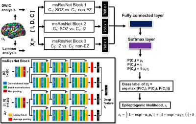 |
A novel ensemble deep learning neural network of multi-scale
deep MRI features to localize the epileptogenic zone in
pediatric epilepsy
Jeong-Won Jeong1,2,
Min-Hee Lee1,2,
Csaba Juhász1,2,
and Eishi Asano1,3
1Pediatrics, Wayne State University, Detroit, MI, United States, 2the Translational Imaging Laboratory, Children's Hospital of Michigan, Detroit, MI, United States, 3Neurology, Children's Hospital of Michigan, Detroit, MI, United States Keywords: Neuro, Epilepsy, Ensemble deep learning, Localization of epileptogenic zone We present a novel ensemble deep learning neural network of multi-scale deep MRI features that can non-invasively localize the epileptogenic zone (EZ) partially overlapping seizure onset zone (SOZ) and irritative zone (IZ) in children with medically intractable epilepsy. The presented network provided high balanced accuracy of 90% to predict SOZ and IZ in 3-fold cross-validation. It also yielded a new MRI marker of epileptogenicity providing a huge effect size between the ground-truth EZ and non-EZ (i.e., Cohen’s d = 2.73), suggesting its high translational value for accurately guiding invasive EEG practice to determine the boundaries of the EZ sites |
| 10:07 |
0053.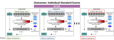 |
Brain Ventricular and Microstructural Correlates of Executive
Dysfunction in Congenital Heart Disease Using Explainable
Machine Learning
Vincent Kyu Lee1,2,
William Thomas Reynolds2,3,
Benjamin Meyers4,
Julia Wallace4,
Daryaneh Badaly5,
Cecilia Lo6,
Ashok Panigrahy1,3,4,
and Rafael Ceschin3,4 1Bioengineering, University of Pittsburgh, Pittsburgh, PA, United States, 2Radiology, UPMC Children's Hospital of Pittsburgh, Pittsburgh, PA, United States, 3Biomedical Informatics, University of Pittsburgh School of Medicine, Pittsburgh, PA, United States, 4Radiology, University of Pittsburgh School of Medicine, Pittsburgh, PA, United States, 5Learning and Development Center, Child Mind Institute, New York, NY, United States, 6Developmental Biology, University of Pittsburgh School of Medicine, Pittsburgh, PA, United States Keywords: Adolescents, Neuro, Congenital Heart Disease Neurodevelopment Machine Learning This study examined cerebrospinal fluid volumes as neuroimaging features and their role in predicting specific executive function impairments among adolescents with congenital heart disease using explainable machine learning models. The findings showed CSF volumes were among the most important predictors of executive function inhibition domain with 3 CSF volumes ranked amongst the top 20, and 4 more CSF volumes among the top 20% of all features. Selective increased lateral ventricular volume in the frontal regions in CHD patients may be secondary to loss of white matter integrity in the uncinate fasciculus (emotional regulation) and subsequently lead to inhibitory dysfunction. |
 Back to Meeting Home
 Back to the Program-at-a-Glance
The International Society for Magnetic Resonance in Medicine is accredited by the Accreditation Council for Continuing Medical Education to provide continuing medical education for physicians.
 Back to Meeting Home
 Back to the Program-at-a-Glance
 View Presentation Video