Oral Session
Advances in RF Coil Arrays
ISMRM & ISMRT Annual Meeting & Exhibition • 03-08 June 2023 • Toronto, ON, Canada

| 15:45 |
1054.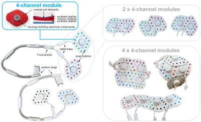 |
First in vivo results with a modular system of flexible coil
arrays for 3 T MRI (ModFlex)
Lena Nohava1,
Michael Obermann1,
Roberta Frass-Kriegl1,
Onisim Soanca1,
and Elmar Laistler1
1High Field MR Center, Center for Medical Physics and Biomedical Engineering, Medical University of Vienna, Vienna, Austria Keywords: RF Arrays & Systems, RF Arrays & Systems Form-fitting radiofrequency coil arrays have the potential to enhance the signal-to-noise ratio, enable faster imaging and improve patient comfort in MRI. We developed a flexible modular coil array system for 3T MRI (ModFlex) with 16 receive channels. In neck, spine, ankle and hip imaging, we measured an SNR gain for 4 out of 6 anatomical target regions and similar SNR for 2 out of 6 as compared to commercial reference coils. The coil’s versatility is beneficial for different use cases with varying subject sizes. |
15:53 |
1055.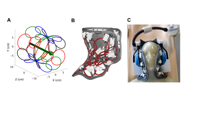 |
The FACE: Flexible Array for Cervical & Extraspinal 3T MR
Imaging
Frederik Abel1,
Ek T. Tan1,
Martijn Lunenburg2,
Carel van Leeuwen2,
Thijs van Hooren2,
Mark van Uden2,
Catalina Arteaga2,
Jana Vincent3,
Fraser Robb3,
Darren R. Lebl1,
and Darryl B. Sneag1
1Hospital for Special Surgery, New York, NY, United States, 2Tesla Dynamic Coils, Zaltbommel, Netherlands, 3GE HealthCare, Aurora, OH, United States Keywords: RF Arrays & Systems, RF Arrays & Systems Conventional cervical coils lack the flexibility to closely conform to the inherently curved neck region, particularly at its head/shoulder junctions. High SNR imaging of the c-spine and extraspinal soft tissues (including small peripheral nerves) relies on close proximity of receive elements to these targeted structures. This study evaluates the performance of a novel, conformal, 23-channel Flexible Array for Cervical & Extraspinal (FACE) 3T MR Imaging with increased flexibility to enhance image quality and SNR compared to conventional coils. SNR measurements on phantoms and high resolution 2D and 3D in vivo c-spine and peripheral nerve neck imaging were performed at 3T. |
16:01 |
1056.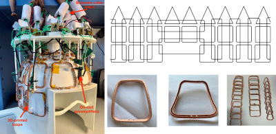 |
A 32-Channel 3D-Printed-Loop Receive Array with Direct
High-Impedance Preamplifiers for Brain Imaging at 7T
Paul-François Gapais1,2,
Michel Luong3,
Eric Giacomini1,
Alexandre Vignaud1,
François Nizery4,
Gabriel Maitre4,
Sajad Hosseinnezhadian2,
Marc Dubois2,
Elodie Georget2,
and Alexis Amadon1
1BAOBAB, Université Paris-Saclay/CEA/Joliot/NeuroSpin, GIF-SUR-YVETTE, France, 2Multiwave Imaging SAS, Marseille, France, Metropolitan, 3DACM, Université Paris-Saclay/CEA/IRFU, GIF-SUR-YVETTE, France, 4LCAP, CEA/DRF/IRFU/DIS, GIF-SUR-YVETTE, France Keywords: RF Arrays & Systems, RF Arrays & Systems, Coil A fully customized 32-channel receive array has been designed, fabricated and evaluated using MR experiments at 7T. The design is made of non-geometrically decoupled loops arranged in two layers of large and small loops. Copper loops are 3D printed from additive manufacturing, preamplifiers are home-built, and an easy implementation procedure is proposed. Receive array performances shows SNR comparable to the Nova Medical reference coil while being superior at the top of the coil and for parallel imaging accelerated in the vertical direction. |
|
16:09 |
1057.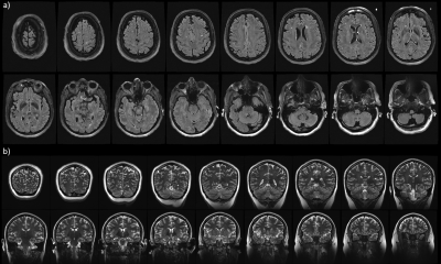 |
An Open 60-channel Tx/ 32-channel Rx RF Coil System for Routine
Use at 7T
Andrea N Sajewski1,
Tales Santini1,
Anthony DeFranco1,
Boris Keil2,
Hecheng Jin1,
Jacob Berardinelli1,
Jinghang Li1,
Cong Chu1,
Tiago Martins1,
and Tamer S Ibrahim1
1University of Pittsburgh, Pittsburgh, PA, United States, 2Institute of Medical Physics and Radiation Protection, TH Mittelhessen University of Applied Sciences, Giessen, Germany Keywords: New Devices, Neuro A 60-channel transmit array for 7T neuro MRI was implemented using the Tic-Tac-Toe coil design. The transmit coil and the receive insert are open in the front to promote patient comfort and reduce claustrophobia. By performing RF shimming on three head models, we optimized the coil for a range of ages and head sizes/shapes for use in sTx mode. Simulations, experimental B1+ maps and in-vivo images demonstrate consistency across subjects, showing extended coverage into the temporal lobe, cerebellum, brainstem and C5-C6 spinal cord segments. High quality images are currently being acquired using this coil in over 30 human studies. |
| 16:17 |
1058.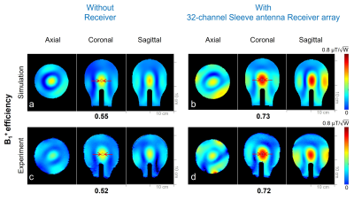 |
Evaluation of Coupling between A 32-channel Sleeve Antenna
Receiver Array to A 16-channel Loop Transmitter for The Human
Head Imaging at 10.5 T
Myung Kyun Woo1,
Lance DelaBarre2,
Matt Waks2,
Steve Jungst2,
Pat Nguyen2,
Russell Lagore2,
Andrea Grant2,
Joo Yoon Jang1,
Alireza Sadeghi-Tarakameh2,
Yigitcan Eryaman 2,
Kamil Ugurbil2,
and Gregor Adriany2 1University of Ulsan, Ulsan, Korea, Republic of, 2Center for Magnetic Resonance Research, Minneapolis, MN, United States Keywords: RF Arrays & Systems, RF Arrays & Systems We evaluated in simulation and experimentally the effects that the insertion of a 32-channel sleeve antenna receiver array has on the B1+ and SAR performance of a 447 MHz/10.5 T 16-channel loop transmitter array. For this we carefully developed accurate models of both the 16-channel loop transmitter and the 32-channel sleeve antenna receiver, compared simulated with experimental B1+ and evaluated the expected SAR efficiency of the 16-channel loop transmitter array with and without the 32-channel sleeve antenna receiver array insert. |
| 16:25 |
1059.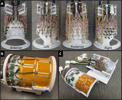 |
128-channel brain imaging array with improved acceleration at
10.5 Tesla
Russell Luke Lagore1,
Andrea Grant1,
Lance DelaBarre1,
Edward J Auerbach1,
Matt Waks1,
Steve Jungst1,
Steen Moeller1,
Jerahmie Radder1,
Alireza Sadeghi-Tarakameh1,
Yigitcan Eryaman1,
Pierre-Francois Van de Moortele1,
Gregor Adriany1,
and Kamil Ugurbil1
1Center for Magnetic Resonance Research, University of Minnesota, Minneapolis, MN, United States Keywords: RF Arrays & Systems, RF Arrays & Systems Described is a 128-channel receive (Rx) array for 10.5T brain imaging, comprised of 120 small Rx loops and 8 of the 16 transmitter elements used as receivers. The coil is compared to another 10.5T 64-Rx array and to 7T 32- and 64-Rx arrays. The principal benefit for the same field strength was improved parallel imaging. Secondarily, important engineering innovations are demonstrated to suppress transmit/receive interactions and optimize transmit efficiency. Modest improvements to peripheral SNR were achieved for the 10.5T 128-Rx over 64-Rx array. A 50% improvement in central SNR was realized with the use of transmitter elements as receivers. |
| 16:33 |
1060.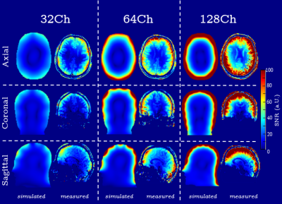 |
Performance Evaluation of a 128-Channel head-only Receiver array
at 7 Tesla
Bernhard Gruber1,2,3,
Jason P. Stockmann1,4,5,
Azma Mareyam1,
Yulin Chang6,
Boris Keil7,
Berkin Bilgic1,4,
Alexander Beckett8,9,
David A. Feinberg8,9,
and Lawrence L. Wald1,4,5
1A.A. Martinos Center for Biomedical Imaging, Department of Radiology, Massachusetts General Hospital, Harvard Medical School, Charlestown, MA, United States, 2High Field MR Center, Center for Medical Physics and Biomedical Engineering, Medical University Vienna, Vienna, Austria, 3BARNLabs, Muenzkirchen, Austria, 4Harvard Medical School, Boston, MA, United States, 5Harvard-MIT Division of Health Sciences and Technology, Cambridge, MA, United States, 6Siemens Medical Solutions USA Inc., Malvern, PA, United States, 7Department of Life Science Engineering, Institute of Medical Physics and Radiation Protection, Mittelhessen University of Applied Sciences, Gießen, Germany, 8Advanced MRI Technologies, Sebastopol, CA, United States, 9Helen Wills Neusoscience Institute, University of California, Berkeley, CA, United States Keywords: RF Arrays & Systems, RF Arrays & Systems The performance of a 128-channel Rx-only 7T brain array was evaluated using simulations and measurements. SNR and g-factor maps show a significant performance increase for highly accelerated imaging in cortical areas from a combination of improved peripheral unaccelerated SNR and g-factor. Measured SNR in cortical areas increased by 42% from 32- to 128-ch and 18% from 64- to 128-ch. The 1/g-factor maps show an improved mean and a tighter distribution, with both effects becoming more pronounced at higher accelerations. At 6x2-fold the 128-channel array has 17.9% g-factor benefit over the 64-ch, and a 48.2% benefit over the 32-ch array. |
| 16:41 |
1061.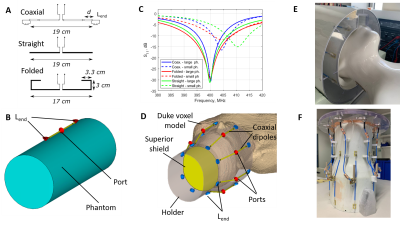 |
Evaluation of Coaxial Dipole Antennas as Transceiver Elements of
Human Head Array for Ultra-High Field MRI at 9.4T
Georgiy Alekseevich Solomakha1,
Dario Bosch 1,2,
Klaus Scheffler1,2,
and Nikolai Ivanovich Avdievich 1
1High-field Magnetic Resonance, Max Planck Institute for Biological Cybernetics, Tuebingen, Germany, 2Department for Biomedical Magnetic Resonance, University of Tübingen, Tuebingen, Germany Keywords: RF Arrays & Systems, RF Arrays & Systems Arrays of dipole antennas were recently introduced as transceiver RF coils for human head imaging at UHF as a simple and robust alternative to loop arrays. Due to the head size, dipoles should be significantly shorter than λ/2 at working frequency. Short dipoles suffer from high SAR and insufficient brain coverage. In addition, since head arrays are usually placed on rigid holders, the resonance frequency of dipoles change drastically with head size variation. In this work, we developed a coaxial dipole array for human head imaging at 9.4T. The developed coil provides whole-brain coverage, low SAR, and low frequency variation. |
| 16:49 |
1062.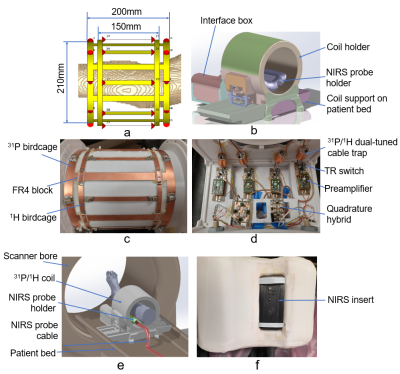 |
3 Tesla 31P/1H Calf Muscle Coil for 1H and 31P MRI / MRS
integrated with NIRS
Bei Zhang1,
Daniel Lowrance1,
David Zaha1,
Manoj Kumar Sarma1,
Michael Douglas Nelson2,
and Anke Henning1
1Advanced Imaging Research Center, UT Southwestern Medical Center, Dallas, TX, United States, 2The University of Texas at Arlington, Arlington, TX, United States Keywords: Non-Array RF Coils, Antennas & Waveguides, Skeletal, 31P/1H Dual-tuned, NIRS In this work, we present a 3T 31P/1H calf coil with two interleaved and co-centered birdcages in one layer to provide homogenous transmit fields and good SNR for both 31P and 1H nuclei. In addition, the coil design allows for integration of a Near-infrared spectroscopy (NIRS) probe for simultaneous NIRS and MRI readouts during exercise. Simulation and phantom experimental results show that the coil provides homogeneous transmit and receive fields. In vivo experiment show that the coil provide good SNR for both 31P MRS and 1H MRI and MRS |
| 16:57 |
1063.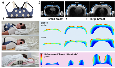 |
In vivo performance of a wearable coil vest for 3 T breast MRI
(BraCoil)
Michael Obermann1,
Lena Nohava1,
Roberta Frass-Kriegl1,
Onisim Soanca1,
Jean-Christophe Ginefri2,
Jacques Felblinger3,
and Elmar Laistler1
1High Field MR Center, Medical University of Vienna, Vienna, Austria, 2Laboratoire d’Imagerie Biomédicale Multimodale Paris Saclay (BioMaps), CEA, CNRS, Inserm, Université Paris-Saclay, Paris-Saclay, France, 3IADI, Inserm, Université de Lorraine, Nancy, France Keywords: New Devices, RF Arrays & Systems Recently, we introduced the “BraCoil”, a wearable coil for prone and supine breast imaging designed to overcome several shortcomings in clinical breast MRI. Here, we present in vivo performance tests on 12 volunteers with different breast volumes ranging from 495 mL (bra size 70A) to 3020 mL (90D). SNR gain up to a factor of three over a commercial breast coil was found. Acceleration factors up to 6x4 can be used with reasonable g-factors. High-quality in vivo images with different contrasts are presented. |
| 17:05 |
1064. |
A radiofrequency coil for infants and toddlers
Kyle M Gilbert1,2,
Emily S Nichols3,4,
Joseph S Gati1,2,
and Emma G Duerden3,4,5
1Centre for Functional and Metabolic Mapping, Western University, London, ON, Canada, 2Department of Medical Biophysics, Western University, London, ON, Canada, 3Applied Psychology, Western University, London, ON, Canada, 4Western Institute for Neuroscience, Western University, London, ON, Canada, 5Department of Pediatrics, Western University, London, ON, Canada Keywords: RF Arrays & Systems, RF Arrays & Systems This abstract presents an adjustable RF coil designed for imaging three-month-old infants to three-year-old toddlers, where motion and variance in head size can be detrimental to image quality. The coil is designed with an open face to allow for the use of camera or motion tracking systems. The tailored RF coil produced higher SNR and lower geometry factors than adult coils. Accelerated protocols can now be combined with prospective motion correction, thereby improving the success rate of imaging infant and toddler populations. |
| 17:13 |
1065.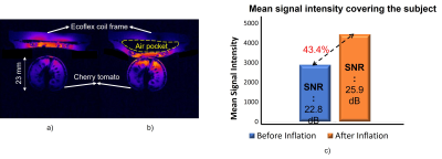 |
Inflatable RF Coil with Liquid Metal for MR Imaging
Sri Kirthi Kandala1,
Kwanjoon Song2,
and Sung-Min Sohn1
1Arizona State University, Tempe, AZ, United States, 2Yonsei University, Gangwon-do, Korea, Republic of Keywords: Non-Array RF Coils, Antennas & Waveguides, New Devices, Liquid metal RF coil In this work, we present a novel inflatable radio-frequency receive coil for Magnetic Resonance Imaging at 7T. A small sample was imaged with and without inflation, and the overall signal-to-noise ratio of the sample was improved by 12.7% in dB with inflation. This coil design will offer many opportunities in the field of endorectal imaging, imaging for irregular sample shapes, and reducing motion artifacts. Inflation also changes the tuning and matching conditions, which compensate for loading effects by precisely adjusting the air volume inside the cavity. |
| 17:21 |
1066.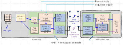 |
Feasibility Study for Wireless RF Coil: Wireless transfer of 8ch
MR received data using body array coil and system clock
Kazuya Okamoto1,
Sojuro Kato2,
Mark Spring3,
Yoshinori Hamamura4,
Daisuke Horio5,
Yu Tanaka5,
Shun Sugimoto5,
and Kazunari Watanabe5
1MRI Systems Development Department, CANON Medical Systems Corporation, Kawasaki, Japan, 2CANON Medical Systems Corporation, Kawasaki, Japan, 3Platform Technology, Canon Medical Research USA Inc., Vernon Hills, IL, United States, 4MRI Systems, Canon Medical Research USA Inc., Mayfield Village, OH, United States, 5Digital Business Platform Development Headquarters, Canon Inc., Tokyo, Japan Keywords: RF Arrays & Systems, RF Arrays & Systems, wireless We prototyped a new acquisition board (NAB) equipped with wireless signal transmission modules, and connected it to the reception RF coil and the scanner system. The configuration enabled that the transmission of the 8-ch reception signal data from the RF coil and the system clock was carried out between boards wirelessly. Finally, two-dimensional SE images were obtained without deterioration in image quality. |
| 17:29 |
1067.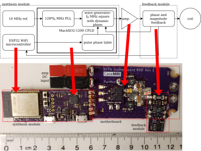 |
Digital Synthesis at the Coil in a WiFi-enabled Modular Switch
Mode RFPA Platform for Gradient-Free Imaging
N Reid Bolding1,
Chris Vaughn2,
Aria Patel1,
Snow Lin1,
Andrew Dupuis1,
William A Grissom2,
and Mark A Griswold1
1Case Western Reserve University, Cleveland, OH, United States, 2Vanderbilt University, Nashville, TN, United States Keywords: RF Arrays & Systems, Low-Field MRI, Gradient Free Imaging We present here the development of a wireless flat amplitude 2 MHz radiofrequency transmitter for under $100, focusing on the pulse synthesis stage. We gave special consideration to simplifications that can be made with high speed digital synthesis and switch mode amplifier topology. This is a component of a distributed transmit receive system, meeting the special requirements of gradient free low field quantitative imaging techniques, such as selective encoding through nutation and fingerprinting (SENF). |
| 17:37 |
1068. |
A modular sparse hemispherical transmit/receive phased array for
microbubble-mediated MR-guided focused ultrasound brain therapy
Ryan M Jones1,
Dallan McMahon1,
Dallas Leavitt1,
Rohan Ramdoyal1,
Kang U Lee1,
Wai M Kan1,
Steven D Yang1,
Yi-Shiuan Chen1,
Chris Adams1,
and Kullervo Hynynen1,2 1Sunnybrook Research Institute, Toronto, ON, Canada, 2University of Toronto, Toronto, ON, Canada Keywords: New Devices, Focused Ultrasound MRI is commonly employed to assess microbubble-mediated focused ultrasound (FUS) treatment outcomes, but is restricted to post-treatment evaluations due to its low temporal resolution. Here, we describe a modular sparse hemispherical transmit/receive phased array for microbubble-mediated FUS brain therapy and simultaneous 3D passive cavitation imaging (PCI) for real-time intraoperative treatment monitoring and control. The device was evaluated via MR-guided FUS experiments in rabbits. The utility of 3D PCI in calibrating FUS exposure levels and predicting MRI-inferred tissue damage volume distributions resulting from high target level sonications was demonstrated. |
 Back to Meeting Home
 Back to the Program-at-a-Glance
The International Society for Magnetic Resonance in Medicine is accredited by the Accreditation Council for Continuing Medical Education to provide continuing medical education for physicians.
 Back to Meeting Home
 Back to the Program-at-a-Glance
 View Presentation Video