Oral Session
New Devices for Intervention & Sensing
ISMRM & ISMRT Annual Meeting & Exhibition • 03-08 June 2023 • Toronto, ON, Canada

Oral Session
New Devices for Intervention & Sensing
| 08:15 |
0744.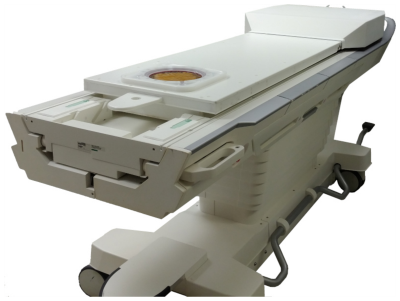 |
A flat fully-populated MR-guided focused ultrasound phased array
body system
Ryan M Jones1,
Yuexi Huang1,
Benjamin BC Lucht2,
Samuel T Gunaseelan1,
Tyler Portelli2,
Elizabeth David3,4,
and Kullervo Hynynen1,4 1Sunnybrook Research Institute, Toronto, ON, Canada, 2Arrayus Technologies Inc., Burlington, ON, Canada, 3Sunnybrook Health Sciences Centre, Toronto, ON, Canada, 4University of Toronto, Toronto, ON, Canada Keywords: Uterus, MR-Guided Interventions Existing commercial body MR-guided focused ultrasound (MRgFUS) systems have limited electronic steering ranges and rely on mechanical translation/rotation to ablate large tissue volumes. Here, we describe a flat fully-populated MRgFUS phased array body system with increased electronic steering capabilities. Following extensive benchtop and pre-clinical testing, the device was evaluated in a pilot clinical trial of MRgFUS for the treatment of uterine fibroids, which demonstrated the feasibilty and safety of the approach. The technology is extensible, stackable, and modular, allowing custom MRgFUS device development tailored to any indication. |
| 08:23 |
0745.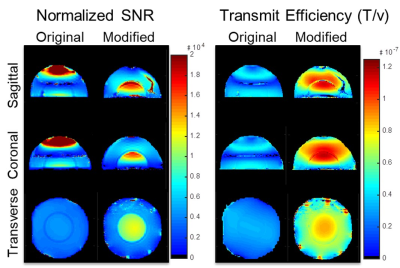 |
Improvement of Experimental SNR in Mock Transcranial MRgFUS
System with Strategic Design of Transfer Medium and Transducer
Ground Plane at 3T
Karthik Lakshmanan1,2,
Ryan Brown1,2,
Giuseppe Carluccio1,2,
and Christopher M Collins1,2
1Radiology, NYU Grossman School of Medicine, New York, NY, United States, 2Center for Advanced Imaging Innovation and Research (CAI2R), New York University, New York, NY, United States Keywords: Non-Array RF Coils, Antennas & Waveguides, RF Arrays & Systems We show that novel modifications to a mock Transcranial MR-guided Focused Ultrasound (TMRgFUS) system show potential to greatly improve MR performance. By using a transfer medium with reduced electric permittivity combined with slots in the transducer array ground plane we measure a 2-3 fold increase in SNR across the middle of a head-phantom portion of a mock TMRgFUS system. |
08:31 |
0746.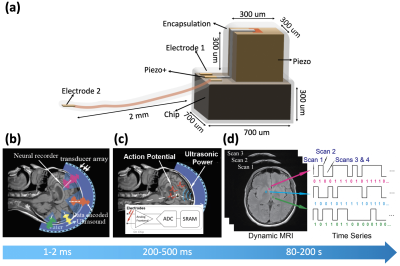 |
The MRDust: An Implantable Neural Interface Powered via Focused
Ultrasound with Data Communication via MR Image Modulation
Biqi Rebekah Zhao1,
Yuhan Wen1,
Alexander Chou1,
Elad Alon1,
Rikky Muller1,
Chunlei Liu1,
and Michael Lustig1
1Department of Electrical Engineering and Computer Sciences, University of California, Berkeley, Berkeley, CA, United States Keywords: New Devices, Neuroscience We propose a new device for neuroscience studies: the MRDust, a sub-mm wireless programmable neural recording mote with on-device memory and compute. It receives power via focused ultrasound, records neural signals in burst mode, and uses a micro-coil to perturb local magnetic fields to achieve data uplink via dynamic MRI signal modulation. We demonstrate proof-of-concept experiments in which digital information is encoded in images of an SE-EPI dynamic sequence, and in which a piezoelectric harvester can harvest enough ultrasonic power to sustain device operation, and receive control signals through amplitude modulation. |
| 08:39 |
0747.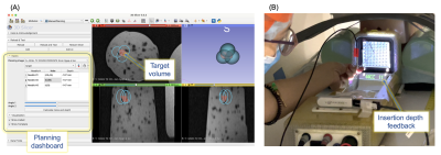 |
Motorized template for MRI-guided focal cryoablation of prostate
cancer
Pedro Moreira1,2,
John P Grimble 3,
Mariana Bernardes1,2,
Nick V Iftimia3,
Kemal Tuncali1,2,
Junichi Tokuda1,2,
and Jesung Park3 1Brigham and Women's Hospital, Boston, MA, United States, 2Harvard Medical School, Boston, MA, United States, 3Physical Sciences Inc., Andover, MA, United States Keywords: Interventional Devices, Prostate MR-guided focal cryoablation of prostate cancer has often been selected as a minimally-invasive treatment option. Placing multiple cryo-needles accurately to form an ablation volume that adequately covers the target volume is crucial for a better oncological/functional outcome. This work presents an MRI-compatible system combining a motorized template grid and insertion depth sensing, which allows the physician to precisely place the cryo-needles into the desired location. Our preliminary results demonstrate the advantages of adding additional degrees of freedom to the template and providing the physician with real-time feedback on the insertion depth. |
| 08:47 |
0748.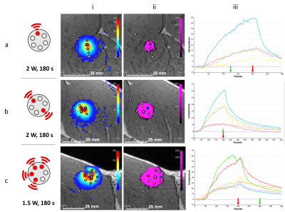 |
Creating in vivo conformal thermal treatments using a
multisource LITT device combined with real time multi-slice
MR-thermometry
Manon Desclides1,2,
Valéry Ozenne1,
Guillaume Machinet3,
Christophe Pierre3,
Stéphane Chemouny2,
and Bruno Quesson1
1University of Bordeaux, CNRS, CRMSB, UMR 5536, IHU Liryc, Bordeaux, France, Metropolitan, 2Certis Therapeutics, Pessac, France, 3ALPhANOV, Talence, France Keywords: Interventional Devices, MR-Guided Interventions, Laser Percutaneous clinical ablation devices usually create a heating pattern with an ellipsoid shape. Output power and duration of application are the only degrees of freedom, which does not allow creating conformal ablation. To overcome this limitation, we designed a Laser Interstitial Thermal Therapy (LITT) device allowing creating various heating patterns. We present here in vivo results in pig muscle monitored by real-time 2D multi-slice MR-thermometry. |
| 08:55 |
0749.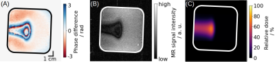 |
Feasibility of MR phase-contrast imaging for proton beam
visualisation in liquid water phantoms
Juliane Peter1,2,3,
Sebastian Gantz1,2,
Aswin Hoffmann1,2,4,
and Jörg Pawelke1,2
1OncoRay – National Center for Radiation Research in Oncology, Faculty of Medicine and University Hospital Carl Gustav Carus, Technische Universität Dresden, Helmholtz-Zentrum Dresden-Rossendorf, Dresden, Germany, 2Helmholtz-Zentrum Dresden-Rossendorf, Institute of Radiooncology – OncoRay, Dresden, Germany, 3Technische Universität Dresden, Dresden, Germany, 4Department of Radiotherapy and Radiation Oncology, Faculty of Medicine and University Hospital Carl Gustav Carus, Technische Universität Dresden, Dresden, Germany Keywords: New Devices, Radiotherapy, Proton Therapy Proton beam-induced convection in water triggered local MRI magnitude signal loss in combined imaging and irradiation experiments performed on a new research prototype in-beam low-field MRI proton radiotherapy device. In this study, the influence of convection on the MRI phase signal was tested. Both mechanical and thermal inhibition of convection in dedicated water phantoms resulted in the absence of MRI phase signatures, which were clearly visible under conditions were convection could develop. Moreover, a change in either convection velocity or Venc sequence motion sensitivity changed the observed phase contrast, confirming the convection-driven phase contrast mechanism. |
| 09:03 |
0750.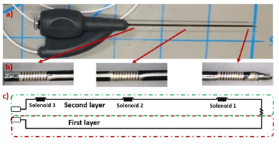 |
Real-time Needle Tracking Under 0.55T MRI Using Current
Controlled B1 Field Artifacts
Dogangun Uzun1,2,
Dursun Korel Yildirim1,
Rajiv Ramasawmy1,
Amanda Potersnak1,
Adrienne Campbell-Washburn1,
Ozgur Kocaturk3,
and Robert J. Lederman1
1National Heart Lung and Blood Institute, National Institutes of Health, Bethesda, MD, United States, 2Institute of Biomadical Engineering, Bogazici University, Istanbul, Turkey, 3Transmural Systems, Andover, MA, United States Keywords: Interventional Devices, Interventional Devices In this work, an interventional device tracking method under high performance 0.55T MRI is presented using a custom designed nitinol needle. Ultra-thin miniature solenoid coil markers were designed and printed on the needle using conductive ink. A negative contrast is created around the needle by applying an alternative current to the markers and hence introducing an offset to the B1 field. A signal generator circuit is designed for supplying and controlling the AC signal and the visibility of the needle is tested under MRI with in-vitro experiments during a real time b-SSFP sequence. |
| 09:11 |
0751.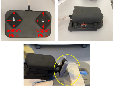 |
eGantryMate: A Piezo-Motor driven lean and flexible Assistance
System for MR-Guided Interventions at 1.5T
Andreas Reichert1,
Ali Caglar Özen1,
Simon Reiss1,
Samantha Hickey1,
Thomas Lottner1,
Niklas Verloh2,
Srdjan Milosavljevic3,
Michael Vogele3,
and Michael Bock1
1Division of Medical Physics, Department of Radiology, University Medical Center Freiburg, Freiburg, Germany, 2Division of Interventional Radiology, Department of Radiology, University Medical Center Freiburg, Freiburg, Germany, 3Interventional Systems GmbH, Kitzbuehel, Austria Keywords: Interventional Devices, Interventional Devices We present a small and flexible piezo-driven assistance system, which can be steered from outside the magnet bore via a control unit. The assistance system is combined with a tracking sequence, which is able to follow in-bore manipulations of a dedicated end effector in real-time. For an initial evaluation, the assistance systems is equipped with a biopsy needle and targeting experiments are performed in a phantom setting. |
| 09:19 |
0752.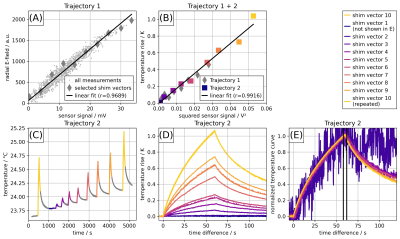 |
Experimental and numerical calibration procedure for RF safety
evaluation of implant-embedded sensors
Johannes Petzold1,
Berk Silemek1,
Lukas Winter1,
Bernd Ittermann1,
and Frank Seifert1
1Physikalisch-Technische Bundesanstalt (PTB), Braunschweig and Berlin, Germany Keywords: Safety, Data Acquisition Embedded sensors in implants that communicate with an MRI system can improve patient safety substantially, in particular when parallel transmission is applied to mitigate the RF-heating risk. Calibration procedures and virtual models are presented in this work to support a rigorous safety assessment of this novel approach to implant safety. Experiments show a good correlation (r>0.96) between sensor signal (voltage at the tip) and field-probe measurements (E-field and temperature) for a custom-built reference implant. These results are further supported by simulations that can be extended to investigate more complex implant heating scenarios in human voxel models. |
| 09:27 |
0753.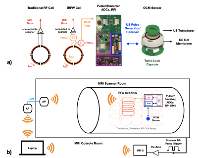 |
Motion Monitoring using a Wireless Ultrasound-Based Sensor and
an Integrated RF/Wireless Coil Array
Devin Willey1,2,
Spencer Lynch3,4,
Olivia Jo Dickinson3,4,
Trong-Kha Truong3,4,
Fraser Robb5,
Allen W Song3,6,
Bruno Madore1,2,
and Dean Darnell3,4 1Harvard Medical School, Boston, MA, United States, 2Radiology, Brigham and Women's Hospital, Boston, MA, United States, 3Medical Physics Graduate Program, Duke University, Durham, NC, United States, 4Brain Imaging and Analysis Center, Duke University, Durham, NC, United States, 5GE Healthcare, Aurora, OH, United States, 6Brain Imaging And Analysis Center, Duke University, Durham, NC, United States Keywords: New Devices, Body A wireless, battery-powered, MR-compatible ultrasound device consisting of an integrated RF/wireless coil, an organ-configuration motion sensor and its associated electronics was used to acquire OCM sensor signals on a healthy volunteer. Signals were obtained that characterized internal motion, which were wirelessly transmitted to the console room. Validation was performed against a real-time MR acquisition. |
| 09:35 |
0754.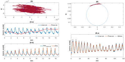 |
Doppler radar signal integrity investigation for in-bore vital
sensing applications
Wonje Lee1,
John Pauly1,
Shreyas Vasanawala1,
and Greig Scott1
1Stanford University, Palo alto, CA, United States Keywords: Hybrid & Novel Systems Technology, RF Arrays & Systems, motion sensing This study investigates complex baseband doppler radar signal integrity with linear and phase demodulations to confirm feasibility for in-bore MRI vital sensing applications. |
| 09:43 |
0755.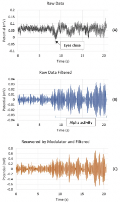 |
Development of a cost-effective, fiber optic-based,
MRI-compatible EEG system: a proof-of-concept study
Michael Potter1,
Emily Holz1,
Lindsay Demblowski1,
Kyle Hunkar1,
Udunna Anazodo2,
Stefan Preble1,
and Iris Asllani1,3 1Rochester Institute of Technology, Rochester, NY, United States, 2McGill University, Montreal, QC, Canada, 3University of Sussex, Brighton, United Kingdom Keywords: Multimodal, fMRI (task based), EEG, EEG recording, MRI compatible EEG A cost-effective, 8-channel, MRI-compatible optical EEG prototype was implemented and tested. The protoype has the potential to be especially suited for low-field MRI applications. The system uses PhotrodeTM technology, a high impedance device that can pick up signals without the need for "wet" contact with the skin. The minimum resolvable voltage of the modulator was ~ 12.5 uV, sufficient for most EEG waves. |
| 09:51 |
0756.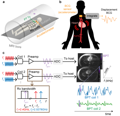 |
Wireless In-Bore Ballistocardiography with 2.4GHz Beat Pilot
Tone (BPT)
Suma Anand1 and
Michael Lustig1
1Electrical Engineering and Computer Sciences, University of California, Berkeley, Berkeley, CA, United States Keywords: Hybrid & Novel Systems Technology, Cardiovascular, RF Arrays and Systems, Motion Correction We previously proposed the Beat Pilot Tone (BPT), a motion sensing method that uses microwave frequencies and preamplifier intermodulation to obtain increased motion sensitivity compared to Pilot Tone (PT). Here, we show that cardiac signals acquired with BPT correlate strongly with displacement ballistocardiograms (dBCG). We compare the BPT signals to dBCG acquired with an accelerometer. Additionally, we show that BPT has greater signal modulation compared to PT in sensing cardiac motion. BPT could be a novel, non-contact, and wireless method for acquiring dBCG signals, offering robustness in setup for patients and rich physiological information complementary to the MRI exam. |
09:59 |
0757. |
Selective Shielding for RF Energy Harvesting Without B1
Interference
Oskar Bjoerkqvist1 and
Klaas P. Pruessmann2
1ETH Zürich, Zurich, Switzerland, 2ETH Zürich, Zürich, Switzerland Keywords: New Devices, New Devices, RF Harvesting; Wireless energy; In this abstract we present a solution for harvesting RF excitation pulses in MRI while suppressing interference caused by the harvesting system, which is otherwise known to be an issue. This approach to RF harvesting offers an alternative for powering circuitry that needs to be within the field of view free from cables and without dedicated energy transmission. |
|
10:07 |
0758.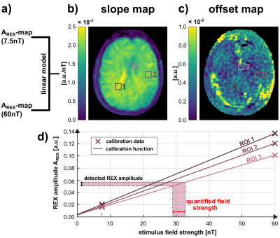 |
Transmission of Rotary Excitation enables Quantification of
Oscillating nT-Fields using a Linear Calibration Model
Petra Albertova1,2,
Maximilian Gram1,2,
Verena Schirmer2,
Martin Blaimer3,
Martin J. Herrmann4,
Matthias Gamer5,
Peter Nordbeck1,6,
and Peter Michael Jakob2
1Department of Internal Medicine I, University Hospital Würzburg, Würzburg, Germany, 2Experimental Physics 5, University of Würzburg, Würzburg, Germany, 3Fraunhofer Institute for Integrated Circuits IIS, Würzburg, Germany, 4Department of Psychiatry, Psychosomatics, and Psychotherapy, University Hospital Würzburg, Würzburg, Germany, 5Department of Psychology, University of Würzburg, Würzburg, Germany, 6Comprehensive Heart Failure Center (CHFC), University Hospital Würzburg, Würzburg, Germany Keywords: Bioeffects & Magnetic Fields, Quantitative Imaging, spin-lock Rotary excitation based MRI, which enables spatially resolved, non-invasive, direct detection of biomagnetic fields, complements the measurement range accessible via MEG since detection is independent of the distance to the body surface. We present an extension of the REX method towards quantitative imaging. A calibration function can be composed from REX data of two measurements of adjustable fields projected onto the tissue by the scanner’s gradient system. In subsequent measurements, the field strength can be inferred from the calibration model. First in vivo proof-of-concept measurements show that the calibration successfully eliminates influences of tissue properties and accurately quantifies oscillatory nT-fields. |
 Back to Meeting Home
 Back to the Program-at-a-Glance
The International Society for Magnetic Resonance in Medicine is accredited by the Accreditation Council for Continuing Medical Education to provide continuing medical education for physicians.
 Back to Meeting Home
 Back to the Program-at-a-Glance
 View Presentation Video