Oral Session
Higher Field, Higher Expectations
ISMRM & ISMRT Annual Meeting & Exhibition • 03-08 June 2023 • Toronto, ON, Canada

| 16:00 |
1412.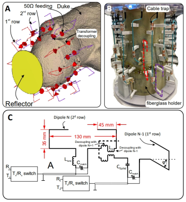 |
Double-Row 16-element Folded-End Dipole Transceiver Array for
Human Whole Brain Imaging at 9.4 T.
Nikolai I Avdievich1,
Anton V Nikulin1,2,
Dario Bosch1,2,
Georgiy Solomakha1,
and Klaus Scheffler1,2
1High-Field MR Center, Max Planck Institute for Biological Cybernetics, Tübingen, Germany, 2Biomedical Magnetic Resonance, University of Tübingen, Tübingen, Germany Keywords: High-Field MRI, RF Arrays & Systems, Dipole antenna Homogeneity and coverage of transmit (Tx) RF coils at ultra-high field (UHF,>7 T) can be improved by 3D RF shimming. This, however, requires using multi-row Tx-arrays. Dipole antennas provide unique simplicity and robustness while offering comparable Tx-efficiency and SNR to conventional loop designs. Single-row UHF dipole Tx-arrays for human head imaging have been previously described. Recently, we developed a novel type of dipole elements, a folded-end dipole, which improved the longitudinal coverage and specific absorption rate (SAR) efficiency. In this work, we developed, constructed, and evaluated a 16-element double-row transceiver folded-end dipole array for human whole-brain imaging at 9.4 T. |
| 16:08 |
1413.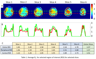 |
Substantially Improved Receive Sensitivity of Human Whole-Brain
MRI at 7T using a High-Permittivity Material (HPM) Slurry-Filled
Helmet
Soo Han Soon1,2,
Matt Waks1,
Hannes M. Wiesner1,
Xiao-Hong Zhu1,
Michael T. Lanagan3,
Qing X. Yang4,
and Wei Chen1,2
1Center of Magnetic Resonance Research (CMRR), Department of Radiology, University of Minnesota, Minneapolis, MN, United States, 2Department of Biomedical Engineering, University of Minnesota, Minneapolis, MN, United States, 3Department of Engineering Science and Mechanics, Pennsylvania State University, University Park, PA, United States, 4Center for NMR Research, Department of Neurosurgery and Radiology, College of Medicine, Pennsylvania State University, Hershey, PA, United States Keywords: High-Field MRI, New Devices, High Permittivity Material (HPM), High Dielectric Constant (HDC) Material Novel methods such as high-permittivity materials (HPM) and metasurfaces have improved RF coil transmission efficiency and receive field (B1-) sensitivity for MRI applications at ultrahigh field. One of the recent studies, which applied ceramic HPM helmet with the permittivity of 100, showed significant improvement in signal-to-noise ratio (SNR). Motivated from the previous studies with various forms of HPM at 3T and 7T, this study introduces an easy and accessible method with an HPM slurry helmet to largely improve imaging quality of human brain MRI at 7T, which could improve B1- field by 57% and SNR by 47%. |
| 16:16 |
1414. |
Simultaneous Head and Cervical Spinal Cord Imaging at 7T with a
16-channel transceiver loop array
Bei Zhang1,
Daniel Lowrance1,
and Anke Henning1
1Advanced Imaging Research Center, UTSouthwestern Medical Center, Dallas, TX, United States Keywords: High-Field MRI, Head & Neck/ENT In this work, we represented 7T simultaneous brain and cervical spinal cord images with diagnostic quality. The images were acquired with a 16-channel transceiver array in a 7T 8-channel parallel transmit system which has RF shimming capability, Specifically, we acquired MP2RAGE and FLAIR images of the head and T2-weighted and GRE images of the cervical spinal cord with parameter settings in sequences for clinical applications in our center. These images show high-resolution anatomical structures and nice contrast of white and gray matters. Moreover, all these images were acquired without changing the table position or repositioning the volunteers during the scan. |
| 16:24 |
1415. |
Highly decoupled 8x2 transceiver array for human brain at 7T (Rx
performance evaluation)
Junghwan Kim1,2,
Changyu Sun1,3,
Chan-Hong Moon4,
Hoby Hetherington1,5,
and Jullie Pan1
1Radiology, University of Missouri, Columbia, MO, United States, 2EECS, University of Missouri, Columbia, MO, United States, 3BBCE, University of Missouri, Columbia, MO, United States, 4Radiology, University of Pittsburgh, Pittsburgh, PA, United States, 5Resonance Research Inc., Billerica, MA, United States Keywords: High-Field MRI, RF Arrays & Systems, Rx array While the Tx performance of the 8x2 transceiver has been shown to achieve excellent amplitude and homogeneity, with its limited coil numbers (16), it is thought to give substantially lower SNR and acceleration in comparison to conventional arrays (8Tx/32Rx). It is recognized however, that the Tx decoupling is also constructive for Rx since the SNR and g-factors benefit from the decreased noise correlation. We evaluated the SNR, g factors, and noise covariance and found that up to an in-plane acceleration value of approximately <=4, the transceiver gives comparable performance to a commercial reference coil. |
| 16:32 |
1416.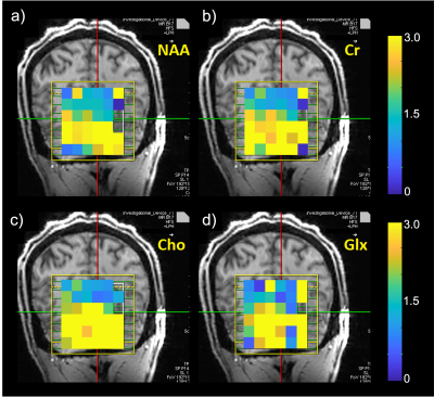 |
Improvement of Brain MRS at 7T Using a Wireless RF Array
Akbar Alipour1,
Gaurav Verma1,
Andrew Frankini1,
Bradley N Delman1,
and Priti Balchandani1
1Radiology, Icahn School of Medicine at Mount Sinai, New York, NY, United States Keywords: High-Field MRI, Spectroscopy Magnetic resonance spectroscopy (MRS) can particularly benefit from substantial enhancement in SNR and spectral resolution at ultra-high field (UHF ≥7T), enabling improved quantification of metabolites. However, at 7T wavelength effects cause a highly inhomogeneous transmit magnetic field in the human brain, with lower transmit efficiency in the posterior-fossa manifesting as signal dropout in this region. Recently, we reported advantages of a surface-applied inductively-coupled radiofrequency array to improve transmit efficiency and signal sensitivity at 7T MRI focusing on cerebellum and inferior temporal lobes. Here we demonstrate the feasibility and effectiveness of in-vivo MRS using the array in human cerebellum at 7T. |
| 16:40 |
1417.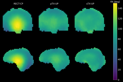 |
Optimized T1 and T2 Weighted Structural Imaging on Clinical 7
Tesla Systems Employing Single Channel GRAPE Universal Pulses
Eberhard Daniel Pracht1,
Daniel Löwen1,
Laurent Lamalle2,
Franck Mauconduit3,
Vincent Gras3,
Nicolas Boulant3,
and Tony Stöcker1,4
1German Center for Neurodegenerative Diseases (DZNE), Bonn, Germany, 2GIGA-CRC in Vivo Imaging, University of Liège, Liège, Belgium, 3Université Paris-Saclay, Commissariat à l’Energie Atomique, CNRS, NeuroSpin, BAOBAB, Paris-Saclay, France, 4Department of Physics and Astronomy, University of Bonn, Bonn, Germany Keywords: High-Field MRI, Brain We present single channel universal GRAPE pulses for optimized T1 and T2 weighted (fluid suppressed) imaging on clinical ultra-high field systems. B0 and B1 inhomogeneities are effectively mitigated while no pulse calculations are necessary during the imaging session. Compared to a parallel transmission set-up SAR estimation is less complex, and with more established history of safe use, it makes this approach a promising tool for clinical applications such as lesion detection. |
| 16:48 |
1418.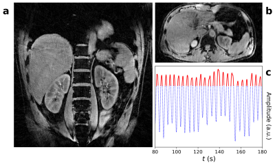 |
Radial Stack-of-Stars Abdominal MRI at 7 Tesla
I T Maatman1,
S Ypma1,
K T Block2,
M C Maas1,
J Schulz1,
and T W J Scheenen1
1Department of Medical Imaging, Radboud University Medical Center, Nijmegen, Netherlands, 2Department of Radiology, NYU Langone Health, New York, NY, United States Keywords: High-Field MRI, Body, Radial MRI Abdominal MRI at 7T is sensitive to transmit field inhomogeneities and motion-induced artifacts. Transmit inhomogeneities have previously been addressed using time-interleaved acquistion of modes (TIAMO), providing uniform flip angles across a large field-of-view in the body. Meanwhile, the radial stack-of-stars sequence has been shown to be well-suited for motion-corrected MRI. In this work, TIAMO and motion-corrected radial MRI were combined to create abdominal images of three volunteers at high spatial resolutions. Results showed homogeneous transmit fields in all volunteers with excellent image quality at very high resolution. However, low-resolution scans suffered from artifacts due to gradient non-linearities and residual motion. |
| 16:56 |
1419.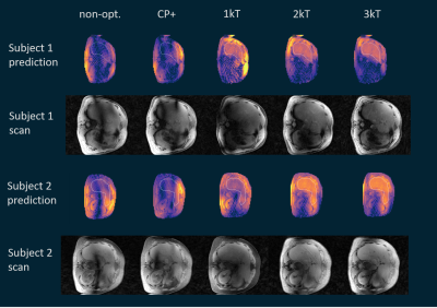 |
In-vivo 3D liver imaging at 7T using kT-point pTx pulses and a
32-Tx-channel whole-body RF antenna array
Johannes Anton Grimm1,2,
Christoph Stefan Aigner3,
Sebastian Dietrich3,
Stephan Orzada1,
Thomas M. Fiedler1,
Armin M. Nagel1,4,
Mark E. Ladd1,2,5,
and Sebastian Schmitter1,3,6
1Medical Physics in Radiology, German Cancer Research Center (DKFZ), Heidelberg, Germany, 2Faculty of Physics and Astronomy, Heidelberg University, Heidelberg, Germany, 3Physikalisch-Technische Bundesanstalt (PTB), Braunschweig and Berlin, Germany, 4Institute of Radiology, University Hospital Erlangen, Friedrich-Alexander-Universität Erlangen-Nürnberg (FAU), Erlangen-Nürnberg, Germany, 5Faculty of Medicine, Heidelberg University, Heidelberg, Germany, 6Center for Magnetic Resonance Research, University of Minnesota, Minneapolis, MN, United States Keywords: High-Field MRI, Liver Using a 32-Tx-channel whole-body RF antenna array at 7T could benefit exciting large body parts as these regions often suffer from flip angle inhomogeneity or dropouts. This study compares static pTx and 3D pTx pulses with a varying number of kT-points to the CP+ mode and non-optimized shim. With static pTx, the flip angle dropouts are reduced, and 2-3 kT-points seemed to deliver the best tradeoff between flip angle homogeneity and RF power. |
| 17:04 |
1420.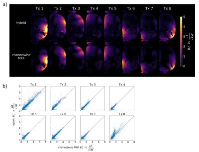 |
MRF-based channel-wise absolute B1+ mapping at low RF power in
the human abdomen at 7T
Max Lutz1,
Christoph Stefan Aigner1,
Sebastian Flassbeck2,3,
Felix Krüger1,
Constance G. F. Gatefait1,
Tobias Schaeffter1,4,5,
and Sebastian Schmitter1,6,7
1Physikalisch-Technische Bundesanstalt, Braunschweig and Berlin, Germany, 2Dept. of Radiology, Center for Biomedical Imaging, New York, NY, United States, 3Center for Advanced Imaging Innovation and Research, New York, NY, United States, 4School of Biomedical Engineering and Imaging Sciences, King's College London, London, United Kingdom, 5Department of Biomedical Engineering, Technical University of Berlin, Berlin, Germany, 6Medical Physics in Radiology, German Cancer Research Center (DKFZ), Heidelberg, Germany, 7Center for Magnetic Resonance Research, University of Minnesota, Minneapolis, MN, United States Keywords: High-Field MRI, High-Field MRI For pTx applications, knowledge of the underlying Tx channel-wise B1+ profile is essential. In this work, we present a hybrid absolute B1+-mapping approach where MRF-based B1+-mapping is utilized to map the channel-wise absolute B1+ profile, providing higher accuracy at low flip angles than standard methods. The hybrid approach is compared to mapping each Tx channel separately using the MRF-based method. Phantom and body in-vivo measurements at 7T show good agreement between the two approaches. |
| 17:12 |
1421.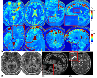 |
Rapid mesoscale 3D whole-brain MRF in the Next-Generation 7T
brain scanner: challenges and advantages
Xiaozhi Cao1,2,
Congyu Liao1,2,
Alexander Beckett3,4,
An Vu5,6,
Samantha Ma7,
Sophie Schauman1,2,
Siddharth Srinivasan Iyer1,8,
Mahmut Yurt1,2,
Elizabeth Tong1,
Adam Kerr2,
David A Feinberg3,4,
and Kawin Setsompop1,2 1Department of Radiology, Stanford university, Stanford, CA, United States, 2Department of Electrical Engineering, Stanford university, Stanford, CA, United States, 3Helen Wills Neuroscience Institute, University of California, Berkeley, CA, United States, 4Advanced MRI Technologies, Sebastopol, CA, United States, 5Department of Radiology, University of California, San Francisco, CA, United States, 6San Francisco Veteran Affairs Health Care System, San Francisco, CA, United States, 7Siemens Medical Solutions USA Inc., Berkeley, CA, United States, 8Department of Electrical Engineering and Computer Science, MIT, Cambridge, MA, United States Keywords: MR Fingerprinting/Synthetic MR, Brain 3D MRF with spiral projection trajectory was implemented on the 7T NexGen scanner to take advantage of its SNR benefit and state-of-the-art gradient system. To achieve high-fidelity and high-efficiency multi-parameter mapping at the mesoscale, novel techniques were developed to overcome several technical challenges in performing this acquisition, including spiral residual gradient compensation, trajectory measurement, water-only excitation RF pulse, B0 correction, B1+ correction and frequency response correction. The proposed technical developments enabled high-quality whole-brain T1, T2, and proton density mapping at 1-mm isotropic resolution in 1 minute, and 560mm isotropic resolution in 4-minutes scan time. |
| 17:20 |
1422.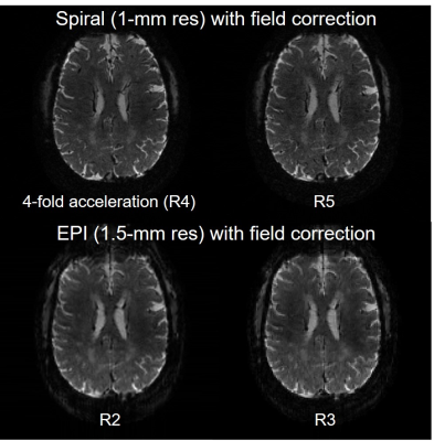 |
Single-shot 2D spiral and echo planar imaging at 7 and 10.5
Tesla with field monitoring and correction
Xiaoping Wu1,
Alexander Bratch1,2,
Jerahmie Radder1,
Andrea Grant3,
Edward Auerbach1,
Pierre‐Francois Van de Moortele1,
Gregor Adriany1,
and Kamil Ugurbil1 1CMRR, Radiology, University of Minnesota, Minneapolis, MN, United States, 2Radiology, Stanford University, Stanford, CA, United States, 3University of Minnesota, Minneapolis, MN, United States Keywords: High-Field MRI, Data Acquisition, Brain imaging Interest in pursuing MRI at ultra-high field (UHF) is increasing owing to increased SNR. However, MRI at UHF presents acquisition challenges due to decreased T2* and worsened field inhomogeneities. Rapid sampling strategies such as spiral and echo planar (EPI) acquisitions have been proposed to help mitigate these challenges. Incorporation of field monitoring to UHF applications has been demonstrated to improve the reconstruction of these images thanks to direct measurement and correction of spatiotemporal phase accrual. Here we demonstrate field-monitoring-enabled spiral and EPI acquisitions using Pulseq and field cameras. |
| 17:28 |
1423.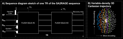 |
Whole-brain 3D B1+ mapping in under 30 seconds:
compressed-sensing accelerated SA2RAGE
Gabriele Bonanno1,2,3,
Tom Hilbert4,5,6,
Patrick Liebig7,
José P. Marques8,
and Tobias Kober4,5,6
1Advanced Clinical Imaging Technology, Siemens Healthineers International AG, Bern, Switzerland, 2Magnetic Resonance Methodology, Institute of Diagnostic and Interventional Neuroradiology, University of Bern, Bern, Switzerland, 3Translational Imaging Center (TIC), Swiss Institute for Translational and Entrepreneurial Medicine, Bern, Switzerland, 4Advanced Clinical Imaging Technology, Siemens Healthineers International AG, Lausanne, Switzerland, 5Department of Radiology, Lausanne University Hospital and University of Lausanne, Lausanne, Switzerland, 6LTS5, École Polytechnique Fédérale de Lausanne (EPFL), Lausanne, Switzerland, 7Magnetic Resonance, Siemens Healthcare GmbH, Erlangen, Germany, 8Donders Institute for Brain Cognition and Behaviour, Radboud University, Nijmegen, Netherlands Keywords: High-Field MRI, RF Pulse Design & Fields, B1+ mapping To fully exploit the strengths of UHF imaging, accurate B1+ mapping is essential, ideally in 3D with sufficient coverage and high dynamic range. However, resulting scan times on the order of minutes are problematic, especially with the prospect of increased clinical use of 7T imaging. We implemented a compressed sensing readout and reconstruction for the SA2RAGE technique yielding <30 s scan time for a whole-brain, 4-mm isotropic resolution B1+ map. We tested different acceleration factors, validated against the GRAPPA-accelerated reference protocol of ~2 min and found <0.05 relative B1+ difference in most regions of the brain. |
| 17:36 |
1424.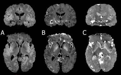 |
First QSM of an ex vivo human brain on the Iseult 11.7T
whole-body system using parallel transmission and virtual coil
reconstruction
Mathieu David Santin1,2,
Isabelle Plu3,
Vincent Gras4,
Michel Luong4,
Edouard Chazel4,
Franck Mauconduit4,
Alexis Amadon4,
Alexandre Vignaud4,
Cecile Lerman4,
and Nicolas Boulant4 1Institut du Cerveau – Paris Brain Institute – ICM, INSERM, CNRS, Sorbonne Université, Paris, France, 2CENIR - Centre for NeuroImaging Research, Sorbonne University, Paris, France, 3Hôpital Pitié-Salpêtrière, AP-HP, Paris, France, 4CEA, CNRS, BAOBAB, NeuroSpin, University of Paris-Saclay, Gif-sur-Yvette, France Keywords: High-Field MRI, Quantitative Susceptibility mapping This work presents the first QSM images obtained on the 11.7T whole body Iseult system, using tailored parallel transmission kT-point pulses and a virtual coil approach for coil combination, on a post mortem brain. |
| 17:44 |
1425.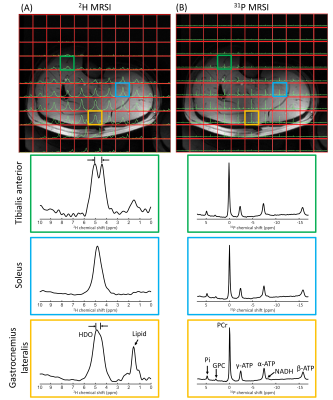 |
Development of a double tuned 2H/31P whole-body birdcage
transmit coil for 2H and 31P MR applications from head to toe at
7T
Ayhan Gursan1,
Mark Gosselink1,
Dimitri Welting1,
Martijn Froeling1,
Hans Hoogduin1,
Evita Wiegers1,
Dennis W.J. Klomp1,
and Jeanine J. Prompers1
1Center for Image Sciences, University Medical Center Utrecht, Utrecht, Netherlands Keywords: Whole Body, Metabolism, Deuterium, Phosphorus Deuterium (2H) and phosphorus (31P) MRS are complementary methods for evaluating tissue metabolism non-invasively in vivo. Combined 2H and 31P MRS would therefore be of interest for various applications. In this work, we developed a double tuned 2H/31P whole-body birdcage transmit coil for 7T, for 2H and 31P MRS with homogeneous excitation over a large field-of-view. The B1+ variation of the whole-body birdcage coil over a body-sized phantom was 14% for 2H and 25% for 31P. Using a two-channel 2H/31P prototype receive array, we obtained high-quality 2H and 31P 3D MRSI data in the brain, liver, and lower-leg muscles. |
| 17:52 |
1426.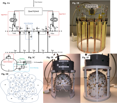 |
An Optimized RF Coil for Sensitive 31P Magnetic Resonance
Spectroscopic Imaging of the Human Brain and Cerebellum at 7T.
Johnny Chris Der Hovagimian1,2,
Pedram Yazdanbakhsh2,3,
Marcus Couch2,3,4,
and David A. Rudko1,2,3
1Department of Biomedical Engineering, McGill University, Montreal, QC, Canada, 2McConnell Brain Imaging Centre, Montreal Neurological Institute and Hospital, Montreal, QC, Canada, 3Department of Neurology and Neurosurgery, McGill University, Montreal, QC, Canada, 4Siemens Healthcare Limited, Montreal, QC, Canada Keywords: High-Field MRI, RF Arrays & Systems An optimized RF coil was constructed for 7T, Phosphorus (31P) Magnetic Resonance Spectroscopic Imaging (MRSI) of the whole brain including the cerebellum. The cerebellum is a region not well covered by most existing 31P coils. The design consisted of an optimized 16-rung high-pass birdcage transmit coil and a 24-channel phased array receive coil affixed to a custom head-shaped former. The coil provided high sensitivity to 31P signals across the whole brain and brainstem. In vivo 31P 3D CSI experiments showed high quality spectra with regional PCr SNR values near the cerebellum comparable to values near the brain centre. |
 Back to Meeting Home
 Back to the Program-at-a-Glance
The International Society for Magnetic Resonance in Medicine is accredited by the Accreditation Council for Continuing Medical Education to provide continuing medical education for physicians.
 Back to Meeting Home
 Back to the Program-at-a-Glance
 View Presentation Video