Oral Session
Low-Field & Point-of-Care/Portable MRI
ISMRM & ISMRT Annual Meeting & Exhibition • 03-08 June 2023 • Toronto, ON, Canada

Oral Session
Low-Field & Point-of-Care/Portable MRI
| 16:00 | 0299.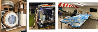 |
On-site construction of a point-of-care low-field MRI system in Africa
Johnes Obungoloch1, Ivan Muhumuza1, Wouter Teeuwisse2, Joshua Harper3, Martin van Gijzen4, Steven Schiff5, Andrew Webb2, and Thomas O'Reilly2 1Mberara University of Science and Technology, Mberara, Uganda, 2Leiden University Medical Center, Leiden, Netherlands, 3Universidad Paraguayo Alemana, Asuncion, Paraguay, 4Delft Institute of Applied Mathematics, Delft, Netherlands, 5Yale University, New Haven, CT, United States Keywords: Low-Field MRI, Low-Field MRI Point-of-care (POC) low-field MRI systems have a large potential to increase the accessibility and sustainability of MRI in low- and middle-income countries (LMICs). An important step in translating scientific developments from high-income countries to LMICs is technology that can be assembled or constructed locally. We describe the construction and testing of a POC system on site in Africa. All components to assemble a 50 mT Halbach magnet based system, together with the necessary tools, were air-freighted from The Netherlands to Uganda. With four instructors and six untrained personnel, the complete project from delivery to first image took approximately 11 days. |
| 16:08 | 0300.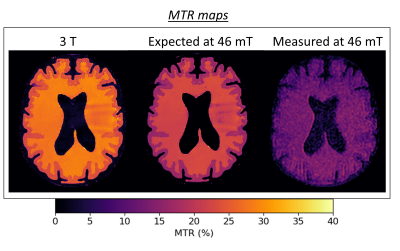 |
T1ρ and magnetization transfer experiments on a point-of-care 46 mT MRI system
Beatrice Lena1, Chloé Najac1, Lena Václavu1, Thomas O'Reilly1, and Andrew Webb1
1Leids Universitair Medisch Centrum, Leiden, Netherlands Keywords: Low-Field MRI, RF Pulse Design & Fields, Saturation RF pulses Contrast mechanisms such as T1ρ and MT are interesting for point-of-care (POC) low field systems, to supplement/augment standard T1- and T2- weighting. However they typically require high B0 homogeneity. Here, we use a high homogeneity Halbach-magnet array POC to perform experiments involving either direct on- or off-resonance pulses, and to calculate T1ρ and MT ratio in tissue-mimicking phantoms. T1ρ showed different white matter (WM)/gray matter (GM) contrast compared to T2. The measured magnetization transfer ratio (MTR) showed higher values in the WM than in the GM. |
16:16 |
0301.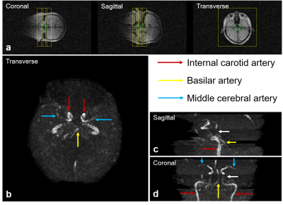 |
Cerebral Time-of-flight Magnetic Resonance Angiography at Ultra-low-field: A Preliminary Study at 0.055 Tesla
Shi Su1,2, Jiahao Hu1,2, Linfang Xiao1,2, Ye Ding1,2, Vick Lau1,2, Yujiao Zhao1,2, Junhao Zhang1,2, Christopher Man1,2, Alex T. L. Leong1,2, and Ed X. Wu1,2
1Laboratory of Biomedical Imaging and Signal Processing, the University of Hong Kong, Hong Kong, China, 2Department of Electrical and Electronic Engineering, the University of Hong Kong, Hong Kong, China Keywords: Low-Field MRI, Brain Recent development of ultra-low-field (ULF) MRI presents opportunities for low-cost and portable brain imaging in point-of-care scenarios or/and low- and mid-income countries. Magnetic resonance angiography (MRA) is an essential part of MR neuroimaging protocols especially for stroke assessment, yet its feasibility at ULF remains unknown. In this study, we explore the time-of-flight MRA at 0.055 Tesla. We demonstrate cerebral MRA using flow-compensated gradient echo sequences, enabling visualization of main cerebral arteries and veins. We envision that usable and quality brain MRA can be potentially achieved at ULF with further sequence/reconstruction optimization and use of intravascular contrast agent. |
| 16:24 | 0302.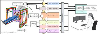 |
Full-scale imaging system for brain MRI and magnetoencephalography
Koos Zevenhoven1, Marko Havu1, Iiro Lehto1, Antti Mäkinen1, Juho Luomahaara2, Petteri Laine3, Mikko Kiviranta2, and Risto J Ilmoniemi1
1Aalto University, Espoo, Finland, 2VTT, Espoo, Finland, 3MEGIN Oy, Espoo, Finland Keywords: Low-Field MRI, New Devices, SQUID, MEG We have built the first full-scale brain scanner performing both MRI and neuromagnetic measurements. Many improvements have been made compared to previous-generation systems. Parts of the system include a helmet-shaped array of 120 superconducting sensors and a pulsed superconducting polarizing coil. An open-geometry coil system provides unobstructed access, and MRI signal acquisition is performed at ultra-low field. The first MEG and MRI data have been successfully obtained with the device. The new prototype is an important milestone on the way towards a commercial implementation. |
| 16:32 | 0303.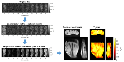 |
Four-fold acceleration of in vivo T1 mapping at 0.1 T using heavily-undersampled Look-Locker data and matrix completion
Marco Fiorito1, Mauro Spreiter1, Mathieu Sarracanie1,2, and Najat Salameh1,2
1Center for Adaptable MRI Technology (AMT Center), Department of Biomedical Engineering, University of Basel, Allschwil, Switzerland, 2AMT Center, Institute of Medical Sciences, School of Medicine, Medical Sciences & Nutrition, University of Aberdeen, Aberdeen, United Kingdom Keywords: Low-Field MRI, Relaxometry Quantitative T1 mapping has proven useful in several clinical applications, nevertheless it is not widely deployed as it generally suffers from long acquisition times. This problem is exacerbated at low magnetic fields, due to inherent lower sensitivity. In spite of more favourable magnetisation properties (e.g., shorter recovery times, higher dispersion), exotic acquisition strategies and reconstruction pipelines are key to make quantitative MRI at low field fast, and compatible with clinical routines. Here, we present a low-rank matrix completion-based reconstruction method applied to heavily undersampled Look-Locker data for accelerated T1 mapping at 0.1 T. |
| 16:40 | 0304.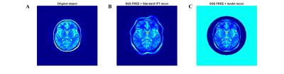 |
Sequential Gradient Superposition (SGS) FREE
Parker JB Jenkins1, Xiaoping Wu2, Gregor Adriany2, and Mike Garwood2
1Biomedical Engineering, University of Minnesota, Minneapolis, MN, United States, 2Radiology, University of Minnesota, Minneapolis, MN, United States Keywords: Low-Field MRI, New Trajectories & Spatial Encoding Methods, RF encoding, Machine Learning, Low Cost MRI Frequency-modulated Rabi Encoded Echoes (FREE) is a RF encoding technique that has the potential reduce the overall costs of MRI by eliminating B0 gradient hardware and infrastructure. However, linear encoding quality has limited its efficacy. To address this problem, we present Sequential Gradient Superposition (SGS) FREE. With SGS-FREE we can decompose the net effective RF gradient into a series of sequentially applied RF gradient sets. This ultimately allows imcreased flexability in linear encoding. This simulation study explores the principle and demonstrates high resolution imaging potential with FREE technology. |
| 16:48 | 0305.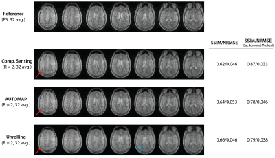 |
Accelerated Imaging at Ultralow Magnetic Fields: A comparative study of traditional and neural-network-based reconstruction approaches
David E. J. Waddington1, Efrat Shimron2, Shanshan Shan1, Neha Koonjoo3, Sheng Shen3, and Matthew S. Rosen3,4,5
1Image X Institute, Faculty of Medicine and Health, The University of Sydney, Sydney, Australia, 2Department of Electrical Engineering and Computer Sciences, UC Berkeley, Berkeley, CA, United States, 3A. A. Martinos Center for Biomedical Imaging, Massachusetts General Hospital, Charlestown, MA, United States, 4Harvard Medical School, Boston, MA, United States, 5Department of Physics, Harvard University, Cambridge, MA, United States Keywords: Low-Field MRI, Image Reconstruction Portable MRI scanners that operate at very low magnetic fields are increasingly being deployed in clinical settings. However, accelerated acquisition and reconstruction methods that boost the quality of low-field MR images are needed to improve the diagnostic accuracy of the modality. Here, we compare leading data-driven and model-driven deep learning frameworks to compressed sensing (CS) for the reconstruction of undersampled ultralow field MRI data, finding that neural network approaches can boost quantitative image reconstruction metrics. |
| 16:56 | 0306.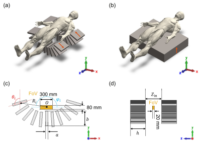 |
Compact and Lightweight Single-sided Inward-outward (IO)-ring permanent magnet array for back imaging
Ting-Ou Liang1, Erping Li2, Wenwei Yu3, and Shao Ying Huang1
1Singapore University of Technology and Design, Singapore, Singapore, 2Zhejiang University, Hangzhou, China, 3Chiba University, Chiba, Japan Keywords: Low-Field MRI, Low-Field MRI, back imaging A compact and lightweight single-sided Inward-outward (IO)-ring permanent magnet array is proposed for back imaging. Its size is 1396×528×1080mm3 and it weighs 924 kg. Within a field-of-view (FoV) of 300×80×20mm3, it has an axial magnetic field with an average strength of 129.8mT, a built-in gradient (averaged at 158mT/m) that corresponds to a radiofrequency (RF) bandwidth of 9.7% and a good linearity with a R2-coefficient of 0.952. It has a compact 5-Gauss zone of 400×320×460mm3. |
| 17:04 | 0307.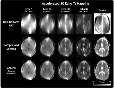 |
Ultra-Low Field Quantitative T2 Mapping
Adam V. Dvorak1, Sharada Balaji1, Megan E. Poorman2, Francesco Padormo2, Rui Pedro A.G. Teixeira2, Melissa Haskell2, Sudarshan Ragunathan2, William J. Hollander3, Alex L. MacKay1,4, Steven C.R. Williams5, Sean C.L. Deoni6, Emil Ljungberg5,7, and Shannon H. Kolind1,4,8 1Physics and Astronomy, University of British Columbia, Vancouver, BC, Canada, 2Hyperfine Inc., Guilford, CT, United States, 3CaliberMRI Inc., Boulder, CO, United States, 4Radiology, University of British Columbia, Vancouver, BC, Canada, 5Department of Neuroimaging, King’s College London, London, United Kingdom, 6Maternal, Newborn & Child Health Discovery & Tools, Bill and Melinda Gates Foundation, Seattle, WA, United States, 7Department of Medical Radiation Physics, Lund University, Lund, Sweden, 8Medicine (Neurology), University of British Columbia, Vancouver, BC, Canada Keywords: Low-Field MRI, Relaxometry We demonstrated quantitative T2 mapping at 0.064 T using a multi-echo spin-echo sequence and CALIPR subspace constrained image reconstruction. CALIPR reconstruction results had minimal evidence of undersampling, providing better artifact suppression and preservation of fine anatomical detail than compressed sensing reconstruction. Phantom validation work was performed using 14 vials doped with known concentrations of MnCl2 and showed a relationship between measured T2 and MnCl2 concentration in agreement with expectations from a mathematical model. In-vivo, we demonstrated short acquisition time (8m:40s or 5m:59s), strong agreement with a highly-sampled reference acquisition, and T2 maps with good contrast between tissue types. |
| 17:12 | 0308.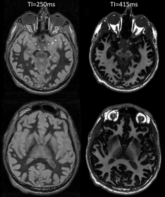 |
Optimizing Deep Brain Grey Matter T1w Imaging at 0.5T
Andrew T Curtis1, Sofia Chavez1, and Jeff Stainsby1
1Research & Development, Synaptive Medical, Toronto, ON, Canada Keywords: Low-Field MRI, Tissue Characterization We report on protocol optimizations to produce white matter nulled images as well as to optimize visualization of subcortical structures. Quantitative T1 mapping of brain tissue at 0.5T estimated the T1 of white and grey matter as 510 and 760 ms respectively. From this an MPRAGE protocol with optimal subcortical matter contrast was found with TI=415ms. Deep brain grey/white matter visualization can be improved with a white matter nulled protocol using TI=250ms. |
| 17:20 | 0309.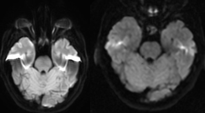 |
MUSE reconstruction of multishot DW-EPI is not limited by SNR at 0.5T
Ally Klassen1, James Rioux2,3, Chris Bowen3, Curtis Wiens4, Sharon Clarke5,6, and Steven Beyea3,7
1Biomedical Engineering, Dalhousie University, Halifax, NS, Canada, 2BIOTIC / Biomedical Translational Imaging Centre, Nova Scotia Health, Halifax, NS, Canada, 3Physics and Atmospheric Science, Dalhousie University, Halifax, NS, Canada, 4Synaptive Medical, Toronto, ON, Canada, 5Diagnostic Radiology, Dalhousie University, Halifax, NS, Canada, 6Diagnostic Radiology, Nova Scotia Health, Halifax, NS, Canada, 7BIOTIC / Biomedical Translational Imaging Centre, IWK Health, Halifax, NS, Canada Keywords: Low-Field MRI, Susceptibility, Susceptibility distortion Multi-shot echo planar imaging (EPI) can be used to reduce spatial distortion and improve spatial resolution in diffusion-weighted imaging; however, patient motion between shots can result in large intra-shot phase differences and increased artifact. It is unknown whether multiplexed sensitivity encoding (MUSE) can be applied to reduce these phase differences in DW-EPI images acquired at low field due to the inherently lower SNR. In this work we evaluate the performance of MUSE in DW-EPI data acquired at 0.5T, in terms of SNR and ghost-to-noise ratio (GNR) as a function of signal averaging, and demonstrate that multi-shot DW-EPI is feasible. |
17:28 |
0310.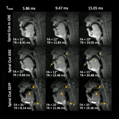 |
Artifact reduction for real-time spiral MRI using out-in sampling at 0.55T
Jeffery Wong1, Prakash Kumar1, Krishna S. Nayak1, and Ye Tian1
1Ming Hsieh Department of Electrical and Computer Engineering, University of Southern California, Los Angeles, CA, United States Keywords: Low-Field MRI, Low-Field MRI, Speech, RT-MRI We demonstrate reduced artifacts for real-time MRI of speech production at 0.55T with a gradient echo sequence using spiral out-in sampling, and constrained image reconstruction. This approach avoids banding artifacts experienced by spiral bSSFP sequences, and exhibits reduced blurring artifact compared to a spiral out gradient echo sequence at the same readout duration. |
| 17:36 | 0311.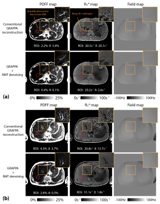 |
Multi-Coil Multi-Contrast Random Matrix Theory-Based Denoising for Liver Fat and R2* Quantification at 0.55T
Shu-Fu Shih1,2, Zhaohuan Zhang1,2, Bilal Tasdelen3, Ecrin Yagiz3, Sophia X. Cui4, Xiaodong Zhong4, Krishna S. Nayak3, and Holden H. Wu1,2
1Radiological Sciences, David Geffen School of Medicine, University of California Los Angeles, Los Angeles, CA, United States, 2Bioengineering, University of California Los Angeles, Los Angeles, CA, United States, 3Ming Hsieh Department of Electrical and Computer Engineering, University of Southern California, Los Angles, CA, United States, 4MR R&D Collaborations, Siemens Medical Solutions USA, Inc., Los Angles, CA, United States Keywords: Low-Field MRI, Low-Field MRI Liver fat and R2* quantification has been extensively validated at 1.5T and 3T. Recently, there is renewed interest in lower-field MRI because of potential advantages such as a larger bore and lower costs. However, it is challenging to acquire images with sufficient signal-to-noise ratio for accurate fat and R2* quantification at 0.55T. Previous random matrix theory (RMT)-based approaches leverage Gaussian noise characteristics to markedly reduce the noise without compromising the tissue signal. In this work, we investigated a multi-coil multi-contrast RMT-based denoising approach which is compatible with parallel imaging and we demonstrated improved liver fat and R2* quantification at 0.55T. |
| 17:44 | 0312.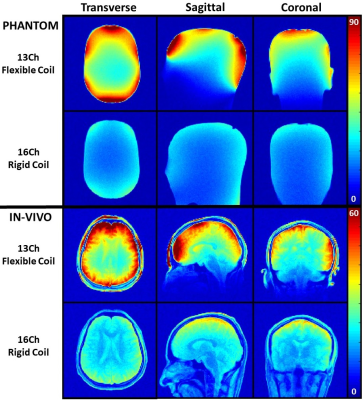 |
A Flexible Coil Based on a Cable Conductor for 0.55T Head Imaging
Bili Wang1, Jerzy Walczyk1, Mary Bruno1, Mahesh Keerthivasan2, Robert Rehner3, and Ryan Brown1
1Radiology, Center for Advanced Imaging Innovation and Research (CAI2R), New York University, Grossman School of Medicine, New York City, NY, United States, 2Siemens Medical Solutions USA Inc., Malvern, PA, United States, 3Siemens Healthcare GmbH, Erlangen, Germany Keywords: Low-Field MRI, Brain Flexible coils have the potential to improve SNR over rigid coils but have been rarely applied to head imaging. We built a flexible 13-channel coil for 0.55T based on RG-223 cable loops arranged with dodecahedral geometry. The coil closely fit the head and provided 30-50% SNR gain in the ventricles and significant image quality improvements over a conventional rigid coil. While the current minimalistic prototype coil showed promising performance, practical advances, such as an insulating layer that can be easily sanitized and a structure to immobilize the head, must be integrated to make the coil user friendly. |
| 17:52 | 0313.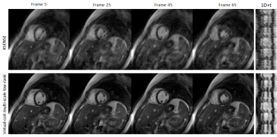 |
Virtual coil concept with multi-scale low-rank for real-time cardiac MR at 0.55T
Gastao Cruz1, Jesse Hamilton1, Evan Cummings1, Yuchi Liu1, Vikas Gulani1, and Nicole Seiberlich1 1Department of Radiology, University of Michigan, Ann Arbor, MI, United States Keywords: Heart, Low-Field MRI Real-time MR applications are often highly undersampled due to the high temporal resolution required. Here, we propose a novel linear forward model combining virtual coils and multi-scale low-rank to enable highly accelerated real-time cardiac MR. The virtual coil concept leverages Hermitian symmetry, whereas multi-scale low-rank attempts to model real-time motion as a combination of subspaces across multiple image scales. Experiments in five healthy subjects demonstrate a considerable improvement in image quality over conventional iterative SENSE, enabling real-time cardiac imaging at 0.55T. |
 Back to Meeting Home
 Back to the Program-at-a-Glance
The International Society for Magnetic Resonance in Medicine is accredited by the Accreditation Council for Continuing Medical Education to provide continuing medical education for physicians.
 Back to Meeting Home
 Back to the Program-at-a-Glance
 View Presentation Video