Oral Session
ML/AI for Acquisition & Reconstruction
ISMRM & ISMRT Annual Meeting & Exhibition • 03-08 June 2023 • Toronto, ON, Canada

Oral Session
ML/AI for Acquisition & Reconstruction
| 08:15 |
0381.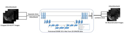 |
Rapid 3D SPirAl Respiratory and Cardiac Self-gated (SPARCS) Cine
Imaging with a DEep learning-based rapid Spiral Image
REconstruction (DESIRE)
Xitong Wang1,
Junyu Wang1,
Shen Zhao1,
Ruixi Zhou2,
Yang Yang3,
and Michael Salerno1
1Stanford Univeristy, Stanford, CA, United States, 2Beijing University of Posts and Telecommunications, Beijing, China, 3Department of Radiology and Biomedical Imaging, University of California, San Francisco, San Francisco, CA, United States Keywords: Machine Learning/Artificial Intelligence, Cardiovascular We developed a rapid 3D self-gated cardiac cine technique, using a variable-density undersampled randomized stack of spiral gradient echo sequence to perform cine evaluation of the whole left ventricle. Our proposed slice-by-slice deep learning-based imaging reconstruction technique for self-gated free-breathing 3D stack of spiral cardiac cine imaging can produce cine images with high temporal (40 ms) and spatial resolution (1.25x1.25x8 mm) within a 20s acquisition time with <1s deep learning inference time. |
| 08:23 |
0382.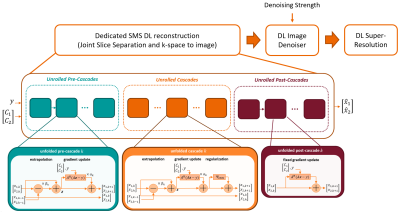 |
Deep Learning Reconstruction for Combined 8-fold Accelerated
Parallel Imaging and Simultaneous Multislice Acquisition
Mahmoud Mostapha1,
Gregor Koerzdoerfer2,
Boris Mailhe1,
Esther Raithel2,
Inge M. Brinkmann2,
Nirmal Janardhanan1,
Maria Ringholz2,
Mariappan S. Nadar1,
and Jan Fritz3
1Siemens Healthineers, Princeton, NJ, United States, 2Siemens Healthcare GmbH, Erlangen, Germany, 3Department of Radiology, NYU Grossman School of Medicine, New York, NY, United States Keywords: Machine Learning/Artificial Intelligence, MSK Combining parallel imaging (PI) and simultaneous multislice (SMS) acceleration realized a clinical 4-fold accelerated 2D TSE MRI of the knee. However, 8-fold acceleration with conventional reconstruction methods suffers from significant image quality degradation. We propose a complete DL approach for combined slice separation and k-space-to-image reconstruction of SMS-PI-accelerated knee MRI with tunable denoising strength and super-resolution image enhancement. The proposed methods enable artifact-free image reconstruction of 8-fold accelerated 2D TSE MR images in multiple planes and with multiple image contrasts. Clinical evaluations suggest equivalence of image quality and detection rates of 8-fold S2P4 DL reconstructions compared to the reference standard. |
| 08:31 |
0383.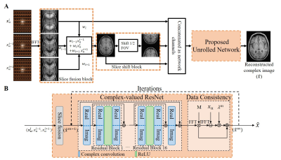 |
Deep Learning based MR Image Reconstruction from Uniformly
Undersampled MR Data
Linfang Xiao1,
Yilong Liu2,
Zhizun Zhang1,
Ruixing Zhu1,
Weijun Chen1,
Zeyao Ma1,
Congying Mao1,
and Ke Wang1
1Hangzhou Weiying Medical Technology Co., Ltd, Hangzhou, China, 2Guangdong-Hongkong-Macau Institute of CNS Regeneration, Key Laboratory of CNS Regeneration (Ministry of Education), Jinan University, Guangzhou, China Keywords: Machine Learning/Artificial Intelligence, Image Reconstruction MR Image reconstruction of uniformly undersampled data often relies on prior information estimated from additional calibration data, leading to compromised acquisition efficiency and flexibility. Here, we propose a joint multi-slice deep learning strategy for MR image reconstruction from uniformly undersampled data with complementary undersampling across adjacent slices. Specifically, we design a slice fusion block to fully exploit the structural and phase similarity in adjacent slices and a slice shift block to further suppress the aliasing artifacts introduced by uniform undersampling. Consequently, the proposed strategy enables accurate MR image reconstruction for both image magnitude and phase without additional calibration information. |
| 08:39 |
0385.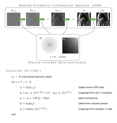 |
Image reconstruction based on a physics-informed reverse
diffusion model trained with magnitude-only data
Tobias Wech1,
Oliver Schad1,2,
and Jonas Kleineisel1
1Department of Diagnostic and Interventional Radiology, University Hospital Würzburg, Würzburg, Germany, 2Experimental Physics 5, University of Würzburg, Würzburg, Germany Keywords: Machine Learning/Artificial Intelligence, Image Reconstruction
We trained a
score-based generative diffusion model with cardiac MR
images, which allows generating new, randomized instances of
the given data distribution. By conditioning each step of
the underlying reverse time stochastic differential equation
with a physics-informed data consistency step, undersampled
MR data can be reconstructed. An initial estimation of the
complex phase, which slowly transfers into the actual phase
of the image, allows to train the diffusion model with
magnitude data only. The approach was evaluated in fast
spiral dynamic cardiac MRI at 1.5T, where it provided
superior SNR-levels compared to alternative acceleration
techniques. |
| 08:47 |
0386.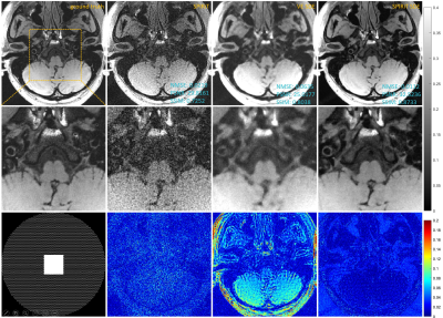 |
SPIRiT Diffusion: SPIRiT-driven Score-Based Generative Modeling
for Vessel Wall imaging
Chentao Cao1,2,
Zhuo-Xu Cui1,
Jing Cheng1,
Sen Jia1,
Hairong Zheng1,
Dong Liang1,3,
and Yanjie Zhu1 1Shenzhen Institutes of Advanced Tech- nology, Chinese Academy of Sciences, Shenzhen, China, 2University of Chinese Academy of Sciences, Beijing, China, 3Pazhou Lab, Guangzhou, China Keywords: Machine Learning/Artificial Intelligence, Machine Learning/Artificial Intelligence, SPIRiT, Diffusion Models, Vessel Wall Imaging Diffusion model is the most advanced method in image generation and has been successfully applied to MRI reconstruction. However, the existing methods do not consider the characteristics of multi-coil acquisition of MRI data. Therefore, we give a new diffusion model, called SPIRiT-Diffusion, based on the SPIRiT iterative reconstruction algorithm. Specifically, SPIRiT-Diffusion characterizes the prior distribution of coil-by-coil images by score matching and characterizes the k-space redundant prior between coils based on self-consistency. With sufficient prior constraint utilized, we achieve superior reconstruction results on the joint Intracranial and Carotid Vessel Wall imaging dataset. |
| 08:55 |
0387.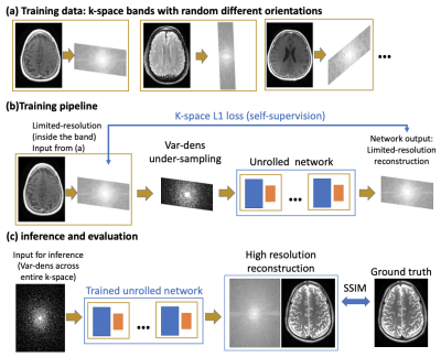 |
K-band: A self-supervised strategy for training deep-learning
MRI reconstruction networks using only limited-resolution data
Han Qi1,
Frederic Wang1,
Alfredo De Goyeneche1,
Michael Lustig1,
and Efrat Shimron1 1Electrical Engineering and Computer Sciences, University of California, Berkeley, Berkeley, CA, United States Keywords: Machine Learning/Artificial Intelligence, Image Reconstruction, MRI reconstruction, self-supervised, deep learning Although Deep learning (DL) techniques are powerful for MRI reconstruction, their development is hindered by the need for large training datasets. We propose a self-supervised method for training DL reconstruction networks using only limited-resolution data, acquired in k-space “bands”, which are generally easier to acquire than variable-density full-resolution data. Although the network is trained using low-resolution data, during inference it can reconstruct high-resolution images. Comprehensive experiments demonstrate that k-band is robust to various acceleration factors, outperforms two other methods trained on low-resolution data, and obtains comparable performance with SSDU and MoDL, while also reducing the need for full-resolution data. |
| 09:03 |
0388.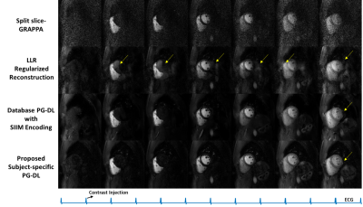 |
Database-Free Zero-Shot Deep Learning Reconstruction for
Highly-Accelerated Free-Breathing Perfusion CMR
Omer Burak Demirel1,2,
Chi Zhang1,2,
Burhaneddin Yaman1,2,
Toygan Kilic1,2,
Steen Moeller2,
Chetan Shenoy3,
Sebastian Weingärtner4,
Tim Leiner5,
and Mehmet Akçakaya1,2
1Electrical and Computer Engineering, University of Minnesota, Minneapolis, MN, United States, 2Center for Magnetic Resonance Research, University of Minnesota, Minneapolis, MN, United States, 3Department of Medicine (Cardiology), University of Minnesota, Minneapolis, MN, United States, 4Delft University of Technology, Delft, Netherlands, 5Department of Radiology, Mayo Clinical, Rochester, MN, United States Keywords: Machine Learning/Artificial Intelligence, Machine Learning/Artificial Intelligence Myocardial perfusion CMR is used to functionally assess coronary artery disease. However, its resolution and coverage remain limited and require rapid imaging .At high accelerations for whole-heart coverage and high spatio-temporal resolution, conventional reconstructions suffer from noise and aliasing artifacts. Physics-guided deep learning (PG-DL) reconstruction has shown improved image quality in fast MRI, but its application to perfusion CMR has been limited due to substantial differences in breathing and contrast uptakes among subjects. In this work, we tackle these challenges by adopting subject-specific self-supervised PG-DL that does not require a training database for simultaneous multi-slice accelerated myocardial perfusion CMR. |
| 09:11 |
0389.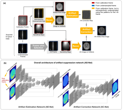 |
Deep Reconstruction Framework with Self-calibration Mechanisms
(DEISM) for Accelerated CEST Imaging
Jianping Xu1,
Tao Zu1,
Yi-Cheng Hsu2,
Yi Sun2,
and Yi Zhang1
1Key Laboratory for Biomedical Engineering of Ministry of Education, Department of Biomedical Engineering, College of Biomedical Engineering & Instrument Science, Zhejiang University, Hangzhou, Zhejiang, China, Hangzhou, China, 2MR Collaboration, Siemens Healthcare Ltd., Shanghai, China, Shanghai, China Keywords: Machine Learning/Artificial Intelligence, Image Reconstruction The widespread clinical adoption of chemical exchange saturation transfer (CEST) imaging has been hampered by its prolonged scan time, due to multiple data acquisitions over the varying saturation offset frequencies. In this work, we utilize the artifact suppression algorithm and propose an effective deep reconstruction framework with self-calibration mechanisms (DEISM). The DEISM method was validated on brain tumor patients at 3T. In conjunction with deep-learning multi-coil image reconstruction and data-driven artifact suppression mechanisms, DEISM can provide reliable reconstructions of highly accelerated CEST data, yielding superior performance compared to state-of-the-art methods. |
| 09:19 |
0390.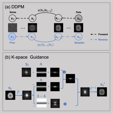 |
MRI reconstruction using DDPM with sparsely sampled k-space as
guidance
Wei Jiang1,
Yang Gao1,
and Hongfu Sun1
1University of Queensland, Brisbane, Australia Keywords: Machine Learning/Artificial Intelligence, Image Reconstruction, diffusion models, compressed sensing, MRI Recovering MR images from partially acquired k-space data can reduce scan time, leading to significant applications. Deep learning approaches have been proposed to train neural networks using under-sampled and full-sampled image pairs in a supervised manner. However, these supervised models usually have poor generalizability when deployed to acquisitions different from the training datasets. Here, we propose an unsupervised Denoising Diffusion Probabilistic Model (DDPM) capable of not only generating random high-fidelity MR images but also reconstructing images corresponding to k-space data from arbitrary acquisition patterns. |
| 09:27 |
0391.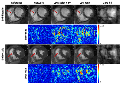 |
Undersampling reconstruction of ferumoxytol-enhanced cardiac
cine MRI using a spatiotemporal neural network
Chang Gao1,2,
Zhengyang Ming1,2,
Kim-Lien Nguyen1,2,
Xiaodong Zhong3,
and John Paul Finn1,2
1Department of Radiological Sciences, University of California Los Angeles, Los Angeles, CA, United States, 2Department of Physics and Biology in Medicine, University of California Los Angeles, Los Angeles, CA, United States, 3MR R&D Collaborations, Siemens Medical Solutions USA, Inc., Los Angeles, CA, United States Keywords: Machine Learning/Artificial Intelligence, Image Reconstruction Ferumoxytol can support high quality SGE cardiac cine with blood-myocardial CNR similar to SSFP cine, but free of off-resonance artifact. Much work is ongoing to accelerate the acquisition of multi-slice, breath held cardiac cine. Deep learning-based reconstruction methods can accelerate the image acquisition and reconstruction but needs a large amount of data to train. When compared with compressed sensing and low rank reconstructions, our network showed sharper images and higher consistency with the reference and significantly better quantitative evaluation metrics. We showed that a network trained with non-contrast images could generalize to accelerated ferumoxytol-enhanced cardiac cine MRI with 10x acceleration. |
| 09:35 |
0392.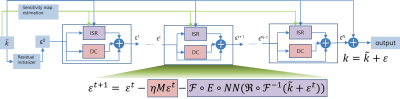 |
An end-to-end residual learning VarNet for under-sampled MRI
reconstruction
Lijun Zhang1 and
Sha Wang1
1Research and Development Center, Canon Medical Systems (China), Beijing, China Keywords: Machine Learning/Artificial Intelligence, Image Reconstruction Model driven deep learning reconstruction methods usually utilize residual learning within single cascade which is composed by a neural network and a data consistency module, here we propose a model based end-to-end residual learning variational network(E2E-ResVarNet), a k-space residual is passed through cascades, then added to acquired under-sampled k-space after the last cascade output. It was demonstrated that the image quality is significantly improved at 4x/6x/8x acceleration factors trained with brain data set. |
| 09:43 |
0393.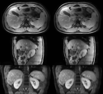 |
MRI-movienet: fast motion-resolved 4D MRI reconstruction
exploiting space-time-coil correlations without k-space data
consistency
Victor Murray1,
Syed Siddiq1,
Ramin Jafari1,
Can Wu1,
and Ricardo Otazo1,2
1Department of Medical Physics, Memorial Sloan Kettering Cancer Center, New York, NY, United States, 2Department of Radiology, Memorial Sloan Kettering Cancer Center, New York, NY, United States Keywords: Machine Learning/Artificial Intelligence, Radiotherapy Motion-resolved 4D MRI enables free-breathing imaging and access to important physiological information. However, long reconstruction times for 4D MRI techniques like XD-GRASP have restricted routine clinical use. Even with unrolled convolutional networks, reconstruction enforcing data consistency in a high-dimensional space is still long. This work presents a deep learning approach named MRI-movienet that exploits spatial-time-coil correlations without enforcing data consistency to enable 2-fold scan acceleration compared to XD-GRASP and 4D reconstruction in less than 2 seconds. MRI-movienet uses the intrinsic separation into static and dynamic components to avoid hallucinations. MRI-movienet high performance will promote 4D MRI for routine clinical use. |
09:51 |
0394.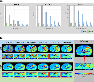 |
Single breath-hold full abdominal T1 mapping using a CNN based
short inversion-recovery sampling technique
Eze Ahanonu1,
Ute Goerke2,
Kevin Johnson3,
Brian Toner4,
Diego Martin5,
Vibhas Deshpande6,
Ali Bilgin1,3,7,
and Maria Altbach3,7
1Department of Electrical and Computer Engineering, The University of Arizona, Tucson, AZ, United States, 2Siemens Healthineers, Tucson, AZ, United States, 3Department of Medical Imaging, The University of Arizona, Tucson, AZ, United States, 4Applied Math Program, The University of Arizona, Tucson, AZ, United States, 5Department of Radiology, Houston Methodist Hospital, Houston, TX, United States, 6Siemens Healthineers, Austin, TX, United States, 7Department of Biomedical Engineering, The University of Arizona, Tucson, AZ, United States Keywords: Machine Learning/Artificial Intelligence, Quantitative Imaging Comprehensive liver evaluation with T1 mapping requires full abdominal coverage with sufficiently high spatial resolution for detection of pathology. Existing methods for abdominal T1 mapping are only able to achieve partial coverage, primarily limited by the breath hold and the time required to sample the T1 recovery curve (T1RC) for accurate T1 estimation. We present a radial Look-Locker T1 mapping framework which utilizes short T1RC sampling combined with deep learning based T1 estimation to achieve full abdominal coverage within a single 20s breath hold period. |
| 09:59 |
0395.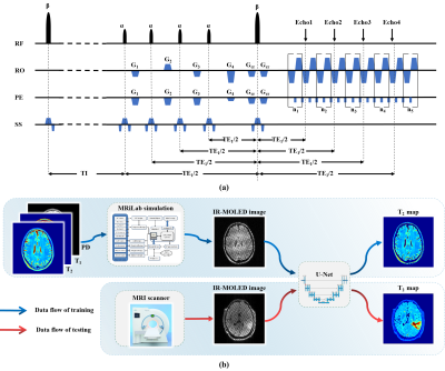 |
Single-shot T2-FLAIR mapping via inversion recovery multiple
overlapping-echo acquisition and deep neural network
reconstruction
Yanhong Lin1,
Qinqin Yang1,
Wenhua Geng1,
Haitao Huang1,
Jianfeng Bao2,
Shuhui Cai1,
Zhong Chen1,
and Congbo Cai1
1Department of Electronic Science, Fujian Provincial Key Laboratory of Plasma and Magnetic Resonance, Xiamen University, Xiamen, China, 2Department of Magnetic Resonance Imaging, The First Affiliated Hospital of Zhengzhou University, Zhengzhou, China Keywords: Quantitative Imaging, Quantitative Imaging T2-weighted imaging via conventional FLAIR sequence can suppress the signal of cerebrospinal fluid (CSF), making it easier for identifying long T2 lesions in the vicinity of the CSF. But it was usually used for qualitative analysis because of its inevitable time-consuming acquisition. In this study, we applied inversion recovery overlapping-echo acquisition together with deep learning-based reconstruction to achieve ultra-fast T2-FLAIR mapping. In vivo results from a healthy volunteer and two glioma patients demonstrate the good accuracy and robustness of our proposed method. |
|
10:07 |
0384.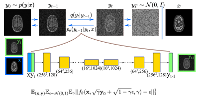 |
Conditional Denoising Diffusion Probabilistic Models for Inverse
MR Image Recovery
Mahmut Yurt1,
Batu Ozturkler1,
Kawin Setsompop1,2,
Shreyas Vasanawala2,
John Pauly1,
and Akshay Chaudhari2,3 1Department of Electrical Engineering, Stanford University, Stanford, CA, United States, 2Department of Radiology, Stanford University, Stanford, CA, United States, 3Department of Biomedical Data Science, Stanford University, Stanford, CA, United States Keywords: Machine Learning/Artificial Intelligence, Machine Learning/Artificial Intelligence High-resolution, multi-contrast magnetic resonance imaging (MRI) protocols are required for accurate clinical diagnoses, but are limited by long scan times. Recovering high-quality, multi-contrast images from low-quality accelerated acquisitions is a promising approach to mitigate this limitation. Prior studies have demonstrated deep-learning for tasks such as contrast synthesis, image super-resolution, and image reconstruction. However, each of these tasks requires different architectures and training paradigms. Motivated by these challenges, we introduce a unified conditional denoising diffusion probabilistic model (DDPM) for inverse MR image recovery. Experiments performed on three image recovery tasks demonstrate that DDPMs achieve superior performance compared to prior state-of-the-art approaches. |
 Back to Meeting Home
 Back to the Program-at-a-Glance
The International Society for Magnetic Resonance in Medicine is accredited by the Accreditation Council for Continuing Medical Education to provide continuing medical education for physicians.
 Back to Meeting Home
 Back to the Program-at-a-Glance
 View Presentation Video