Oral Session
Validation of Quantitative Imaging Techniques
ISMRM & ISMRT Annual Meeting & Exhibition • 03-08 June 2023 • Toronto, ON, Canada

Oral Session
Validation of Quantitative Imaging Techniques
13:45 |
1253.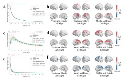 |
Association analysis of age-dependent changes in R1map at the
brain region level with gene expression patterns
Xiang Chen1 and
Xiaoyong Zhang1 1Fudan University, Shanghai, China Keywords: Quantitative Imaging, Quantitative Imaging Quantitative MRI can observe biologically distinct microstructural processes that shows great potential in aging research. In this work, we studied the trajectories of R1map changes in aging brain and found R1map regions with significant changes in different age groups (young, middle-aged, and elderly groups). Graph theoretical analysis of covariance networks revealed global clustering coefficient of youth group is higher than that of middle-aged and elderly groups. Association analysis of spatial gene expression patterns with changes in R1map at the brain region level reveal that the Kyoto Encyclopedia of Genes and Genomes (KEGG) pathway is mainly enriched in various neurodegenerative diseases. |
| 13:53 |
1254.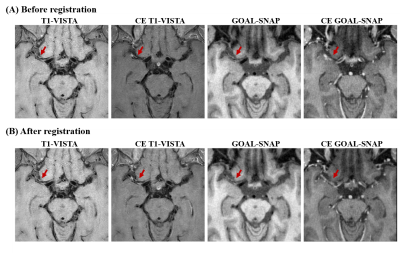 |
In-vivo T1 mapping for quantitative evaluation of intracranial
atherosclerotic plaques
Jiaqi Dou1,
Xiaoming Liu2,3,
Yajie Wang1,
Ziming Xu1,
Jing Wang2,3,
and Huijun Chen1
1Center for Biomedical Imaging Research, Tsinghua University, Beijing, China, 2Department of Radiology, Union Hospital, Tongji Medical College, Huazhong University of Science and Technology, Wuhan, China, 3Hubei Province Key Laboratory of Molecular Imaging, Wuhan, China Keywords: Quantitative Imaging, Atherosclerosis T1 mapping could provide more reliable quantitative measurements of plaques than traditional T1w MR images. In this study, the feasibility of in vivo quantitative T1 mapping of intracranial plaque had been demonstrated for the first time using a 3D SNAP with golden angle radial k-space sampling (GOAL-SNAP) sequence. Symptomatic patients showed significantly lower intraplaque T1 values than asymptomatic patients (1695.21 ± 493.23 vs. 2241.76 ± 300.28 ms, p=0.045). Quantifying plaque T1 values and enhancement through 3D GOAL-SNAP T1 mapping images is promising for characterizing the symptomatic plaques, serving as one potential tool for further plaque analysis. |
| 14:01 |
1255.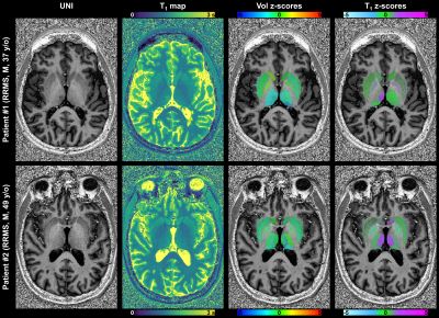 |
Subject-specific detection of macro- and micro-structural
alterations of deep gray matter nuclei using submillimeter 7T
quantitative MRI
Gian Franco Piredda1,2,3,
Alexandre Cabane4,5,
Samuele Caneschi1,6,7,
Tom Hilbert1,6,7,
Gabriele Bonanno8,9,10,
Thomas Troalen11,
Jean-Philippe Ranjeva4,5,
Ludovic de Rochefort4,5,
David Seiffge12,
Martina Goeldlin12,
Robert Hoepner12,
Roland Wiest9,13,
Piotr Radojewski9,13,
Tobias Kober1,6,7,
Arnaud Le Troter4,5,
and Bénédicte Maréchal1,6,7
1Advanced Clinical Imaging Technology, Siemens Healthineers International AG, Lausanne, Switzerland, 2Human Neuroscience Platform, Fondation Campus Biotech Geneva, Geneva, Switzerland, 3CIBM-AIT, École Polytechnique Fédérale de Lausanne (EPFL), Lausanne, Switzerland, 4Aix Marseille Univ, CNRS, CRMBM, Marseille, France, 5AP-HM, CHU Timone, Pôle d'Imagerie Médicale, CEMEREM, Marseille, France, 6Department of Radiology, Lausanne University Hospital and University of Lausanne, Lausanne, Switzerland, 7LTS5, École Polytechnique Fédérale de Lausanne (EPFL), Lausanne, Switzerland, 8Advanced Clinical Imaging Technology, Siemens Healthineers International AG, Bern, Switzerland, 9Translational Imaging Center (TIC), Swiss institute for Translational and Entrepreneurial Medicine, Bern, Switzerland, 10Magnetic Resonance Methodology, Institute of Diagnostic and Interventional Neuroradiology, University of Bern, Bern, Switzerland, 11Siemens Healthcare SAS, Saint-Denis, France, 12Department of Neurology, University Hospital Bern, Inselspital, University of Bern, Bern, Switzerland, 13Support Center for Advanced Neuroimaging, Institute for Diagnostic and Interventional Neuroradiology, Inselspital, University of Bern, Bern, Switzerland Keywords: Quantitative Imaging, Tissue Characterization, Ultra-high field MRI High-resolution 7T MRI allows to directly visualize deep gray matter nuclei (DGN), especially within the thalamus, and building reference ranges of volumes and relaxation times for these structures is of clinical relevance. Methods to automatically segment DGN at 7T have been recently proposed. In this study, we segmented DGN from a cohort of 132 healthy subjects scanned with the MP2RAGE at 7T to obtain both T1-weighted images and T1 maps. Reference ranges of volumes and T1 values were established and proved valuable in revealing both macro- and micro-structural tissue alterations in selected cases of patients with neurodegeneration. |
|
14:09 |
1256.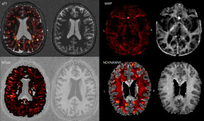 |
Personalized quantitative MRI multiparameter abnormality maps
provide correlates of disability in multiple sclerosis patients
Xinjie Chen1,2,3,
Sabine Schädelin1,2,3,
Po-Jui Lu1,2,3,
Mario Ocampo-Pineda1,2,3,
Matthias Weigel 1,2,3,4,
Muhamed Barakovic1,2,3,
Esther Ruberte1,2,3,
Alessandro Cagol1,2,3,
Bénédicte Maréchal5,
Tobias Kober5,
Jens Kuhle2,3,
Ludwig Kappos2,3,
Lester Melie-Garcia1,2,3,
and Cristina Granziera1,2,3
1Translational Imaging in Neurology (ThINK) Basel, Department of Biomedical Engineering, Faculty of Medicine, University Hospital Basel and University of Basel, Basel, Switzerland, 2Department of Neurology, University Hospital, Basel, Switzerland, 3Research Center for Clinical Neuroimmunology and Neuroscience Basel (RC2NB), University Hospital Basel and University of Basel, Basel, Switzerland, 4Division of Radiological Physics, Department of Radiology, University Hospital Basel, Basel, Switzerland, 5Advanced Clinical Imaging Technology, Siemens Healthineers International AG, Lausanne, Switzerland Keywords: Quantitative Imaging, Multiple Sclerosis We performed an extensive assessment of the clinical relevance of a method that we had previously developed, which provides personalized quantitative MRI abnormality maps of individual multiple sclerosis (MS) patients. Specifically, we assessed the relationships between quantitative T1 (qT1), myelin water fraction (MWF), neurite density index (NDI), magnetization transfer saturation (MTsat) abnormality maps and clinical disability in a cohort of 102 MS patients and 98 healthy subjects. We found that qT1 and NDI alterations in white matter lesions were strongly related to patients' clinical disability, supporting the use of those personalized maps for patient stratification and follow-up in clinical practice. |
|
14:17 |
1257.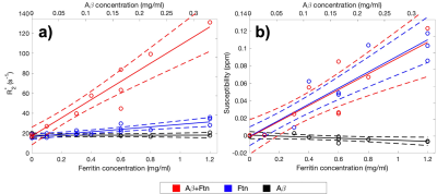 |
The impact of amyloid-β and ferritin on ultra-high-field R2* and
quantitative susceptibility mapping
Jierong Luo1,
James Everett2,
Jane Donnelly1,3,
Festus Slade1,4,5,
Neil Telling2,
and Joanna F Collingwood1,5
1School of Engineering, University of Warwick, Coventry, United Kingdom, 2School of Pharmacy and Bioengineering, Keele University, Stoke-on-Trent, United Kingdom, 3Feinberg School of Medicine, Northwestern University, Chicago, IL, United States, 4Department of Chemistry, University of Warwick, Coventry, United Kingdom, 5Warwick Centre for Doctoral Training in Analytical Science, University of Warwick, Coventry, United Kingdom Keywords: Quantitative Imaging, Contrast Mechanisms, MR Microscopy, Quantitative Susceptibility Mapping, R2*, Iron, Ferritin, Amyloid plaques, Alzheimer's Disease Amyloid plaques, an established hallmark of Alzheimer's disease, are often demonstrably associated with iron deposits post-mortem. Previous T2*-weighted and phase information from ultra-high-field MR microscopy and clinical MRI has shown the potential to detect amyloid deposits in vivo and ex vivo. We investigated the relative contributions to contrast from amyloid-β (Aβ) aggregates with and without ferritin-bound iron in vitro, for R2* and quantitative susceptibility maps (QSM) with 86 μm isotropic resolution at 9.4T. We also demonstrated the quantitative signal evolution with the formation of amyloid aggregates, and the correlation of the signals with the Aβ and ferritin content. |
| 14:25 |
1258.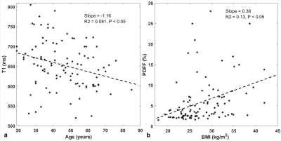 |
Sex-specific changes and association in multiparametric MRI
measurement at 3T in adult livers
Chia-Ying Liu1,
Chikara Noda2,
Rob van der Geest3,
Bruno Triaire4,
Yoshimori Kassai4,
David A Bluemke5,
and Joao Lima2 1Canon Medical Systems Corporation, Ellicott City, MD, United States, 2Division of Cardiology, School of medicine, Johns Hopkins University, Baltimore, MD, United States, 3Department of Radiology, Leiden University Medical Center, Leiden, Netherlands, 4Canon Medical Systems Corporation, Tochigi, Japan, 5Department of Radiology, University of Wisconsin, Madison, WI, United States Keywords: Quantitative Imaging, Liver, parametric mapping; PDFF Imaging biomarkers derived from multiparametric MRI have been investigated for the evaluation of diffuse liver disease. We aimed to determine the sex-specific correlation of MRI parameters with age and BMI, and to evaluate the association between multiparametric MRI parameters. 100 study participants without known hepatic disease were prospectively enrolled. 3 T MRI including T1, T2 and T1ρ mapping and proton density fat fraction (PDFF) and R2* maps were acquired. Multiparametric MRI measures have sex-specific age and BMI dependency. Relaxometry mapping indices could be associated with PDFF. PDFF was significant associated with R2* and T1ρ, but not to T1 or T2. |
| 14:33 |
1259.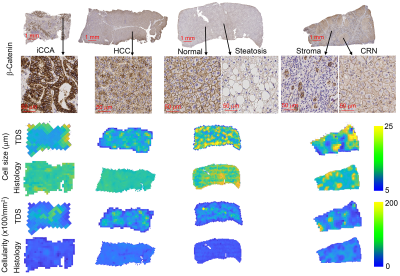 |
Histological Validation of MR Cell Size and Cellularity Imaging
with Human Liver Specimens
xiaoyu jiang1,
John Gore2,
and Junzhong Xu3
1Radiology, Vanderbilt University Medical Center, nashville, TN, United States, 2Vanderbilt University Medical Center, nashville, TN, United States, 3Vanderbilt University Medical Center, Brentwood, TN, United States Keywords: Quantitative Imaging, Liver, microstructure; diffusion; inflammation The size and cellularity of hepatocytes, their variations and changes over time, are fundamental characteristics of liver tissues, and measurements of cell sizes and cellularities may have high clinical significance but currently can be obtained only by liver biopsy. We quantified the microstructures of human liver specimens with different liver diseases, including normal liver tissues, cirrhosis, steatosis, hepatocellular carcinoma (HCC), cirrhotic regenerative nodules (CRN), and intrahepatic cholangiocarcinoma (iCCA), using MR cell size imaging ex vivo. The accuracy of MR-derived cell sizes and cellularities were evaluated by comparisons to histology. |
| 14:41 |
1260.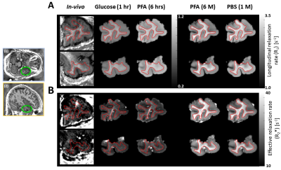 |
Diffusion and relaxometry study of an excised temporal lobe from
a drug-resistance epilepsy patient using in vivo and ex vivo MRI
Francisco Javier Fritz1,
Jan Malte Oeschger1,
Ora Ohana2,
Thomas Sauvigny3,
and Siawoosh Mohammadi1
1Institute for Systems Neuroscience, University Medical Center Hamburg-Eppendorf, Hamburg, Germany, 2Institute of Molecular and Cellular Cognition, University Medical Center Hamburg-Eppendorf, Hamburg, Germany, 3Department of Neurosurgery, University Medical Center Hamburg-Eppendorf, Hamburg, Germany Keywords: Quantitative Imaging, Ex-Vivo Applications, In-vivo to ex-vivo translation; Diffusion MRI; Relaxometry To learn and validate MRI-based microstructure models, MRI from fixed ex-vivo tissue samples can be compared with its histological counterpart. However, translating these models to in-vivo MRI requires thorough characterisation of MR-contrast changes between in-vivo and fixed ex-vivo measurements (e.g., due to fixation). By preforming this characterisation on an epilepsy patient’s freshly excised temporal lobe, we found that diffusion parameters changed strongly whereas relaxometry parameters remained almost unchanged. Our findings provide a missing link between in-vivo and fixed ex-vivo MRI that in future can facilitate in-vivo application of MRI-based microstructure mapping like estimating iron and myelin from relaxometry parameters. |
|
14:49 |
1261.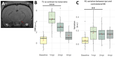 |
Quantitative multiparametric MRI describes neuromelanin-linked
pathogenesis in the AAV-hTyr rat model of Parkinson’s disease
Jean-Baptiste Pérot1,
Mathieu D Santin1,2,
Anthony Ruze1,2,
Lucas Soustelle3,
Sana Rebbah1,
Laura Mouton1,
Romain Valabregue1,2,
Miquel Vila4,
and Stéphane Lehéricy1,2
1Sorbonne Université, Institut du Cerveau - Paris Brain Institute - ICM, Inserm, CNRS, APHP, Hôpital de la Pitié Salpêtrière, Paris, France, 2Center for NeuroImaging Research (CENIR), Paris, France, 3Aix Marseille Univ, CNRS, CRMBM, Marseille, France, 4Neurodegenerative Diseases Research Group, Vall d'Hebron Research Institute (VHIR), Autonomous University of Barcelona, Barcelona, Spain Keywords: Quantitative Imaging, Multi-Contrast Imaging of the neuromelanin (NM) has recently developed as a relevant biomarker of Parkinson’s disease (PD). Here, we present a longitudinal, quantitative, multiparametric study on the AAV-hTyr rat model of PD with NM accumulation in the right substantia nigra. Our protocol allowed to obtain NM-sensitive image, as well as R1, R2* and qMT in a single session. Longitudinal acquisition on AAV-hTyr rats showed inverted U-shaped curve of NM-MRI contrast-to-noise ratio, suggesting NM accumulation followed by neurodegeneration. R1, R2*, MPF and motor symptoms support this hypothesis. Our work may help to understand the pathogenesis of this PD model and identify biomarkers. |
| 14:57 |
1262.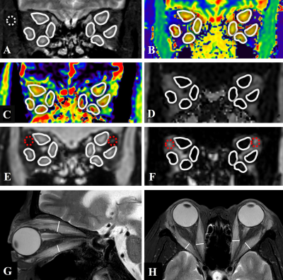 |
Evaluation of Activity of Graves’ Orbitopathy by Multiparameter
Orbital MRI
Xinyi Gou1,
Xiuying Zhang2,
Jianxiu Lian3,
Xiaofang Xu3,
Zhenyu Piao4,
Lingli Zhou2,
Jingyi Cheng1,
Chuhan Chen1,
Lei Chen1,
Ke Jiang3,
Jin Cheng1,
Linong Ji2,
and Nan Hong1 1Department of Radiology, Peking University People’s Hospital, Beijing, China, 2Department of Endocrinology, Peking University People’s Hospital, Beijing, China, 3Philips Healthcare, Beijing, China, 4Department of Ophthalmology, Peking University People’s Hospital, Beijing, China Keywords: Quantitative Imaging, MR Value The evaluation in activity of Graves’ orbitopathy (GO) has important clinical significance for the treatment decision making and prognosis prediction for GO patients. Some previous studies have a limited comprehensive consideration that GO could involve almost entire orbital region, leading to complex changes in MR quantitative parameter, such as T1, T2, and fat fractions. In this study, we established a combined model including MRI quantitative parameters concerning multiple tissues of eyes and multiple sequences to distinguish active GO. And it was better than using a certain parameter alone to evaluate GO activity. |
| 15:05 |
1263.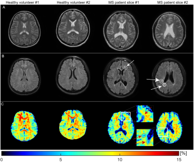 |
Fast macromolecular proton fraction imaging based on spin-lock
Jian Hou1,
Yurui Qian1,
Baiyan Jiang2,
Xiang Fan3,
Winnie Chiu-Wing Chu1,
Tiffany Y. So1,
and Weitian Chen1
1Department of Imaging and Radiology, The Chinese University of Hong Kong, Hong Kong SAR, Hong Kong, 2Illuminatio Medical Technology Limited, Hong Kong SAR, Hong Kong, 3Peking University Shenzhen Hospital, Shenzhen, China Keywords: Quantitative Imaging, Magnetization transfer Macromolecular proton fraction (MPF) represents the relative amount of semi-solid macromolecules involved in magnetization transfer with free water protons. In this work, we reported a novel MPF quantification method based on spin-lock for rapid MPF mapping. The total scan time for 3D brain MPF measurement can be achieved within five minutes. We demonstrated the proposed method via simulation, phantom and in vivo experiments. |
|
15:13 |
1264.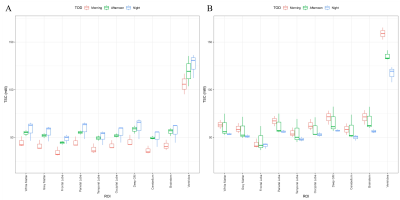 |
Time-of-Day Analysis of Brain Sodium TSC Maps
Cameron E Nowikow1,2,
Paul Polak3,
and Michael D Noseworthy1,2,4,5
1School of Biomedical Engineering, McMaster University, Hamilton, ON, Canada, 2Imaging Research Centre, St. Joseph's Healthcare, Hamilton, ON, Canada, 3Department of Radiology, Children's Hospital Colorado Anschutz Medical Campus, Aurora, CO, United States, 4Electrical and Computer Engineering, McMaster University, Hamilton, ON, Canada, 5Department of Radiology, McMaster University, Hamilton, ON, Canada Keywords: Quantitative Imaging, Non-Proton, Sodium, Variability, Brain, Circadian, Human Previous investigations into brain sodium imaging have avoided the potential of variability due to circadian effects. Thus, in a pilot study we investigated whether time-of-day contributes to tissue sodium concentration (TSC) variance. Three TSC maps were acquired from 7 subjects, at three different times of day (8:00, 16:00, 22:00). Each TSC map was segmented into 10 ROIs before being analyzed using ANOVA with SNR and signal linewidth as added covariates. Time-of-day was a significant source of variance as was spectral linewidth. Between subject variance and SNR were not significant factors in the model. |
| 15:21 |
1265.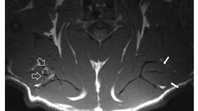 |
Assessment of Rotator Cuff Muscle Fat and Fibrosis Using
Quantitative UTE Magnetization Transfer Imaging and Modeling
Arya A Suprana1,2,
Qingbo Tang1,3,
Elisabeth Orozco3,4,
Hyungseok Jang1,
Saeed Jerban1,
Jiang Du1,2,5,
Yajun Ma1,
and Eric Y Chang1,5
1Department of Radiology, University of California, San Diego, La Jolla, CA, United States, 2Department of Bioengineering, University of California, San Diego, La Jolla, CA, United States, 3Research Service, VA San Diego Healthcare System, San Diego, CA, United States, 4Department of Orthopedic Surgery, University of California, San Diego, La Jolla, CA, United States, 5Radiology Service, Veterans Affairs San Diego Health Care System, San Diego, CA, United States Keywords: Quantitative Imaging, Muscle At present, there is a lack of available tools to non-invasively evaluate rotator cuff muscle fibrosis. Prior studies have suggested that magnetization transfer measurements may be useful for quantifying collagen when acquired with an ultrashort echo time technique. However, the increased presence of fat after muscle injury may confound these measurements. In this study, a rat model of chronic massive rotator cuff tearing was used with UTE-T1 and UTE-MT mapping without and with fat suppression to show that muscle collagen and fibrosis could be quantified. |
| 15:29 |
1266.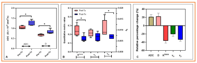 |
Evaluation of tumor physiology after single-fraction irradiation
in a mouse model of pancreatic ductal adenocarcinoma using mpMRI
Ramesh Paudyal1,
James Rusell1,
Eve LoCastro1,
Carl C. Lekaye1,
Joseph O. Deasy1,
John L. Humm1,
and Amita Shukla-Dave1,2
1Medical Physics, Memorial Sloan Kettering Cancer Center, New York, NY, United States, 2Radiology, Memorial Sloan Kettering Cancer, New York, NY, United States Keywords: Quantitative Imaging, Preclinical Pancreatic ductal adenocarcinoma (PDAC) is the leading cause of cancer deaths worldwide. Quantitative multiparametric magnetic resonance imaging (mpMRI) allows for measuring the tumor's physiological characteristics, such as cellularity and vascularity/permeability. This study aimed to evaluate tumor physiology using mpMRI after single-fraction irradiation in a mouse model. The results demonstrated the changes in the functional status of mpMRI metrics. They were validated with in vivo histology markers of tumor perfusion (Hoechst 33342) and tissue morphology (Hematoxylin and eosin staining). |
| 15:37 |
1267.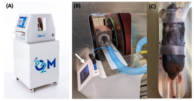 |
Whole Body Mouse EPR Oxygen Imaging of Implanted Beta Cell
Replacement Devices
Mrignayani Kotecha1,
Navin Viswakarma1,
Safa Hameed1,
Eliyas Siddiqui1,
Feya Epel1,
Cherie Stabler2,
Minglin Ma3,
and Boris Epel4
1Oxygen Measurement Core, O2M Technologies, LLC, Chicago, IL, United States, 2Department of Biomedical Engineering, University of Florida, Gainesville, IL, United States, 3Cornell University, Ithaca, NY, United States, 4Radiation and Cellular Oncology, The University of Chicago, Chicago, IL, United States Keywords: Quantitative Imaging, Electron Paramagnetic Resonance, Oxygen Imaging, EPR imaging, Type I diabetes, cell encapsulation devices Beta-cell replacement therapy remains the only approach with a clinical proof-of-concept that demonstrates long-term insulin independence can be achieved in type 1 diabetic (T1D) patients. The major challenge for beta cell replacement devices is to keep cells viable by avoiding hypoxia until vascularization is established. We hypothesize that by performing oxygen imaging and controlling the oxygenation of the devices early on, we can achieve better survival of beta cells and, consequently, better T1D reversal. EPROI is a quantitative oxygen imaging method that was used for obtaining pO2 maps in three different beta cell replacement devices in this study. |
 Back to Meeting Home
 Back to the Program-at-a-Glance
The International Society for Magnetic Resonance in Medicine is accredited by the Accreditation Council for Continuing Medical Education to provide continuing medical education for physicians.
 Back to Meeting Home
 Back to the Program-at-a-Glance
 View Presentation Video