Oral Session
Late Breaking
ISMRM & ISMRT Annual Meeting & Exhibition • 03-08 June 2023 • Toronto, ON, Canada

| 16:00 | 0284.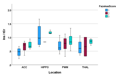 |
An investigation of the glial biomarker myo-Inositol as a marker of brain inflammation in elderly patients with and without delirium.
Franklyn A Howe1, Daniel Richardson2, Lauren Binnie3, Uzma Khan4, Philip Rich4, Daniel HJ Davis5, Atticus H Hainsworth2, and Jeremy Isaacs3
1Neurosciences Research Section, St George's, University of London, London, United Kingdom, 2Neurosciences Research Section, St George's, University of London, LONDON, United Kingdom, 3Dept Neurology, St George's University Hospital Foundation Trust, LONDON, United Kingdom, 4Neuroradiology, St George's University Hospital Foundation Trust, LONDON, United Kingdom, 5MRC Unit for Lifelong Health and Ageing, University College London, LONDON, United Kingdom
Myo-Inositol is sometimes considered a marker of brain inflammation and studies indicate mI (mI/tCr) increases with age up to the 6th decade. We performed 1H MRS in four brain regions of very elderly hospitalised patients (n=25, aged 67-90 yrs) of which 12 had delirium. Elevated mI/tCr was associated with increasing Fazekas score for white matter damage (p<0.001) in all regions. In addition mI/tCr and mI/NAA showed negative correlation with age (p<0.001). There were no significant differences between patients with or without delirium. Decreased mI/tCr due to reduced glial cell numbers or dysfunction may coincide with increased mI/tCr associated with inflammation.
|
| 16:08 | 0285.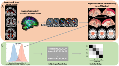 |
The sequence of regional structural disconnectivity due to chronic active and inactive multiple sclerosis lesions
Ceren Tozlu1, Emily Olafson1, Keith Jamison1, Emily Demmon2, Ulrike Kaunzner2, Melanie Marcille2, Nicole Zinger2, Nara Michaelson2, Neha Safi2, Thanh Nguyen1, Susan Gauthier2, and Amy Kuceyeski1 1Department of Radiology, Weill Cornell Medicine, NYC, NY, United States, 2Department of Neurology, Weill Cornell Medicine, NYC, NY, United States
In this study, we showed that structural disconnectivity due to T2 FLAIR lesions in the ventral attention and subcortical networks, in particular within the supramarginal and putamen, occurs earlier in the sequence of disability (i.e., mild to severe) in multiple sclerosis. We also showed that paramagnetic rim lesion (PRL)-based structural disconnectivity in motor-related regions occurs earlier in the disability event sequence, followed by non-PRL-based structural disconnectivity in the caudate and postcentral gyrus. T2 FLAIR lesion-based structural disconnectivity in subcortical regions, including the thalamus, occurs earliest in the sequence of cognitive impairment (i.e. preserved to impaired).
|
| 16:16 | 0286.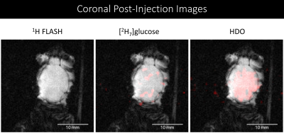 |
Metabolic Imaging of Alzheimer’s Disease Related Inflammation Using Deuterium MRI
Matthew E Merritt1, Mario Chang1, Tara Hawkinson1, Anna Rushin1, Vikram Kodibagkar2, James Bankson3, and Ramon Sun1 1Biochemistry and Mol. Bio., University of Florida, Gainesville, FL, United States, 2School of Biological and Health Systems Engineering, Arizona State University, Tempe, AZ, United States, 3Imaging Physics, MD Anderson Cancer Center, Houston, TX, United States
Alzheimer’s Disease (AD) is characterized by increased inflammation and increased glucose utilization as detected by FDG-PET. Using a new deuterium MRI method based on a [D7]glucose tracer, we can image the brain of a mouse model of AD with in plane resolution approaching 1 mm. Mass spectrometry imaging of the same brains shows characteristic changes in protein associated glycans that correlate well with immunohistochemical staining for inflammation. We believe this new research pipeline can provide powerful new insights into AD pathophysiology.
|
| 16:24 | 0287.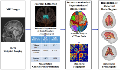 |
Depression Related Structural Changes Based on 3D Brain MRI and Neural Fingerprint Assisted Segmentation
Liya Wang1, Peijia Xu1, Ting Xue1, Xiuliang Deng2, Jiayi Wu1, and Xingkui Xue3 1Radiology, Affiliated Longhua People's Hospital, the Third School of Clinical Medicine, Southern Medical University, Shenzhen, China, 2Clinical Psychology, Affiliated Longhua People's Hospital, the Third School of Clinical Medicine, Southern Medical University, Shenzhen, China, 3Central lab, Affiliated Longhua People's Hospital, the Third School of Clinical Medicine, Southern Medical University, Shenzhen, China
This study focused on the slight quantitative changes of brain structures based on the high-resolution brain MRI and automatic segmentation with neural fingerprint technology for whole-brain of depressive patients. We found the volume changes of specific regional sub-structures, which were related to emotion and spirit, such as the cingulate gyrus, cuneiform lobe, hippocampus, et al before clinical diagnosis. Depression revealed the gender difference. The male depressions were more sensitive to hippocampus, while female’s were more sensitive to entorhinal cortex. It might indicate ovarian hormone fluctuations modulate women’s susceptibility to stress, brain structure/function, and inflammatory activity and reactivity.
|
| 16:32 | 0288.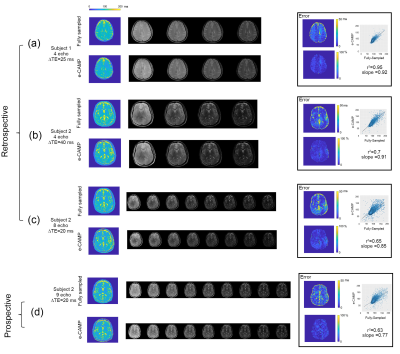 |
A novel method to calculate standardized quantitative maps from a single T2-weighted image: A feasibility study.
Nahla M H Elsaid1, Hemant D Tagare1,2, and Gigi Galiana1,2 1Radiology and Biomedical Imaging, Yale School of Medicine, New Haven, CT, United States, 2Biomedical Engineering, Yale University, New Haven, CT, United States
T2w imaging could assist in the early diagnosis of multiple sclerosis (MS). However, these scans are inherently qualitative, and variable intensities in the images depend on hardware and acquisition parameters. On the other hand, standardized quantitative T2 maps can serve as a sensitive biomarker of MS and the longitudinal assessment of the disease progression. In this abstract, we present the first results demonstrating that calculating T2 maps from vendor T2w sequence is feasible, which could allow for an earlier diagnosis of MS disease.
|
| 16:40 | 0289.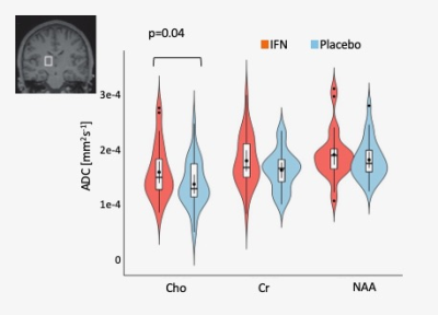 |
Using Diffusion-weighted MR spectroscopy to quantify age- and inflammation-associated changes in glial morphology in-vivo
Eva Periche-Thomas1, Itamar Ronen2, Claire MacIver1, Dmitri Sastin1, Jonathan Underwood1, James Coulson1, Helena Leach1, Francesca Branzoli3, Mara Cercignani1, and Neil Harrison1
1CUBRIC, Cardiff University, Cardiff, United Kingdom, 2CISC, Brighton and Sussex Medical School, Brighton, United Kingdom, 3ICM-Paris Brain Institute, Paris, France Age-associated cognitive decline and neurodegenerative disorders have been linked to altered immune function and inflammation, and particularly to the role of microglia. Current methods for imaging (micro)glia in humans are limited to TSPO-PET, a costly and invasive technique with poor cellular specificity. Here we acquired diffusion weighted (DW) MRS in 15 young and 15 older participants, each scanned twice, after Interferon-1beta or placebo. Results supported our prior hypotheses of selective effects of Interferon-1beta on the apparent diffusion coefficient of thalamic Choline (p<0.040), providing support for DW-MRS as a novel in-vivo method for quantifying glial morphology. |
| 16:48 | 0290.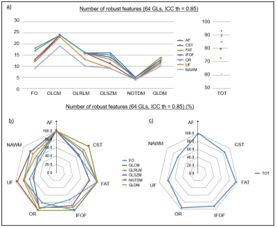 |
Robustness analysis of QSM radiomic features in patients with multiple sclerosis
Cristiana Fiscone1, Leonardo Rundo2, Alessandra Lugaresi1,3, David Neil Manners1, Kieren Allinson4, Elisa Baldin5, Raffaele Lodi1,6, Caterina Tonon1,6, Claudia Testa6,7, Mauro Castelli8, and Fulvio Zaccagna9,10,11
1Department of Biomedical and Neuromotor Sciences, University of Bologna, Bologna, Italy, 2Department of Information and Electrical Engineering and Applied Mathematics, University of Salerno, Salerno, Italy, 3UOSI Riabilitazione Sclerosi Multipla, IRCCS Istituto delle Scienze Neurologiche di Bologna, Bologna, Italy, 4Department of Histopathology, Cambridge University Hospitals NHS Foundation Trust, Cambridge, United Kingdom, 5Epidemiology and Statistics Unit, IRCCS Istituto delle Scienze Neurologiche di Bologna, Bologna, Italy, 6Functional and Molecular Neuroimaging Unit, IRCCS Istituto delle Scienze Neurologiche di Bologna, Bologna, Italy, 7Department of Physics and Astronomy, University of Bologna, Bologna, Italy, 8NOVA Information Management School, Universidade NOVA de Lisboa, Lisbon, Portugal, 9Department of Imaging, Cambridge University Hospitals NHS Foundation Trust, Cambridge, United Kingdom, 10Department of Radiology, University of Cambridge, Cambridge, United Kingdom, 11Radcliffe Department of Medicine, Investigative Medicine Division, University of Oxford, Oxford, United Kingdom
This work evaluates the reliability of radiomic features in healthy controls and patients with multiple sclerosis using MR Quantitative Susceptibility Mapping (QSM), to guide the identification of future potential biomarkers. To ensure reproducibility, we assessed the non-lesioned parenchyma. Feature robustness was evaluated against the number of grey levels and echo times, measuring the Intraclass Correlation Coefficient (ICC). More than 65% of features were robust; different outcomes between regions were interpreted considering their anatomical characteristics (e.g. fibres’ orientation), confirmed by radiomic measurements (e.g. entropy). In future work, we are going to assess characterization and classification potential of those measurements.
|
| 16:56 | 0291.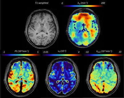 |
A comparison of Gadolinium- and water-based blood-brain barrier dysfunction measures in patients with small vessel disease
Michael S Stringer1, Cameron Manning1, Xingfeng Shao2, Hedok Lee3, Antoine Vallatos4, Hattie Lord1, Carmen Arteaga1, Una Clancy1, Daniela Jaime Garcia1, Maria Valdes Hernandez1, Stewart Wiseman1, Rachel Locherty1, Francesca Chappell1, Rosalind Brown1, Fergus N Doubal1, Ian Marshall1, Helene Benveniste3,5, Michael J Thrippleton1, Danny JJ Wang2, and Joanna M Wardlaw1 1Centre for Clinical Brain Sciences and UK Dementia Research Institute, University of Edinburgh, Edinburgh, United Kingdom, 2Laboratory of FMRI Technology (LOFT), USC Mark & Mary Stevens Neuroimaging and Informatics Institute, Keck School of Medicine, University of Southern California, Los Angeles, CA, United States, 3Department of Anesthesiology, Yale School of Medicine, Yale University, New Haven, CT, United States, 4Glasgow Experimental MRI Centre, School of Psychology and Neuroscience, University of Glasgow, Glasgow, United Kingdom, 5Department of Biomedical Engineering, Yale School of Medicine, Yale University, New Haven, CT, United States
Clinically relevant non-invasive blood-brain barrier (BBB) function imaging techniques are needed to monitor the role of neuroinflammation and endothelial cell dysfunction, particularly in small vessel disease (SVD). Diffusion-weighted ASL measures water exchange rate (kw), a promising endogenous BBB function metric, but has not been assessed in sporadic SVD. We measured kw in SVD patients to explore associations with established Gadolinium-based contrast agent (GBCA) BBB permeability measures and SVD severity. We found only limited associations between kw and GBCA metrics, although patients with more severe SVD tended to have higher kw, reflecting that the methods may probe different mechanisms.
|
| 17:04 | 0292.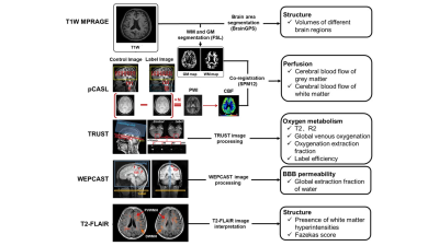 |
Short-term changes in brain structure, perfusion and oxygen metabolism in young adults infected with Omicron: A case control study using MRI
Rui Shen1, Jiachen Liu1, Chenyang Zhao2, Ning Xu1, Shuwan Yu1, Huiyu Qiao1, Zihan Ning1, Hualu Han1, Zixuan Lin3, Hanzhang Lu4, Haiyan Ding1, and Xihai Zhao1
1Center for Biomedical Imaging Research, Department of Biomedical Engineering, School of Medicine, Tsinghua University, Beijing, China, 2Department of Radiology, the Affiliated BenQ Hospital of Nanjing Medical University, Nanjing, China, 3College of Biomedical Engineering & Instrument Science, Zhejiang University, Hangzhou, China, 4Department of Biomedical Engineering, School of Medicine, Johns Hopkins University, Baldimore, MD, United States
We investigated the short-term changes in brain structure, perfusion and oxygen metabolism in young adult patients infected with SARS-COV-2 Omicron but with mild symptoms in China using multimodal MRI. The clinical and brain MR imaging characteristics were compared between patient group (n=98) and healthy control group(n=50). Our findings suggest that there are no significant short-term changes in brain structure, perfusion and oxygen metabolism determined by multimodal MR imaging in young adult patients infected with SARS-CoV-2 Omicron. The potential trend of decline in the volume of cerebral nucleus in the patient group needs further investigation.
|
| 17:12 | 0293.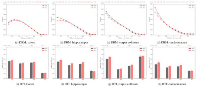 |
Diffusion Bubble Model: A Novel Method For Detecting Neuroinflammation in Mouse Brain With Sanfilippo Syndrome
Erjun Zhang1,2,3, Irene Londono2, Jérémie Fouquet4, Alexey Pshezhetsky5,6, Benjamin De Leener1,2,3,7, and Gregory Lodygensky2,8 1Institute of Biomedical Engineering, Polytechnique Montreal, Montreal, QC, Canada, 2CHU Sainte-Justine University Hospital Center, University of Montreal, Montreal, QC, Canada, 3NeuroPoly Lab, Institute of Biomedical Engineering, Polytechnique Montreal, Montreal, QC, Canada, 4Cerebral Imaging Centre, Douglas Research Center, McGill University, Montreal, QC, Canada, 5Centre Hospitalier Universitaire Sainte-Justine Research Center, University of Montreal, Montreal, QC, Canada, 6Department of Anatomy and Cell Biology, McGill University, Montreal, QC, Canada, 7Department of Computer Engineering and Software Engineering, Polytechnique Montreal, Montreal, QC, Canada, 8Department of Pharmacology, University of Montreal, Montreal, QC, Canada
The Diffusion Bubble Model (DBM) is a new method for detecting neuroinflammation in the brains of mice with Sanfilippo Syndrome. This rare and debilitating disorder primarily affects children and causes progressive neurodegeneration. DBM utilizes diffusion spectrum derived from dMRI to detect brain injuries such as inflammation. It can work in both white and gray matter with limited number of diffusion directions. The study found that brain injuries with inflammation had reduced the fraction of the slow diffusion component and increasing the fraction of the fast diffusion component. The findings may establish non-invasive biomarkers for detecting and evaluating neuroinflammation diseases.
|
| 17:20 | 0294. |
Tracking evolution of multiple sclerosis lesions during medication switch: a quantitative multiparametric 7T MRI approach
Myrte Strik1, Warda T Syeda1, Yasmin Blunck1,2, Vivien Li3,4,5, Rebecca Glarin1, Scott C Kolbe6, Bradford A Moffat1, Leigh A Johnston1,2, Elaine Lui1,7,8, and Trevor J Kilpatrick3,4,5
1Radiology, Melbourne Brain Centre Imaging Unit, University of Melbourne, Melbourne, Australia, 2Biomedical Engineering, University of Melbourne, Melbourne, Australia, 3Neuroimmunology and Remyelination Laboratory, Florey Institute for Neuroscience and Mental Health, Melbourne, Australia, 4Florey Department of Neuroscience and Mental Health, University of Melbourne, Melbourne, Australia, 5Neurology, Royal Melbourne Hospital, Melbourne, Australia, 6Research Partnerships & Translation, RMIT University, Melbourne, Australia, 7Radiology, University of Melbourne, Melbourne, Australia, 8Radiology, Royal Melbourne Hospital, Melbourne, Australia
Multiple sclerosis patients may need to switch disease modifying treatments due to various reasons, with risk of recurrent disease activity during transition period. Identifying patients at risk is challenging. In this pilot study we used a non-conventional quantitative multi-modal 7T MRI approach to assess evolution of lesions when switching medication. We observed altered diffusion, susceptibility and T1 values three months after stopping, and these metrics returning to baseline values three months after starting new medication. Multi-modal quantitative MR techniques at ultra-high field MRI may be more sensitive to detect subtle changes over time to potentially better inform underlying disease activity.
|
| 17:28 | 0295.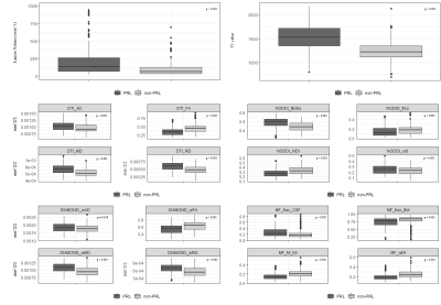 |
ADVANCED DIFFUSION IMAGING METHODS TO ASSESS THE MICROSTRUCTURE OF MULTIPLE SCLEROSIS LESIONS
Anna Stölting1, Colin Vanden Bulcke1,2, Benoit Macq2, and Pietro Maggi1
1Neuroinflammation Imaging Lab, Université catholique de Louvain, Brussels, Belgium, 2ICTEAM Institute, Université catholique de Louvain, Louvain-la-Neuve, Belgium Paramagnetic Rim Lesions (PRL) in Multiple Sclerosis (MS) are associated with increased disability and axonal damage. We examined PRL microstructure using Track Density Imaging (TDI) and four diffusion MRI (dMRI) models, including Microstructure Fingerprinting (MF). When compared to non-PRL, PRL showed larger size, higher T1-values, lower neurite density index and fibre volume fraction on dMRI (p < 0.001) and reduced track density on TDI (p < 0.0001), suggesting impaired axonal density/integrity in PRL. Results obtained with the novel MF model are in line with those obtained with other dMRI models, indicating its potential for studying MS lesion pathology. |
| 17:36 | 0296.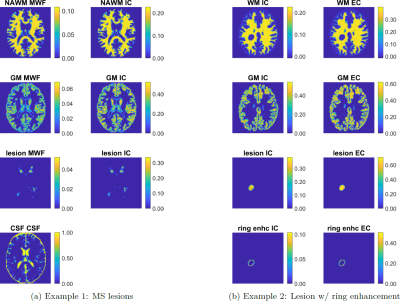 |
MicroCoP: digital Microstructure Correlation Phantoms for benchmarking of multicomponent MRI methods
Sebastian Endt1,2,3,4, Carolin M Pirkl3, Marco Palombo4, and Marion I Menzel1,2,3
1AImotion Bavaria, Technische Hochschule Ingolstadt, Ingolstadt, Germany, 2Technical University of Munich, Munich, Germany, 3GE HealthCare, Munich, Germany, 4Cardiff University Brain Research Imaging Center (CUBRIC), Cardiff University, Cardiff, United Kingdom
We present MicroCoP, a flexible tool for the generation of digital Microstructure Correlation Phantoms that can be used to benchmark multicomponent MRI reconstruction methods. The phantoms provide ground-truth distributions for the MR tissue parameters of your choice. These can be fed into forward simulations and thus provide a pair of ground-truth distributions and resulting signal evolutions to thoroughly evaluate multicomponent MRI reconstruction methods.
|
| 17:44 | 0297.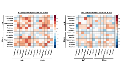 |
Short-term susceptibility correlations are sensitive to disrupted iron dynamics in multiple sclerosis
Jack Reeves1, Fahad Salman1, Michael G Dwyer1,2, Niels Bergsland1, Bianca Weinstock-Guttman3, Sarah Muldoon4, Robert Zivadinov1,2, and Ferdinand Schweser1,2
1Buffalo Neuroimaging Analysis Center, Buffalo, NY, United States, 2Center for Biomedical Imaging at the Clinical Translational Science Institute, University at Buffalo, Buffalo, NY, United States, 3Department of Neurology, Jacobs Comprehensive MS Treatment and Research Center, Buffalo, NY, United States, 4Department of Mathematics, University at Buffalo, Buffalo, NY, United States
Deep gray matter (DGM) iron dyshomeostasis has been linked to neuroinflammation in multiple sclerosis (MS). In the present work, we hypothesized that a novel correlation analysis of magnetic susceptibility co-fluctuations between DGM regions reveals short-term (<1 year) MS-related iron dyshomeostasis that is undetectable with conventional ROI-based methods. Our results showed the correlations-based analysis was more sensitive to MS vs HC group differences than the standard ROI-based approach. A simulation indicated that the MS-related differences may be due to neuroinflammatory-based disruption to normal aging-related iron accumulation. Our methodology may be applied to study neuroinflammatory diseases at short timescales not previously possible.
|
| 17:52 | 0298.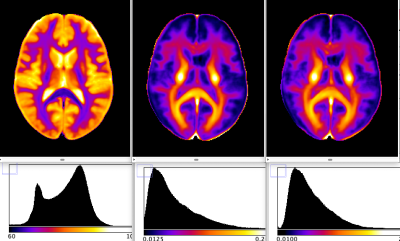 |
A novel atlas of absolute myelin water content: the baseline for studying inflammatory demyelinating diseases
Ana-Maria Oros-Peusquens1, Zaheer Abbas1, Erhan Genc2,3, Christoph Franz2,3, and N. Jon Shah4,5,6 1INM-4, Research Centre Juelich, Juelich, Germany, 2Department of Psychology and Neurosciences, Leibniz Research Centre for Working Environment and Human Factors (IfADo), Dortmund, Germany, 3Faculty of Psychology, Institute for Cognitive Neuroscience, Department of Biopsychology, Ruhr University Bochum, Bochum, Germany, 4JARA-BRAIN - Translational Medicine, Aachen, Germany, 5Department of Neurology, RWTH Aachen University, Aachen, Germany, 6INM-11, JARA, Forschungszentrum Jülich, Juelich, Germany
Quantitative brain atlases can be combined in multiparametric characterisations of the same template voxel with a number of microstructure-sensitive quantities, potentially enabling earlier and more specific characterisation of changes caused by disease. We combine in this study two quantitative atlases, absolute water content and myelin water fraction, to generate a novel atlas describing absolute myelin water content. This is relevant as a baseline for investigating changes in inflammatory diseases involving both demyelination and brain edema. The accompanying relaxometric parameters R2* and R2 are combined in an atlas of the reversible relaxation rate R2’. Fibre tract-specific distributions and correlations are found.
|
 Back to Meeting Home
 Back to the Program-at-a-Glance
The International Society for Magnetic Resonance in Medicine is accredited by the Accreditation Council for Continuing Medical Education to provide continuing medical education for physicians.
 Back to Meeting Home
 Back to the Program-at-a-Glance
 View Presentation Video